
CRUSH SYNDROM and SUDECK'S ATROPHY
Reading File
Now let me search the medical library for both topics in parallel.
Finding Sources
Finding Sources
Finding Sources
Finding Sources
Now let me read the key sections from the most authoritative sources in parallel:
Reading File
Reading File
Reading File
Reading File
Reading File
Now let me get the Pye's section on both topics, and the Rosen's Emergency Medicine section on crush injury, plus the Rockwood section on CRPS:
Reading File
Reading File
Reading File
Reading File
Finding Sources
I now have comprehensive information from multiple authoritative sources. Let me also fetch the CRPS skin changes image and the crush injury image for the response:
Loading Image
Loading Image
I now have all the information needed to compose a detailed, well-structured answer. Here it is:
CRUSH SYNDROME
Definition
Crush syndrome is the systemic manifestation that results from prolonged crushing of muscle tissue and the subsequent release of toxic intracellular contents into the circulation when blood flow is restored (reperfusion). It was first described in victims trapped during the London Blitz in World War II.
- Bailey and Love's Short Practice of Surgery, 28th Ed.
- Rosen's Emergency Medicine, Concepts and Clinical Practice
Pathophysiology
When a large volume of muscle is crushed and then released, a reperfusion injury occurs. The key mechanisms are:
- Rhabdomyolysis - massive muscle destruction releases myoglobin, potassium, phosphate, and uric acid into the circulation.
- Myoglobinuria - myoglobin precipitates in renal tubules (especially in acid urine), causing tubular obstruction and acute tubular necrosis (ATN) - the main cause of renal failure.
- Fluid sequestration - many litres of fluid are trapped in the crushed compartment, causing hypovolemia and renal vasoconstriction further worsening ischaemia.
- Electrolyte disturbances - hyperkalaemia (from dying cells), hypocalcaemia (Ca²⁺ influx into damaged muscle), hyperphosphataemia, and metabolic acidosis (anion-gap type) all develop.
- Vasoactive mediators released from crushed tissue contribute to systemic effects.
- DIC (disseminated intravascular coagulation) can also occur.
The severity increases with the extent of body area crushed. Under extreme conditions crush syndrome can develop within 1 hour; typically it takes 4-6 hours. It occurs in 3-20% of earthquake victims and up to 40% of survivors of multi-storey building collapses.
Causes / Settings
- Earthquakes, mining accidents, battlefield casualties
- Industrial crush injuries
- Prolonged immobility (e.g., drug-induced unconsciousness on a hard floor)
- Vascular occlusion causing large tissue ischaemia (e.g., femoral artery occlusion)
- Untreated compartment syndrome
- Burns (over large surface areas)
Clinical Presentation
Early (on extrication):
- Patient may appear stable initially but deteriorates rapidly after release
- Lacerations, fractures, bruising at the crush site
- Compartment syndrome features
Systemic (after release):
- Hypovolemic shock (fluid sequestration/third spacing)
- Oliguria or anuria - dark, cola/tea-coloured urine (myoglobinuria)
- Dysrhythmias due to severe hyperkalaemia and metabolic acidosis
- Acute kidney injury (AKI) / acute renal failure
Delayed (days later):
- Acute renal failure requiring dialysis
- ARDS (acute respiratory distress syndrome)
- Sepsis
- Ischaemic organ injury
- Multi-organ failure
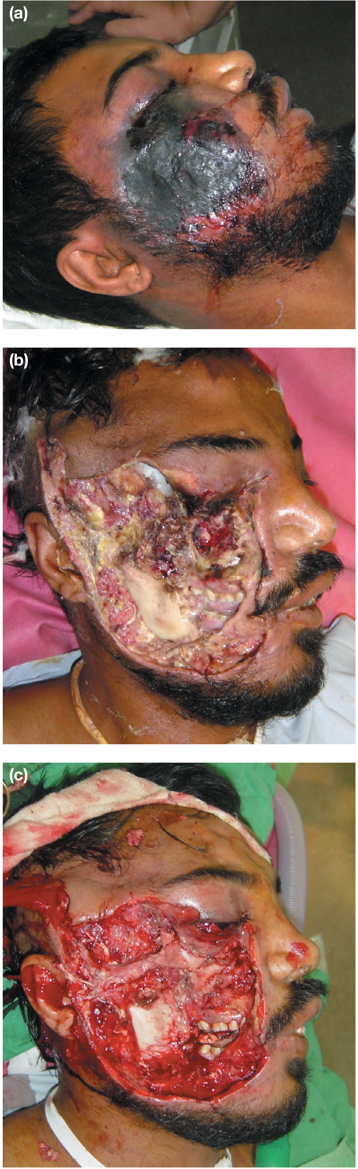
Treatment
The golden rule: begin treatment BEFORE extrication.
Pre-hospital / Field
- Aggressive IV fluid resuscitation (normal saline 1000-1500 mL/h in adults) should begin as soon as the victim is found, before release, to prevent tubular myoglobin precipitation
- Continuous cardiac monitoring (even in confined spaces) for hyperkalaemia-induced dysrhythmias
- Manage hyperkalaemia: insulin + glucose, calcium gluconate, ion exchange resins, β-agonists
- Urinary catheter to measure urine output (target: forced diuresis of up to 8 L/day)
Hospital Management
- Continue aggressive fluid loading (up to 12 L/day in an average adult)
- Mannitol - reduces the reperfusion injury component and promotes osmotic diuresis
- Urinary alkalinisation - sodium bicarbonate to keep urinary pH > 6.5, preventing myoglobin precipitation in tubules (mannitol-alkaline diuresis up to 8 L/day until urine is clear of myoglobin)
- Early fasciotomy - decompresses muscle compartments, prevents severe limb loss
- Caution: A late fasciotomy (>12 hours of entrapment) is likely to cause a massive release of myoglobin and risk introducing infection into dead tissue - it should NOT be performed
- Intensive care with close fluid balance monitoring
- Renal dialysis if AKI develops (anticipating recovery of renal function over several days)
- Amputation of a non-viable limb may prevent renal damage from further myoglobin release
Summary (Bailey & Love's, p. 480)
- Arises as a result of reperfusion
- Acute kidney injury and renal failure from myoglobinuria is a complication
- A late fasciotomy may make things worse, not better
SUDECK'S ATROPHY (Complex Regional Pain Syndrome Type I)
Definition and Nomenclature
Sudeck's atrophy is now classified under Complex Regional Pain Syndrome Type I (CRPS-I), formerly called Reflex Sympathetic Dystrophy (RSD). Other older names include:
- Shoulder-hand syndrome
- Causalgia (CRPS Type II - with a definable nerve lesion)
- Transient osteoporosis
- Acute atrophy of bone
The unifying CRPS classification requires: regional pain and sensory changes following a noxious event, with pain disproportionate to the inciting injury, plus vasomotor/sudomotor/trophic changes.
- Bradley and Daroff's Neurology in Clinical Practice
- Pye's Surgical Handicraft, 22nd Ed.
- Andrews' Diseases of the Skin
Inciting Events
- Fractures (especially around the wrist/distal radius) - 25% of CRPS cases
- Soft tissue injury - ~40% of cases
- Myocardial infarction - ~12%
- Almost any fracture in adults
- Minor trauma, surgery, immobilization
Pathophysiology
The exact cause is unknown. Current understanding:
- An abnormal reflex arc following the sympathetic nervous system pathway, modulated by cortical centres
- Decreased sympathetic outflow to the affected limb
- Autonomic manifestations previously attributed to sympathetic overactivity are now thought to be due to catecholamine hypersensitivity in peripheral tissues
- Significant emotional disturbance and psychological stress at onset are common precipitating factors
- The pain creates a vicious circle: pain → disuse → further bony atrophy → more pain
Clinical Features and Stages
CRPS progresses through three classical stages:
| Stage | Duration | Features |
|---|---|---|
| Stage I (Acute) | Weeks-months | Diffuse burning/throbbing/aching pain, hypersensitivity to touch or cold, localised oedema, vasomotor disturbance (altered skin colour and temperature) |
| Stage II (Dystrophic) | 3-6 months | Progressive soft tissue oedema, skin and periarticular soft tissue thickening, muscle wasting |
| Stage III (Atrophic) | Months-years | Limitation of movement, frozen shoulder, digital contractures, waxy trophic skin changes, brittle ridged nails, severe demineralisation on X-ray |
Characteristic signs on examination:
- Diffuse severe non-segmental pain with cyanosis or mottling
- Increased sweating (hyperhidrosis) and shiny skin
- Swollen, non-articular tissue
- Coldness to touch
- Hypersensitivity to pinprick
- Associated myofascial trigger points and shoulder tendonitis
- Radiograph: diffuse, patchy, "moth-eaten" type osteoporosis (hallmark of Sudeck's)
Motor impairment (weakness, tremor, dystonia) may be present but is NOT required for diagnosis.
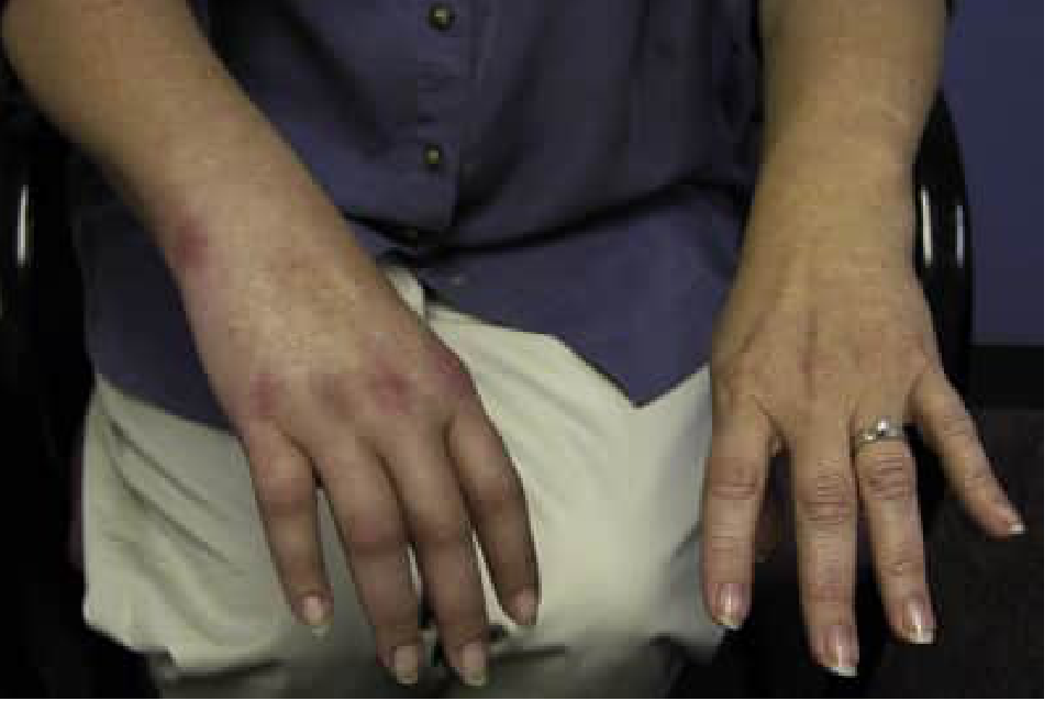
Diagnosis
- Primarily clinical (no single objective diagnostic test for CRPS Type I)
- Bone scintigraphy - most sensitive in Stage I, less useful in later stages
- Autonomic testing - resting sweat output + quantitative sudomotor axon reflex test: 94% sensitive, 98% specific; excellent predictor of response to sympathetic block
- X-ray - patchy periarticular osteoporosis in later stages
- Stellate ganglion block - useful both diagnostically and therapeutically
Treatment
Non-pharmacological
- Vigorous physiotherapy and active movement - the cornerstone; improvement occurs over many months despite pain
- Psychological support (significant emotional component)
Pharmacological
- Bisphosphonates - prevent bone resorption and help with pain control
- Calcium channel blockers (nifedipine, verapamil) - for vasomotor symptoms
- Alpha-blockers (prazosin, phenoxybenzamine)
- Guanethidine (sympatholytic)
- Beta-blockers (propranolol)
- Antidepressants
- Thyrocalcitonin (calcitonin) injections - some evidence of shortening the time course (Pye's, p. 157)
Interventional
- Stellate ganglion block (or regional sympathetic block) - therapeutic and diagnostic; can be repeated if successful
- Sympathectomy - reserved for progressive disease in patients who previously responded to sympathetic block
Key Differences and Comparison
| Feature | Crush Syndrome | Sudeck's Atrophy (CRPS-I) |
|---|---|---|
| Nature | Systemic/metabolic emergency | Regional neurogenic pain syndrome |
| Mechanism | Reperfusion + rhabdomyolysis | Abnormal sympathetic reflex |
| Key organ affected | Kidneys (AKI/ARF) | Bone + skin + joints (affected limb) |
| Timing | Acute (hours after injury) | Subacute to chronic (weeks-months post-injury) |
| X-ray finding | - | Patchy "moth-eaten" osteoporosis |
| Key lab finding | Myoglobinuria, hyperkalaemia, metabolic acidosis | None specific |
| Treatment priority | Aggressive IV fluids + forced diuresis | Physiotherapy + sympathetic blockade |
| Surgery | Early fasciotomy (if <12h) | Sympathectomy (severe progressive cases) |
Sources: Bailey and Love's Short Practice of Surgery 28th Ed. | Rosen's Emergency Medicine | Bradley and Daroff's Neurology in Clinical Practice | Pye's Surgical Handicraft 22nd Ed. | Andrews' Diseases of the Skin
This is a shared conversation. Sign in to Orris to start your own chat.