
Can you tell me the updated boston formulas for acid base disorders?
The Boston rules (also called the Boston criteria) for acid-base compensation are a set of expected compensatory responses for each primary disorder. Here's a quick summary from memory:
- Metabolic acidosis: Expected pCO2 = 1.5 x [HCO3] + 8 (±2) -- Winter's formula
- Metabolic alkalosis: Expected pCO2 = 0.7 x [HCO3] + 21 (±2)
- Respiratory acidosis (acute): HCO3 rises by 1 mEq/L per 10 mmHg rise in pCO2
- Respiratory acidosis (chronic): HCO3 rises by 3.5 mEq/L per 10 mmHg rise in pCO2
- Respiratory alkalosis (acute): HCO3 falls by 2 mEq/L per 10 mmHg fall in pCO2
- Respiratory alkalosis (chronic): HCO3 falls by 5 mEq/L per 10 mmHg fall in pCO2
Searching sources...
Reading File
Searching the Web
Boston rules formulas acid base disorders compensation updated
Now let me search the medical library for acid-base compensation content:
Finding Sources
Finding Sources
Reading File
Reading File
Let me also fetch the acid-base nomogram image and look for Brenner & Rector's table:
Loading Image
Finding Sources
I now have excellent data from multiple authoritative textbooks. Let me compile the full answer.
Here is a complete, textbook-verified summary of the Boston Rules for acid-base compensation:
Boston Rules for Acid-Base Compensation
The Boston rules are six bedside formulas derived from in vivo titration experiments (originally by Schwartz & Relman, 1963 NEJM) that predict the expected compensatory response to each primary acid-base disorder. If the measured value differs from the expected, a mixed disorder is present.
Metabolic Disorders (Respiratory Compensation)
| Primary Disorder | Expected Compensation Formula |
|---|---|
| Metabolic Acidosis | PaCO2 = (1.5 × [HCO3-]) + 8 ± 2 (Winter's formula) |
| OR: PaCO2 decreases 1.2 mmHg per 1 mEq/L fall in HCO3- | |
| OR: Expected PaCO2 ≈ last two digits of pH (quick bedside rule) | |
| Minimum PaCO2 achievable: ~10-15 mmHg | |
| Metabolic Alkalosis | PaCO2 = 0.7 × (HCO3- - 24) + 40 ± 2 |
| OR: PaCO2 increases 0.7 mmHg per 1 mEq/L rise in HCO3- |
Note: A small variation exists in the literature. Some sources write the metabolic alkalosis formula as: expected PaCO2 = (0.7 × HCO3-) + 20. Both are equivalent when baseline HCO3- is assumed to be 24 mEq/L.
Respiratory Disorders (Renal Compensation)
| Primary Disorder | Duration | Expected Compensation |
|---|---|---|
| Respiratory Acidosis | Acute | HCO3- increases 1 mEq/L per 10 mmHg rise in PaCO2 |
| Chronic | HCO3- increases 3.5-4 mEq/L per 10 mmHg rise in PaCO2 | |
| Respiratory Alkalosis | Acute | HCO3- decreases 2 mEq/L per 10 mmHg fall in PaCO2 |
| Chronic | HCO3- decreases 4-5 mEq/L per 10 mmHg fall in PaCO2 |
The chronic respiratory acidosis value is cited as 3.5 mEq/L in some sources and 4.0 mEq/L in others (including Symptom to Diagnosis 4e and Brenner & Rector's The Kidney). The range 3.5-4 is acceptable at the bedside.
Key Principles
- Compensation is never complete - it brings pH toward normal but never fully to 7.40. If pH is normal with an abnormal HCO3- or PaCO2, suspect a mixed disorder.
- If compensation is greater or less than expected, a second primary disorder is present - not just compensation.
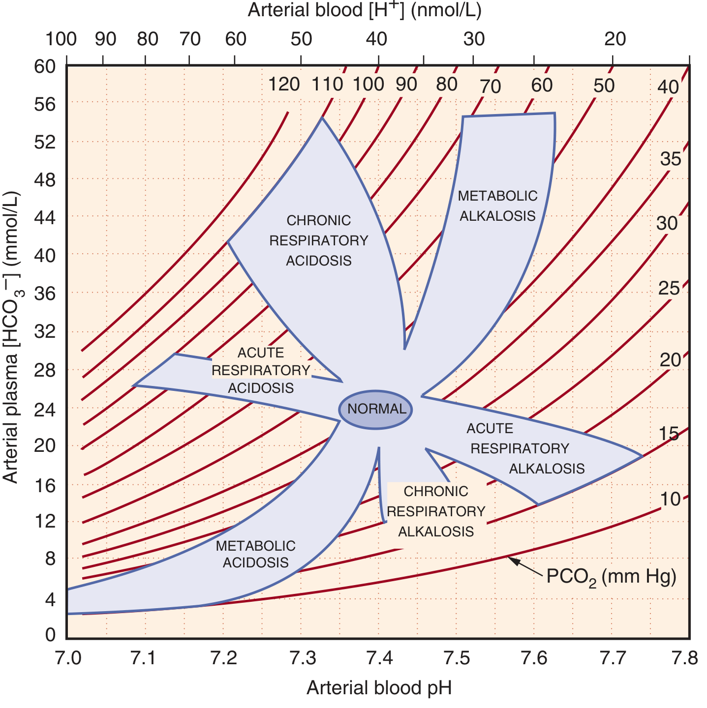
- The acid-base nomogram (shown below from Brenner & Rector) visualizes the 95% confidence bands for each simple disorder. Values falling outside the shaded zones imply a mixed disturbance.
Quick Memory Aid
| Disorder | What compensates | Rule of thumb |
|---|---|---|
| Met. acidosis | Lungs blow off CO2 | CO2 ~ 1.5 x HCO3 + 8 |
| Met. alkalosis | Lungs retain CO2 | CO2 rises ~0.7 per HCO3 rise |
| Resp. acidosis (acute) | Buffers (immediate) | HCO3 up 1 per 10 CO2 |
| Resp. acidosis (chronic) | Kidneys retain HCO3 | HCO3 up 3.5-4 per 10 CO2 |
| Resp. alkalosis (acute) | Buffers (immediate) | HCO3 down 2 per 10 CO2 |
| Resp. alkalosis (chronic) | Kidneys excrete HCO3 | HCO3 down 4-5 per 10 CO2 |
Sources:
- Symptom to Diagnosis: An Evidence Based Guide, 4e (Tables 4-3 and 4-4)
- Brenner & Rector's The Kidney, 2-Volume Set (Table 16.1, Fig. 16.5)
- Harrison's Principles of Internal Medicine, 22e (Winter's equation)
- Frameworks for Internal Medicine (metabolic alkalosis formula)
- Deranged Physiology - Boston vs Copenhagen methods
This is a shared conversation. Sign in to Orris to start your own chat.