
Circadian rhythm full topic mbbs
circadian rhythm physiology MBBS complete notes sleep wake cycle suprachiasmatic nucleus
circadian rhythm suprachiasmatic nucleus diagram sleep wake cycle
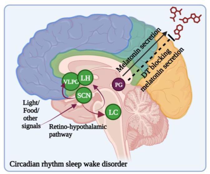
An anatomical diagram of a sagittal section of the human brain illustrating the neurobiological pathways of circadian rhythm regulation. The illustration identifies the Suprachiasmatic Nucleus (SCN) as the master clock, receiving external zeitgebers such as light and food via the retino-hypothalamic pathway. Associated hypothalamic and brainstem structures labeled include the Ventrolateral Preoptic Nucleus (VLPO), Lateral Hypothalamus (LH), and Locus Coeruleus (LC). The diagram highlights the functional relationship between the SCN and the Pineal Gland (PG). It shows that melatonin secretion from the PG is modulated by circadian signaling. Furthermore, the graphic depicts a clinical intervention or physiological state where dopamine (DT) blocks melatonin secretion, which is relevant in the context of circadian rhythm sleep-wake disorders (CRSWDs) and Parkinson's disease. The color-coded cortical regions and deep brain nuclei emphasize the integrated nature of sleep-wake cycles and hormonal output.
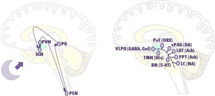
Two sagittal brain schematics illustrating the neuroanatomical pathways of sleep-wake regulation. The left diagram shows the circadian rhythm control system, centered on the suprachiasmatic nucleus (SCN). It depicts neural signaling from the SCN to the paraventricular nucleus (PVN) and preganglionic sympathetic neurons (PSN) in the spinal cord, which subsequently stimulate the pineal gland (PG) for melatonin production. The right diagram illustrates the 'flip-flop switch' of the ascending arousal system. It highlights the inhibitory relationship between the sleep-promoting ventrolateral preoptic nucleus (VLPO), utilizing GABA and galanin, and monoaminergic/cholinergic arousal centers. Labeled nuclei include the tuberomammillary nucleus (TMN, histamine), raphe nuclei (RN, serotonin), locus coeruleus (LC, noradrenaline), perifornical area (PeF, orexin), ventral periaqueductal gray (vPAG, dopamine), and the pedunculopontine and laterodorsal tegmental nuclei (PPT/LDT, acetylcholine). The diagram demonstrates the mutual inhibition between the VLPO and the arousal system that dictates transitions between sleep and wakefulness.
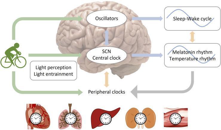
A pathophysiology diagram illustrating the mechanisms of circadian rhythm entrainment by light and physical exercise. The central feature is a human brain model highlighting the Suprachiasmatic Nucleus (SCN) as the 'Central Clock' and secondary 'Oscillators.' These central structures regulate physiological sine-wave rhythms, including the sleep-wake cycle, melatonin secretion, and body temperature. The diagram shows external zeitgebers, specifically light perception/entrainment and physical exercise (represented by a cyclist), providing input to the SCN and peripheral clocks. The 'Peripheral Clocks' are depicted through anatomical illustrations of the heart, lungs, liver, kidneys, and skeletal muscle, each containing a clock face symbol. Bidirectional arrows indicate complex feedback loops between central brain regions, systemic rhythms, and peripheral organ systems, demonstrating how exercise and light synchronize multi-organ biological clocks. This visual is designed for medical education regarding chronobiology and the physiological impact of lifestyle interventions on human homeostasis.
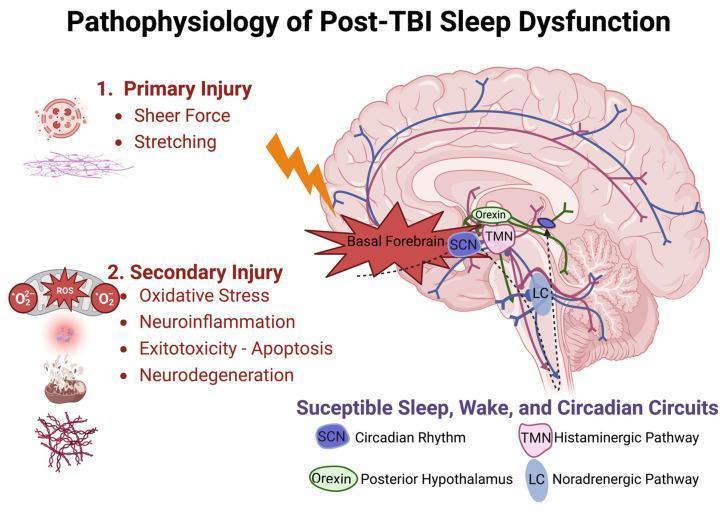
A pathophysiology diagram illustrating the mechanisms of sleep dysfunction following Traumatic Brain Injury (TBI). The central element is a sagittal anatomical diagram of the human brain, highlighting 'Susceptible Sleep, Wake, and Circadian Circuits.' Key neuroanatomical structures indicated include the Suprachiasmatic Nucleus (SCN) for circadian rhythm, the Tuberomammillary Nucleus (TMN) for the histaminergic pathway, Orexin neurons in the posterior hypothalamus, and the Locus Coeruleus (LC) for the noradrenergic pathway. Blue and purple axonal projections are shown extending from these nuclei toward the cortex and brainstem. The diagram categorizes TBI into two phases: 1) Primary Injury, characterized by mechanical 'Sheer Force' and 'Stretching' at the skull base and basal forebrain, and 2) Secondary Injury, involving biochemical cascades such as Oxidative Stress (reactive oxygen species), Neuroinflammation, Excitotoxicity/Apoptosis, and chronic Neurodegeneration. This visual serves as an educational model for how traumatic mechanical disruption and subsequent cellular injury impair the regulatory networks of the sleep-wake cycle.
Circadian Rhythm - Complete MBBS Notes
1. Definition and Terminology
| Term | Meaning |
|---|---|
| Zeitgeber | "Time-giver" - external cue that synchronizes the clock (e.g., light, meals, exercise) |
| Entrainment | Daily resetting of the internal clock to external cues |
| Free-running rhythm | Rhythm that runs without external cues (~24.1 h in humans) |
| Phase advance | Clock shifts earlier than normal |
| Phase delay | Clock shifts later than normal |
| Chronotype | Individual preference for morning ("lark") vs. evening ("owl") activity |
2. The Master Clock: Suprachiasmatic Nucleus (SCN)
- Location: Anterior hypothalamus, bilaterally, just above the optic chiasm, adjacent to the third ventricle
- Size: ~20,000 neurons
- Evidence: Destruction of the SCN renders animals completely arrhythmic. SCN grafts can partially restore rhythmicity in SCN-lesioned animals
- Core (VIP-predominated): receives retinal input; mediates light entrainment
- Shell (AVP-producing): projects to hypothalamic and thalamic targets
3. Light Entrainment Pathway
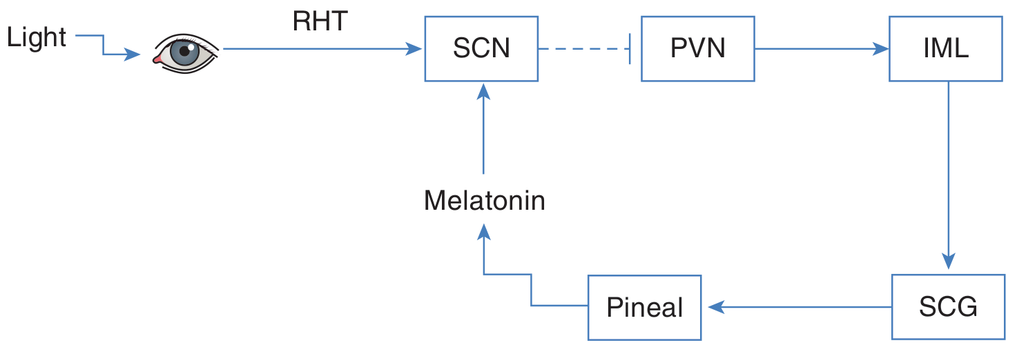
Melanopsin and ipRGCs
- A special subset of retinal ganglion cells (intrinsically photosensitive RGCs) contain melanopsin, a photopigment
- These cells are sensitive to blue light (~480 nm)
- They signal ambient light levels (luminance), not image formation
- They also mediate the pupillary light reflex
- Even in rod/cone-blind mice, circadian entrainment occurs via these cells
4. Molecular Clock Mechanism
Core Negative Feedback Loop
CLOCK + BMAL1 (dimerize)
        ↓  [Transcriptional activators - daytime]
Transcription of Per (PER1, PER2, PER3) and Cry (CRY1, CRY2) genes
        ↓
PER and CRY proteins accumulate → form PER-CRY dimers
        ↓  [Enter nucleus - circadian night]
Inhibit CLOCK-BMAL1 complex → suppress their own transcription
        ↓
PER-CRY proteins slowly degraded (by casein kinase 1ε/δ-mediated phosphorylation → ubiquitination)
        ↓
CLOCK-BMAL1 disinhibited → next cycle begins (~24 h)
Secondary Loop (stabilizing)
- ROR proteins activate BMAL1 transcription
- REV-ERB proteins repress BMAL1 transcription
- These are targets of CLOCK-BMAL1 and are negatively regulated by PER and CRY
- This loop stabilizes and fine-tunes the ~24 h period
Key point: Cell autonomy
5. Melatonin: The Hormone of Darkness
- Source: Pineal pinealocytes
- Synthesis: Serotonin → (N-acetyltransferase) → N-acetylserotonin → (HIOMT) → Melatonin
- Route: Secreted into blood and CSF
- Pattern: High during darkness (peaks ~2 AM), suppressed during daylight
- Receptors in SCN: MT1 and MT2 (GPCRs)
- MT1 activation: inhibits adenylyl cyclase → promotes sleepiness
- MT2 activation: stimulates phosphoinositide hydrolysis → synchronizes light-dark cycle
- Artificial light at night: Delays melatonin secretion → disrupts sleep
- Clinical use: Melatonin and melatonin agonists (e.g., ramelteon) entrain circadian rhythms; useful for jet lag, shift work, non-24-hour sleep-wake disorder
6. SCN Efferent Pathways and Physiological Outputs
- Subparaventricular zone (SPZ) → dorsomedial hypothalamus (DMH)
- DMH → wake-promoting regions: locus coeruleus, lateral hypothalamic nucleus (orexin neurons), ventral tegmental area, dorsal raphe
- SCN → ventrolateral preoptic nucleus (VLPO) → promotes sleep
| Hormone | Circadian Pattern |
|---|---|
| Cortisol (ACTH) | Peak in early morning (4-10 AM); 75% of daily production |
| Melatonin | Peak at ~2 AM, suppressed in daytime |
| Growth hormone | Peak in first hour of NREM sleep |
| TSH | Nocturnal peak |
| Testosterone | Morning peak |
| Body temperature | Lowest at ~4 AM, peaks late afternoon |
7. Sleep-Wake Cycle Regulation: The Two-Process Model
Process S - Homeostatic Sleep Pressure
- Builds during wakefulness; driven by adenosine accumulation in the brain
- Caffeine works by blocking adenosine receptors
- Dissipates during NREM sleep
Process C - Circadian Drive
- Generated by the SCN
- Opposes sleep pressure during daytime → maintains wakefulness
- Falls in evening → allows sleep onset
- The "wake maintenance zone" occurs a few hours before habitual sleep time (when circadian wake drive is still high)
- VLPO neurons (GABA/galanin) - sleep-promoting; inhibit arousal centers
- Arousal centers (locus coeruleus/NE, raphe/5-HT, TMN/histamine, VTA/dopamine, orexin/LH) - mutually inhibit VLPO
- Orexin neurons stabilize wakefulness by reinforcing arousal centers
- Loss of orexin = narcolepsy

8. Phase Response to Light
| Time of light exposure | Effect on clock |
|---|---|
| Daytime | No effect |
| Just after dark | Phase delay (clock shifts later) |
| Just before dawn | Phase advance (clock shifts earlier) |
9. Circadian Rhythm Disorders
A. Delayed Sleep Phase Disorder (DSPD)
- Unable to sleep until very late (e.g., 2-4 AM); cannot wake until 10 AM-12 PM
- Sleep quality normal once asleep
- Common in adolescents ("night owls")
- Treatment: Bright light in morning, evening melatonin, chronotherapy
B. Advanced Sleep Phase Disorder (ASPD)
- Sleep onset very early (7-9 PM); waking very early (3-5 AM)
- Genetic basis: PER2 gene mutation (autosomal dominant) → familial advanced sleep phase syndrome (FASPS)
- Common in elderly
- Treatment: Bright light in evening
C. Non-24-Hour Sleep-Wake Disorder
- Circadian clock not reset daily; period >24 h → progressive drift (a few minutes later each day)
- Common in blind people who lack functional melanopsin-containing RGCs
- Also seen in severe TBI
- Treatment: Tasimelteon (MT1/MT2 agonist)
D. Irregular Sleep-Wake Disorder
- Complete disorganization of the sleep-wake cycle into 3+ irregular episodes per 24 h
- Associated with dementia, brain injury, hypothalamic lesions
- Total sleep normal but fragmented
E. Shift Work Disorder
- 15-25% of workforce; ~10-32% of shift workers develop SWD
- Internal clock conflicts with work schedule
- Consequences: excessive sleepiness at work, insomnia during sleep periods, increased risk of cardiometabolic disease, cancer, GI disorders, mood disorders
- Treatment: Modafinil (for wakefulness), melatonin (to shift sleep timing), bright light therapy
F. Jet Lag
- Transient mismatch after rapid transmeridian travel
- Clock adjusts ~1 hour/day (eastward travel more difficult than westward because it requires phase advance)
10. Clinical Correlations and Exam Points
Cortisol/ACTH Circadian Rhythm
- ACTH secreted in irregular bursts; most frequent in early morning
- 75% of daily cortisol production between 4 AM - 10 AM
- Begins before awakening (not caused by stress of waking)
- If "day" is experimentally lengthened, the adrenal cycle also lengthens but peak ACTH still occurs during the period of sleep
- Biologic clock located in suprachiasmatic nuclei (Ganong's)
Mutations in Clock Genes
| Mutation | Phenotype |
|---|---|
| PER2 (phosphorylation site) | Familial advanced sleep phase |
| Casein kinase 1-delta | Familial advanced sleep phase |
| Knockout of PER or CRY | Arrhythmia (no circadian rhythm) |
Diseases and Circadian Disruption
- Depression: Circadian dysregulation common; morning bright light therapy helps reset SCN
- Bipolar disorder: Circadian rhythm disturbances prominent
- Alzheimer's disease: SCN neurodegeneration → irregular sleep-wake rhythm
- Cancer: Shift work classified as probable carcinogen (IARC Group 2A) - disrupted clock disrupts cell cycle regulation
- Metabolic syndrome: Circadian misalignment causes insulin resistance, obesity
Pharmacological Targets
| Drug | Mechanism | Indication |
|---|---|---|
| Melatonin | MT1/MT2 agonist | Jet lag, insomnia, circadian disorders |
| Ramelteon | MT1/MT2 agonist (selective) | Insomnia, sleep onset |
| Tasimelteon | MT1/MT2 agonist | Non-24-hour sleep-wake disorder |
| Agomelatine | MT1/MT2 agonist + 5-HT2C antagonist | Depression with circadian dysregulation |