
Explanation of boot shaped heart in tetrology of heart on radiography
boot shaped heart tetralogy of Fallot chest X-ray radiography
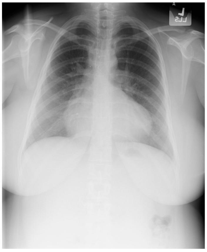
This diagnostic posteroanterior (PA) chest X-ray demonstrates the characteristic findings of Tetralogy of Fallot in a 21-year-old female patient. The image shows a well-centered view with adequate inspiratory effort. The primary finding is an increased cardiothoracic ratio with a distinctive 'boot-shaped' heart, also known as 'coeur en sabot.' This morphology is characterized by an upturned cardiac apex, resulting from significant right ventricular hypertrophy, and a concave pulmonary artery segment. The right heart border is notably prominent, indicating right atrial enlargement. Despite the history of surgical repair (including VSD closure and RV outflow tract reconstruction), these structural changes remain visible. The pulmonary vasculature appears normal, and the lung fields are clear without evidence of opacities, focal lesions, or pleural effusions. Sternal wires and surgical markers from prior cardiac procedures may be visible in the midline. The image serves as a classic educational example of chronic cardiac remodeling in congenital heart disease.
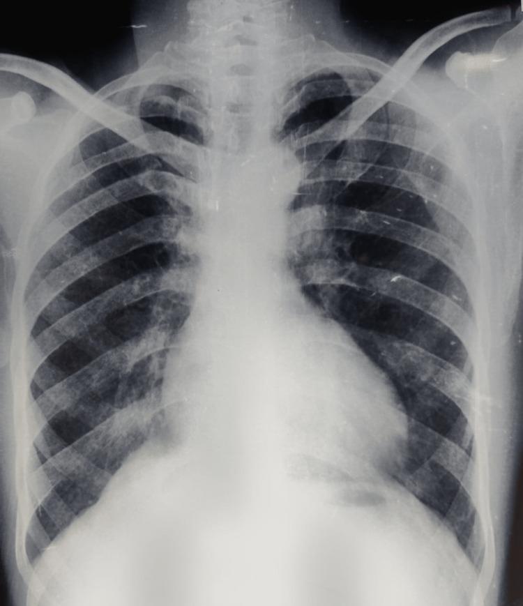
A posteroanterior (PA) chest X-ray of an adult male demonstrating classic radiological features associated with Tetralogy of Fallot (TOF). The cardiac silhouette exhibits mild cardiomegaly and a characteristic 'boot-shaped' heart (coeur en sabot). This morphology is defined by an upturned, rounded cardiac apex, indicating right ventricular hypertrophy, and a concave pulmonary artery segment (pulmonary conus). The lung fields appear relatively oligemic, reflecting decreased pulmonary blood flow typical of right-to-left shunting or pulmonary stenosis. No acute pulmonary infiltrates or pleural effusions are visible. The costophrenic angles are clear. This diagnostic image is intended for medical students and clinicians to identify visual markers of cyanotic congenital heart disease and right ventricular pressure overload in an adult clinical presentation.
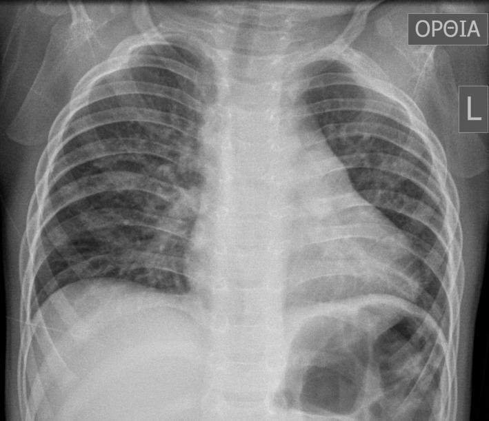
This diagnostic image is an anteroposterior (AP) chest X-ray of a pediatric patient, specifically a 16-month-old male. The image demonstrates a classic 'boot-shaped' heart (coeur en sabot), a hallmark radiographic sign of Tetralogy of Fallot. The cardiac silhouette shows an upturned apex due to right ventricular hypertrophy and a concave main pulmonary artery segment. The lung fields exhibit increased, patchy opacities and markings, particularly prominent in the right lung, which may suggest compensatory pulmonary blood flow or associated bronchial anomalies. The mediastinum appears slightly shifted, and the overall lung volume is clear enough to evaluate the bronchovascular markings. This radiograph serves as a primary educational example of congenital heart disease (CHD) manifestations in pediatric radiology, illustrating the morphological changes to the heart and the secondary effects on pulmonary vascular distribution.
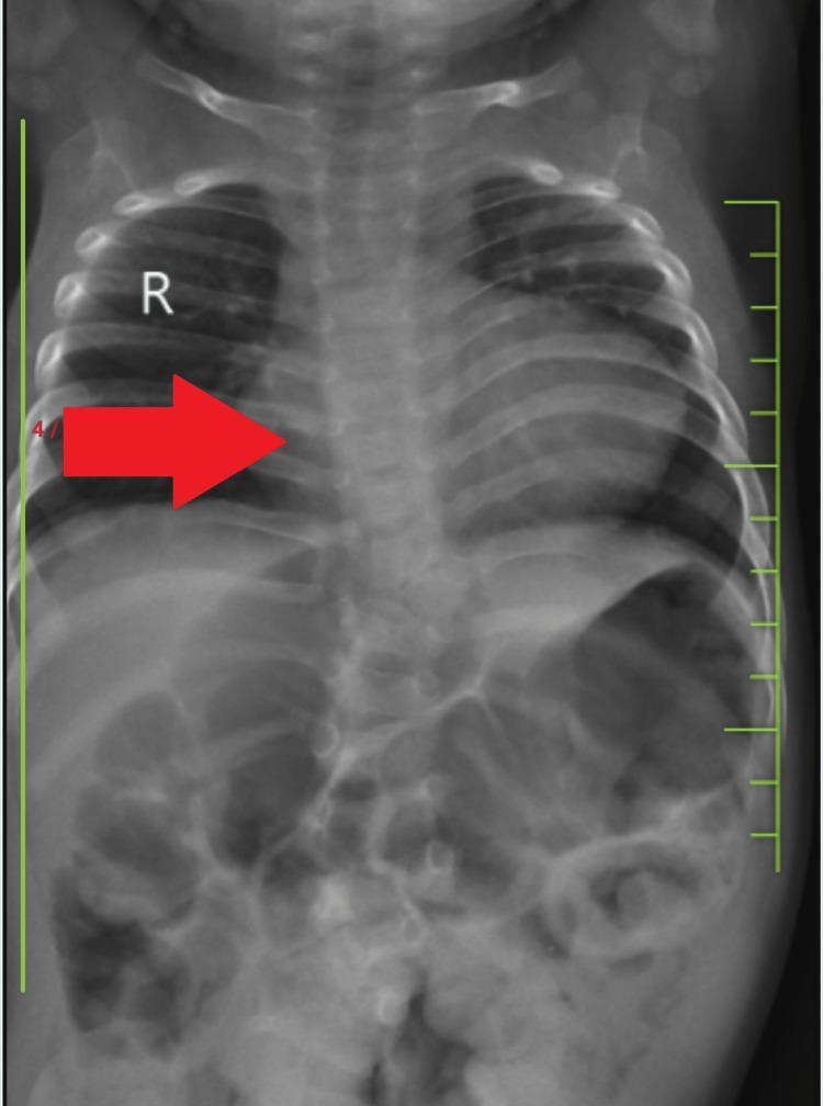
This diagnostic image is a posteroanterior (PA) chest X-ray of a pediatric patient demonstrating classic radiographic signs of Tetralogy of Fallot (TOF). The most prominent finding is a 'boot-shaped' heart (coeur en sabot), characterized by an upturned cardiac apex due to right ventricular hypertrophy and a concave pulmonary artery segment (pulmonary conus). The lung fields appear relatively oligemic, consistent with decreased pulmonary blood flow. The mediastinum is centrally located, and the diaphragm is well-defined, with the right hemidiaphragm slightly superior to the left. The skeletal structures show visible ribs and a vertebral column that appears to have mild alignment irregularities, potentially correlating with lumbar kyphoscoliosis mentioned in clinical context. The lower portion of the radiograph captures the upper abdomen, showing normal bowel gas patterns. This imaging is a classic educational example for pediatric cardiology and radiology, illustrating the morphological changes in the heart and great vessels associated with cyanotic congenital heart disease.
Boot-Shaped Heart (Coeur en Sabot) in Tetralogy of Fallot
What Is Tetralogy of Fallot?
| Component | Description |
|---|---|
| 1. Right Ventricular Outflow Tract (RVOT) Obstruction | Pulmonary stenosis (subvalvular, valvular, or supravalvular) |
| 2. Large, Unrestrictive VSD | Misaligned ventricular septal defect |
| 3. Overriding Aorta | Aorta sits astride the VSD, receiving blood from both ventricles |
| 4. Right Ventricular Hypertrophy (RVH) | Secondary to high pressure load from RVOT obstruction |
Why Does the Heart Look Like a Boot?
1. Concave Pulmonary Artery Segment (the "leg of the boot")
- The RVOT obstruction (pulmonary stenosis) causes hypoplasia of the main pulmonary artery and pulmonary conus
- On a PA chest X-ray, the normal convex bulge of the main pulmonary artery along the upper left heart border is absent - instead it appears flat or concave
- This creates the "waist" or indentation at the top-left border of the heart - the open top of the boot
2. Right Ventricular Hypertrophy (the "toe of the boot")
- Chronic pressure overload causes the right ventricle to hypertrophy massively
- The hypertrophied RV pushes the cardiac apex upward and to the left
- On X-ray, the apex appears elevated, rounded, and tilted upward rather than pointing inferiorly as normal
- This elevated, rounded apex forms the "toe" of the boot
Chest X-Ray Findings in TOF (Summary)
| Finding | Explanation |
|---|---|
| Boot-shaped (coeur en sabot) silhouette | Concave PA segment + upturned RVH apex |
| Normal heart size | TOF is a pressure-overload disease, which causes less cardiomegaly than volume overload |
| Decreased pulmonary vascular markings | Reduced pulmonary blood flow due to RVOT obstruction and right-to-left shunting through VSD |
| Right-sided aortic arch | Present in ~25% of TOF cases |
| Right ventricular hypertrophy on ECG | Right axis deviation, RVH pattern |
The heart size in TOF is normal - this is an important distinguishing point. The boot shape is about contour, not enlargement. - ROSEN's Emergency Medicine
Classic Chest X-Ray (from textbook, Fig. 165.4)
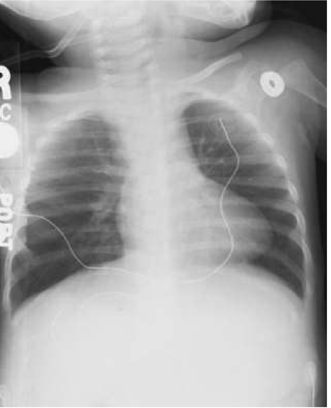
Real PA Chest X-Ray Examples
Mnemonic: Classic Congenital Heart Silhouettes
| Silhouette | Condition |
|---|---|
| Boot-shaped (coeur en sabot) | Tetralogy of Fallot |
| Egg-on-a-string | Transposition of the Great Arteries |
| Snowman / Figure-of-eight | Total Anomalous Pulmonary Venous Return (TAPVR) |