

Find the diagnosis and treatment of this condition
molluscum contagiosum buttocks excoriated papules children
clustered crusted excoriated papules buttocks gluteal cleft child diagnosis
Clinical Impression
- Excoriated insect bite reaction / papular urticaria
- Molluscum contagiosum with secondary inflammation/excoriation (molluscum dermatitis)
- Impetiginized eczema or contact dermatitis
- Herpes simplex (grouped erosions) - less likely given the papulonodular, non-vesicular pattern
Diagnosis - How to confirm
- Look for classic burrows (thin, grayish, tortuous lines) elsewhere on the body - finger webs, wrists, axillae, areolae, periumbilical area, genitals.
- Dermoscopy or skin scraping with microscopy (mineral oil prep) to identify mites, eggs, or fecal pellets ("scybala") confirms the diagnosis; a scraping from a nodule itself is often negative since it represents a hypersensitivity reaction rather than active infestation.
- Ask about intense nocturnal pruritus and whether close contacts/household members are also itching.
Treatment
- Topical permethrin 5% cream applied to the entire body from neck down (scalp/face too in infants), left on 8-12 hours, repeated in 7 days - standard first-line therapy (Dermatology, 2-Vol Set 5e; Goldman-Cecil Medicine).
- Alternative topical: malathion 0.5% lotion/gel, applied similarly.
- Oral ivermectin 200 mcg/kg as a single dose, repeated in 7-14 days (not ovicidal, so a second dose is required). A Cochrane review found oral ivermectin as effective as topical permethrin (Red Book 2021; Harrison's).
- Treat all household members/close contacts simultaneously, even if asymptomatic.
- Wash bedding, clothing, and towels used in the preceding 3 days in hot water and dry on high heat, or seal unwashable items in a plastic bag for 72 hours.
- Antihistamines or a short course of low-potency topical corticosteroids for pruritus and post-scabetic nodules/itch, which can persist for weeks after successful eradication (this is a hypersensitivity phenomenon, not treatment failure).
- Treat secondary bacterial infection (impetiginization) if crusting/erosions show signs of infection (topical or oral antibiotics as needed).
- Persistent nodules unresponsive to antipruritics can be treated with intralesional corticosteroids.
Recent evidence check
scabies AND treatment
Note on recent evidence
Summary
- Topical permethrin 5% cream over the entire body (neck down in adults, including scalp/face in infants), left on 8-12 hours, repeated at day 7-14, OR
- Oral ivermectin 200 mcg/kg as a single dose, repeated in 7-14 days (not ovicidal, so the second dose is required)
- Treat crusted/severe scabies with combined topical permethrin plus repeated oral ivermectin doses
- Treat all household/close contacts simultaneously
- Decontaminate bedding/clothing (hot wash/dry, or bag for 72 hours)
- Antihistamines or mild topical steroids for residual post-scabetic itch/nodules, which can persist for weeks after mites are eradicated
- Watch for and treat secondary bacterial infection of excoriated/crusted areas if present
Find clinical images of scabies burrows and other common presentations
scabies burrows finger web spaces
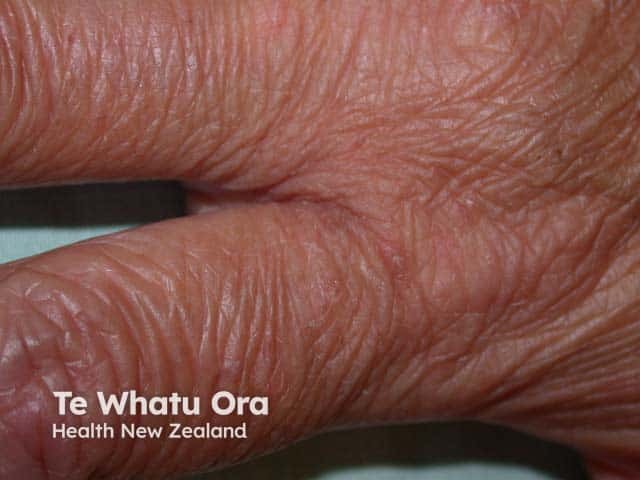
Clinical photography of interdigital skin lesions on a finger, illustrating features characteristic of scabies. Modality: clinical photography; technique: macro close-up; magnification approximately 2–4x; field of view focused on the web spaces of the digits. Location: integumentary system, primarily the interdigital regions at the finger joints. Visual findings: fine scaling (scaling), papules, excoriations, and mild erythema within the web spaces; the skin appears dry and fissured in places. Burrows may be present but are not clearly resolved in this image. The appearance supports a Scabies infestation by Sarcoptes scabiei, with pruritus often intense, particularly at night, and a habitual scratching pattern. Secondary bacterial colonization or infection can occur in scratched sites. This image captures diagnostic features without invasive sampling, enabling immediate clinical correlation. Diagnostic significance: interdigital papules and scale strongly suggest scabies in the appropriate clinical context; differential diagnoses include eczema, contact dermatitis, dyshidrotic dermatitis, tinea manuum, and bacterial folliculitis. Clinical correlation: assess for pruritus, nocturnal activity, and close contacts; consider topical permethrin 5% or oral ivermectin in treatment; emphasize hygiene measures and environmental decontamination. Uses: dermatology education, teledermatology, and clinical decision support for suspected parasitic infestations. This image supports educational use in medical training and exam preparation.
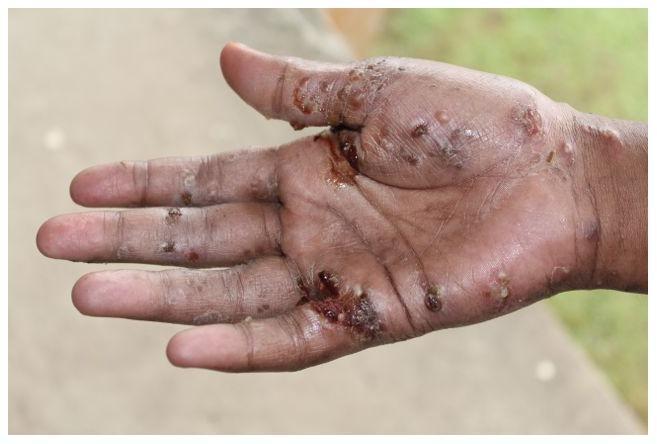
This clinical photograph displays the palmar surface and interdigital spaces of a hand with significant dermatological pathology. The primary findings include widespread papules, nodules, and burrows characteristic of Sarcoptes scabiei infestation. There is prominent evidence of secondary bacterial infection (impetiginization), manifested as honey-colored and dark hemorrhagic crusting, pustules, and focal ulcerations. The distribution is densest in the web spaces, particularly between the thumb and index finger, and along the proximal phalanges. Visible skin changes include erythema, inflammatory thickening, and xerosis. This image serves as a clinical example of neglected scabies and its common complications, such as secondary infection by Staphylococcus aureus or Streptococcus pyogenes, which can lead to serious systemic sequelae if left untreated. The presentation is typical for endemic regions where public health burdens of tropical skin diseases are high.
nodular scabies buttocks
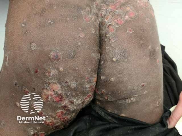
Clinical photograph of the buttocks and gluteal region showing multiple firm nodules with ulceration and crusted surfaces against dark skin. The lesions are localized to the intergluteal and perineal skin extending over the buttock folds, with surrounding erythema and excoriation. The nodules are erythematous-to-brown, dome-shaped, some with central pits or shallow ulcers, and overlying crusts consistent with secondary bacterial infection. The appearance is typical for nodular scabies in which the inflammatory nodules result from a host hypersensitivity reaction to Sarcoptes scabiei antigens. Crusting, scale, and fissuring are evident, indicating chronic scratching and secondary impetiginization. Notable features include clustered lesions, bilateral symmetry on the buttocks, and variable depth of ulceration; there may be surrounding edema in some areas. This image emphasizes cutaneous scabies manifestations beyond classical burrows, highlighting nodular scabies as a pruritic, proliferative eruption that can be misdiagnosed as dermatitis or folliculitis. Diagnostic significance lies in recognizing nodular scabies with secondary infection to guide therapy: topical scabicide (permethrin 5%), systemic ivermectin if extensive, and antibiotics for secondary infection. Potential clinical use cases include dermatology education, differential diagnosis exercises for nodular scabies, telemedicine consultations, and competency assessment of recognition of scabies-related skin lesions in darkly pigmented skin. The image supports teaching about disease manifestations, treatment implications, and infection control measures.
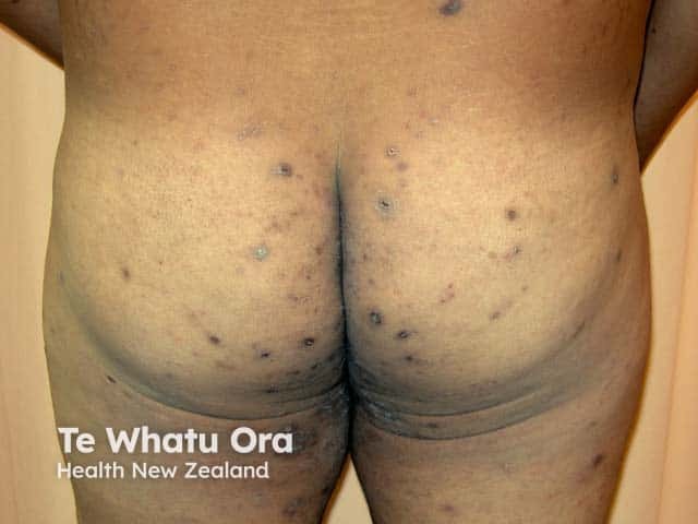
Imaging modality: Dermatologic clinical photograph; Technique: Standard color photography with even lighting; The image captures the bilateral gluteal region from a posterior perspective. Numerous small, dark-brown to violaceous nodules and papules are scattered across the buttocks, extending into the intergluteal fold. Lesions are discrete, well-circumscribed, dome-shaped, and firm on palpation, with no apparent ulceration. Distribution is bilateral and predominantly involves gluteal skin, with occasional spread toward adjacent posterior thighs. Morphology features include nodular/papular texture, surface hyperpigmentation in darker skin tones, and subtle shading around lesion margins. The clinical impression is nodular scabies on the buttocks, consistent with a hypersensitivity reaction to Sarcoptes scabiei infestation. This pattern complements classic burrow lines in other body areas, though burrows may be sparing in this region. The image is educational for dermatology and infectious diseases, illustrating scabies-associated nodules as a chronic, non-vesicular eruption. Relevance spans differential diagnosis with dermatitis, prurigo nodularis, folliculitis, and insect bites, and supports clinical correlation with pruritus history, exposure risk, and household contact tracing. Potential clinical use cases include tele-dermatology assessment, treatment response monitoring, patient education, and public health screening during scabies outbreaks. The image thereby serves as a reference for nodular scabies located on gluteal skin.
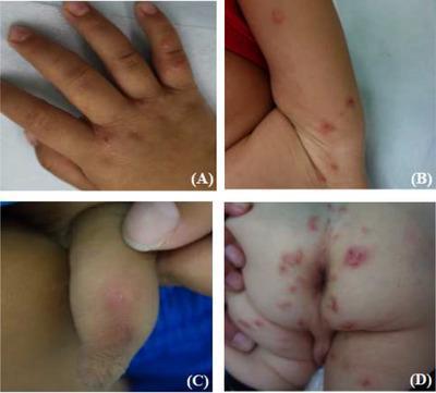
This composite clinical photograph illustrates the diverse dermatological manifestations of scabies in a pediatric patient across four anatomical regions. Panel A shows the dorsal hand and interdigital spaces featuring erythematous papules, papulovesicles, and distinct blisters. Panel B displays the axillary region with scattered red papules and inflammatory nodules. Panel C depicts the genital area (penis) presenting with characteristic pruritic red nodules, where subtle linear, whitish 'tunnels' or burrows—pathognomonic for Sarcoptes scabiei infestation—are visible on the nodular surfaces. Panel D captures the buttocks and perianal area showing extensive polymorphic lesions, including widespread erythema, inflammatory papules, and nodules interspersed with secondary lesions such as excoriations (scratches), shallow ulcers, and hemorrhagic scabs resulting from chronic pruritus. The collection serves as an educational reference for identifying both primary sarcoptic lesions and secondary infections or irritations common in childhood scabies cases.
crusted scabies hands hyperkeratotic
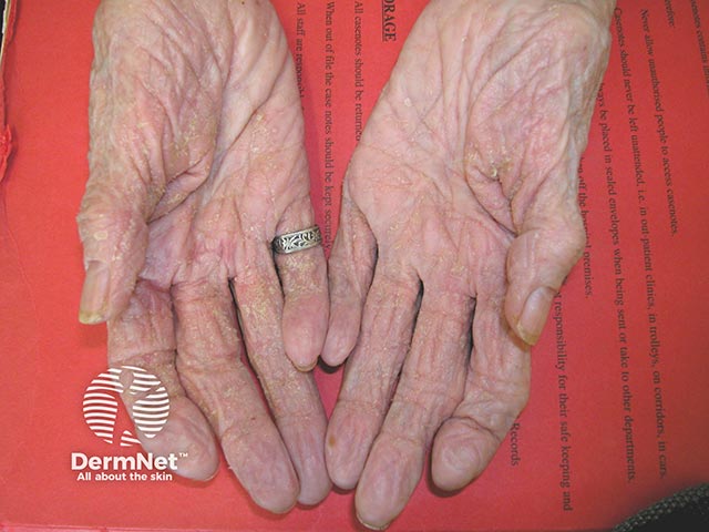
This entry is a high-resolution clinical photograph of bilateral palmar skin illustrating early crusted scabies (Norwegian variant) on palms. Modality: clinical photography; technique: frontal palmar view, close-up capture, natural lighting; specimen: human palmar epidermis with thick hyperkeratotic crusts; anatomical localization: volar surfaces of the hands, including interdigital folds; imaging perspective: anterior exposure, head-on view. Visual features: extensive hyperkeratotic crusting, parchment-like scales, fissuring along palmar creases, mild erythema, and diffuse dryness; there is marked crusted plaque formation with irregular margins, more prominent on thenar and hypothenar eminences. Pathology: infestation by Sarcoptes scabiei var. hominis manifested as crusted scabies; clinical correlate with high mite burden; risk of secondary bacterial infection. Diagnostic significance: hallmark of crusted scabies; highly contagious; necessitates isolation, contact tracing, and prompt antiparasitic therapy (permethrin, ivermectin) and wound care; monitor for secondary infection; educate patient and household contacts. Potential clinical use cases: dermatology teaching, infectious disease illustration, rapid bedside recognition, differential diagnosis workflow for diffuse palm crusting; differential includes eczema, psoriasis, tinea manuum, palmoplantar keratoderma. Synonyms: Norwegian scabies, crusted scabies; keywords: palms, hand dermatosis, hyperkeratosis, crusted lesions, skin infection risk. Consider concomitant bacterial superinfection management and patient education on transmission prevention and skin hygiene. Follow-up recommended after therapy.
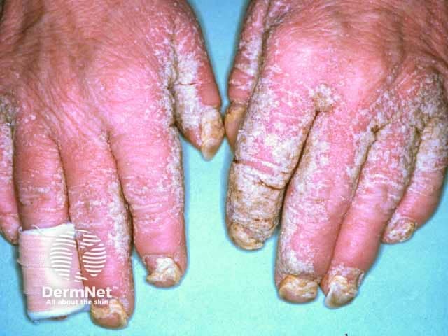
Imaging modality: Clinical photography of the hands, high-resolution frontal view capturing dorsal and palmar surfaces and both nails. The scene shows bilateral, symmetric involvement of the hands with thick, adherent crusts over erythematous, fissured skin. On the dorsal and palmar surfaces, extensive hyperkeratotic plaques exhibit chalky to yellow-white crusts, scale, and superficial erosions. Nails demonstrate dystrophy characterized by thickening, onycholysis, and irregular plate contour; periungual involvement is evident. The morphology is consistent with crusted (Norwegian) scabies, a high-burden infestation by Sarcoptes scabiei, often seen in immunocompromised or elderly hosts and associated with heavy mite burden and high contagion risk. The photograph highlights the diagnostic relevance of cutaneous involvement in crusted scabies and underscores potential secondary infections. Notable differential considerations include chronic dermatitis, psoriasis with nail changes, and tinea manuum; however, the presence of dense crusting with nail dystrophy in both hands favors crusted scabies in the appropriate clinical context. This image is clinically valuable for education and reference in dermatology, infectious disease, and hand diseases; useful in teaching crusted scabies recognition, infection control imperatives, and triage for rapid, combined systemic and topical therapy. Clinically, this dataset supports rapid retrieval for case-based learning, differential diagnosis discussions, and imaging-based diagnostic pattern recognition for scabies crusted infestations.
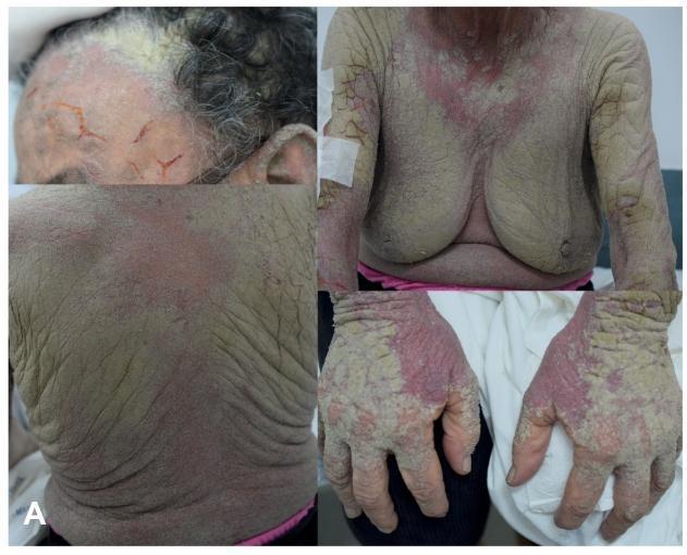
A composite clinical photograph showing four panels of a patient with Norwegian (crusted) scabies. The visual findings demonstrate disseminated, greenish-gray, hyperkeratotic plaques and thick crusts extensively involving the scalp, trunk (chest and back), upper extremities, and hands. Key dermatological features include significant skin thickening, a fissured texture, and underlying confluent erythema. The most severe crusting is evident on the dorsum of the hands and fingers, typical of high mite burden areas. The scalp panel shows prominent scaling and crusted lesions extending into the hairline. On the trunk and arms, the lesions present as broad, scaly patches with deep cutaneous folds. This clinical presentation illustrates the classic manifestations of crusted scabies, often associated with immunosuppression or neglected infestation, leading to extreme hyperkeratosis and secondary bacterial infection risk. The educational focus is on the morphology and distribution of specialized scabies infestations in a clinical setting.
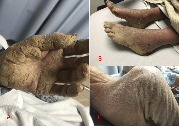
Clinical photograph showing a tripartite display of dermatological manifestations. Panel A illustrates the palmar surface of a left hand covered in thick, yellowish-brown hyperkeratotic plaques with deep, painful-looking fissures and a psoriasiform texture. Panel B displays the bilateral lower extremities with similar hyperkeratotic and crusted lesions predominantly affecting the soles and dorsal aspects of the feet. Panel C depicts the posterior thorax presenting with diffuse, fine, white flaky scales and erythematous patches. Collectively, these features represent a severe generalized dermatosis. The clinical presentation of extensive crusted, fissured, and scaly skin across multiple anatomical regions—hands, feet, and trunk—is characteristic of Crusted (Norwegian) Scabies, particularly in the context of immunocompromise or uncontrolled diabetes. The images demonstrate the transition from heavy keratotic crusting on acral surfaces to widespread scaling on the torso.
scabies genital penile papules
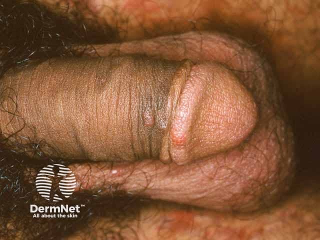
This high-resolution clinical photograph depicts the external genitalia focusing on the penile shaft with adjacent prepuce. The skin shows multiple erythematous papules, small, discrete, firm-topped lesions, some with faint perifollicular erythema. The distribution is localized to the genital skin, without obvious vesicles or crusts in the shown field, although scabies can produce papules and burrows in the groin and penile region. The image is captured as a standard dermatologic clinical photograph (macro view) under ambient lighting, with a frontal exterior view of the penis. The appearance is compatible with common scabies–related dermatitis, especially in sexually active adults, where mites or their products provoke an inflammatory papular eruption. Visualization of burrows may be limited in a single image. Clinically, this pattern should be correlated with intense nocturnal pruritus, involvement of interdigital spaces elsewhere, and contact history. Differential diagnoses include contact dermatitis, folliculitis, genital herpes, molluscum contagiosum, lichen simplex chronicus, and less likely psoriasis. The finding has diagnostic significance for initiating topical scabicides and seeking partner treatment; supports a targeted physical exam and potential dermoscopic or skin scrape if available. This image should be cataloged with dermatology, sexual health, infectious disease, and primary care databases for educational and research use. For teaching.
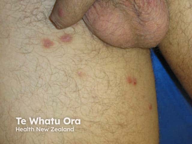
Imaging modality: Clinical photography. Technique: High-resolution external view with frontal close-up of the genital region and proximal thighs under standard ambient lighting; no dermoscopy or magnification. Anatomical location: penile foreskin and adjacent proximal thigh skin. Visual features: multiple erythematous papules and small nodules on the foreskin and inner thigh; some lesions show shallow central pits, mild scaling, and subtle surface edema. The lesions are non-vesicular in this view; color ranges from pink to red; surface is slightly excoriated with superficial capillary prominence. Distribution: localized to genito-femoral skin with bilateral involvement of proximal thighs. Inferred pathology: lesions compatible with nodular scabies and cutaneous hypersensitivity reaction to Sarcoptes scabiei mites; burrows are not clearly visible in a single clinical photograph. Diagnostic significance: supports diagnosis of cutaneous scabies or nodular scabies in a male patient and warrants antiparasitic treatment and contact tracing. Clinical correlation: presents with intense nocturnal pruritus and history of exposure to an infested individual; secondary bacterial infection risk if scratched. Management implications: topical permethrin 5% or oral ivermectin per guidelines, treat all household close contacts, wash clothing and bedding. Differential considerations: folliculitis, contact dermatitis, tinea cruris, eczema, sexually transmitted skin infections. Educational use: clinical recognition of scabies nodules on genital skin.
infant scabies rash whole body
1. Burrows / Interdigital Involvement (classic finding)
2. Nodular Scabies (buttocks/groin - matches the case discussed earlier)
3. Genital Scabies
4. Pediatric Composite (multiple sites in one child)
5. Crusted (Norwegian) Scabies - severe/hyperinfested variant
Quick summary of presentations
| Presentation | Typical site | Key feature |
|---|---|---|
| Classic burrows | Finger webs, wrists | Thin grayish tortuous lines, papules, scale |
| Nodular scabies | Buttocks, groin, axilla | Firm red-brown/violaceous nodules, persistent itch |
| Genital scabies | Penis, scrotum, labia | Papules/nodules, easily mistaken for STI |
| Infant/pediatric scabies | Widespread incl. face/scalp/palms/soles | More diffuse, vesiculopustular |
| Crusted (Norwegian) scabies | Hands, scalp, trunk | Thick hyperkeratotic crusts, highly contagious, seen in immunosuppressed |