
If you were a student and the examiner asked you about Anterior compartment of Thigh how would you answer to score full marks.
Reading File
Now let me search the medical library for the anterior compartment of the thigh.
Finding Sources
Finding Sources
Finding Sources
Finding Sources
I have three great sources. Let me read the most relevant ones in parallel - Gray's Anatomy for Students (THIGH section), Imaging Anatomy (Anterior Thigh Muscles), and THIEME Atlas.
Reading File
Reading File
Excellent. Now let me read the deeper Gray's content (anterior compartment muscles, femoral triangle, femoral nerve, femoral artery) and the THIEME atlas for muscle details.
Finding Sources
Reading File
Let me read the femoral triangle section from Gray's and the anterior compartment section from the THIEME Atlas:
Reading File
Reading File
Now let me search for the adductor canal and femoral nerve sections in Gray's, and also get the muscles table:
Finding Sources
Finding Sources
Reading File
Reading File
Now let me search for the muscles of the anterior compartment (quadriceps, sartorius, iliopsoas) and the profunda femoris artery from Gray's:
Finding Sources
Finding Sources
Reading File
Reading File
I now have comprehensive data from multiple authoritative textbooks. Let me also fetch one of the key anatomical images to include:
Loading Image
Loading Image
I now have all the information needed for a complete, exam-level answer. Let me compile the full response.
Anterior Compartment of the Thigh - Complete Exam Answer
1. Introduction and Overview
The thigh is the region of the lower limb between the hip and knee joints. It is divided into three compartments by intermuscular septa that extend between the posterior aspect of the femur and the fascia lata (the thick deep fascia that completely invests the thigh):
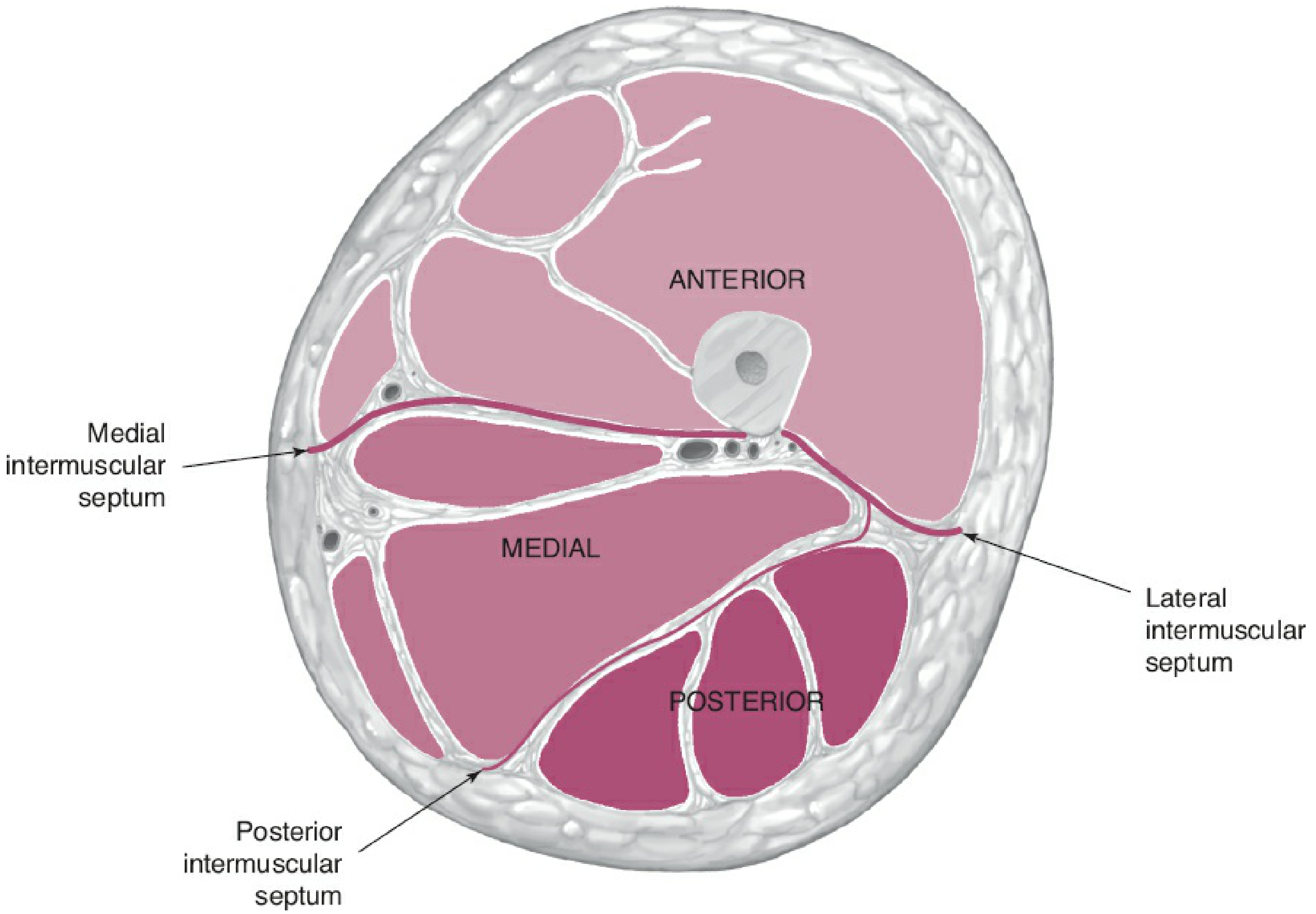
The three compartments are:
- Anterior - extensors of the knee (femoral nerve)
- Medial - adductors of the thigh (obturator nerve)
- Posterior - extensors of the hip, flexors of the knee (sciatic nerve)
2. Boundaries of the Anterior Compartment
The anterior compartment is bounded by:
- Anteriorly/laterally: fascia lata
- Posteriorly/medially: medial and lateral intermuscular septa connecting fascia lata to the linea aspera of the femur
- Superiorly: communicates with the abdomen through the gap between the inguinal ligament and pelvic bone (the lacuna musculorum and lacuna vasorum)
3. Contents - Muscles
The anterior compartment contains 6 muscles (or 7 if you count the four heads of quadriceps individually):
A. Quadriceps Femoris (the large extensor mass)
The most powerful muscle group - composed of four parts that share a common insertion.
| Part | Origin | Insertion | Nerve | Action |
|---|---|---|---|---|
| Rectus femoris | Anterior inferior iliac spine (straight head); acetabular rim (reflected head) | Tibial tuberosity via patellar ligament | Femoral (L2-L4) | Extends knee; flexes hip |
| Vastus medialis | Medial lip of linea aspera; distal intertrochanteric line | Tibial tuberosity + medial patellar retinaculum | Femoral (L2-L4) | Extends knee; prevents lateral patellar dislocation (VMO) |
| Vastus lateralis | Lateral lip of linea aspera; lateral greater trochanter | Tibial tuberosity + lateral patellar retinaculum | Femoral (L2-L4) | Extends knee |
| Vastus intermedius | Anterior femoral shaft | Tibial tuberosity via patellar ligament | Femoral (L2-L4) | Extends knee |
| (Articularis genus) | Distal fibers of vastus intermedius from anterior femur | Suprapatellar recess of knee capsule | Femoral | Retracts synovial capsule during extension (prevents pinching) |
All four converge into the quadriceps tendon → encapsulates the patella → continues as the patellar ligament → inserts into the tibial tuberosity.
B. Sartorius
- Origin: Anterior superior iliac spine (ASIS)
- Insertion: Upper medial surface of tibia (as part of the pes anserinus with gracilis and semitendinosus)
- Nerve: Femoral nerve (L2, L3)
- Action: Flexes, abducts, and laterally rotates the thigh at the hip; flexes the knee (the "tailor's muscle" - crosses the thigh obliquely)
- Importance: Forms the lateral border of the femoral triangle and the roof of the adductor canal
C. Iliopsoas (passing through, not originating in thigh)
- Origin: Iliacus (iliac fossa) + Psoas major (T12-L5 vertebral bodies/transverse processes)
- Insertion: Lesser trochanter of femur
- Nerve: Femoral nerve + direct branches from lumbar plexus (L1-L3)
- Action: Primary flexor of the hip; lateral rotation of thigh
- Note: Enters the thigh through the muscular compartment lateral to the iliopectineal arch
D. Pectineus
- Origin: Pectineal line (pecten pubis) of the pubis
- Insertion: Pectineal line of femur (between lesser trochanter and linea aspera)
- Nerve: Femoral nerve (L2, L3); may also receive a branch from obturator nerve
- Action: Adducts and flexes thigh
- Note: Forms part of the floor of the femoral triangle; classified by some authors as a medial compartment muscle due to dual innervation
4. Femoral Triangle
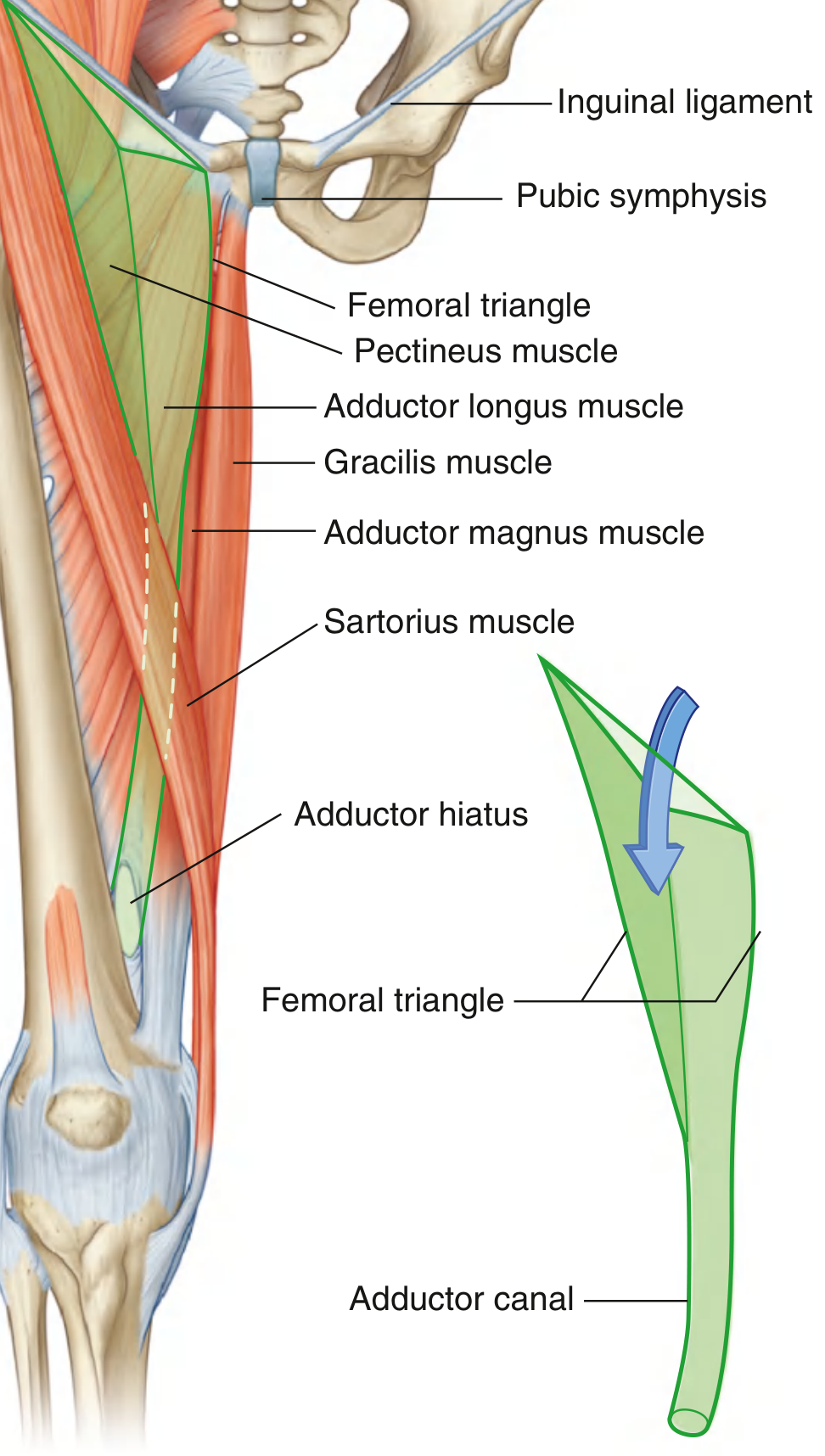
A wedge-shaped depression at the top of the thigh, crucial for access to major neurovascular structures.
Boundaries
| Base (superiorly) | Inguinal ligament |
| Lateral border | Medial margin of sartorius |
| Medial border | Medial margin of adductor longus |
| Floor | Laterally - iliopsoas; medially - pectineus and adductor longus |
| Roof | Fascia lata + cribriform fascia (over the saphenous opening) |
| Apex | Where sartorius crosses adductor longus; continues as adductor canal |
Contents (lateral to medial: mnemonic NAVEL)
- N - Femoral Nerve (most lateral; outside femoral sheath)
- A - Femoral Artery
- V - Femoral Vein
- E - Empty space (femoral canal)
- L - Lymphatics (deep inguinal lymph nodes; Cloquet's/Rosenmüller's node is the most superior)
The femoral artery is palpable just below the inguinal ligament, midway between the ASIS and pubic symphysis (the mid-inguinal point).
Femoral Sheath
A funnel-shaped fascial sleeve (derived from transversalis fascia anteriorly and iliac fascia posteriorly) enclosing three compartments:
- Lateral - femoral artery
- Intermediate - femoral vein
- Medial - femoral canal (contains lymphatics and fat; the opening superiorly is the femoral ring, a potential site for femoral hernias)
The femoral nerve is NOT enclosed within the femoral sheath - it lies lateral to it.
5. Adductor Canal (Canal of Hunter / Subsartorial Canal)
An intermuscular tunnel in the middle third of the thigh that carries the femoral vessels from the femoral triangle to the popliteal fossa.
Boundaries
| Wall | Structure |
|---|---|
| Anterolateral | Vastus medialis |
| Posterior | Adductor longus (proximally), adductor magnus (distally) |
| Medial (roof) | Sartorius + vastoadductor membrane |
Contents
- Femoral artery
- Femoral vein
- Saphenous nerve (branch of femoral nerve)
- Nerve to vastus medialis
- Descending genicular artery (terminal branch; pierces the vastoadductor membrane)
The femoral artery and vein exit through the adductor hiatus (gap in adductor magnus) to become the popliteal artery and vein. The saphenous nerve and descending genicular artery pierce the vastoadductor membrane and pass medially to the knee.
6. Nerve Supply - Femoral Nerve
- Root value: L2, L3, L4 (posterior divisions of the lumbar plexus)
- Origin: Within the substance of psoas major in the abdomen
- Course: Descends between psoas and iliacus; enters femoral triangle lateral to the femoral artery (outside the femoral sheath); divides into anterior and posterior divisions about 4 cm below the inguinal ligament
- Motor branches: All muscles of the anterior compartment (iliopsoas, pectineus, sartorius, and all four parts of quadriceps femoris)
- Sensory branches:
- Intermediate and medial cutaneous nerves of the thigh (anterior thigh skin)
- Saphenous nerve (longest branch, L3-L4) - runs through the adductor canal, emerges at the knee, supplies medial leg and medial foot down to the ball of the great toe
Femoral nerve injury (e.g., from psoas abscess, hematoma, or hip surgery) causes:
- Loss of knee extension (quadriceps paralysis) - patient cannot walk up stairs
- Loss of hip flexion (iliopsoas weakness)
- Absent knee jerk reflex
- Sensory loss over anterior thigh and medial leg
7. Blood Supply
Femoral Artery
- Continuation of the external iliac artery below the inguinal ligament
- Enters femoral triangle at the mid-inguinal point
- Passes through the adductor canal and exits as the popliteal artery at the adductor hiatus
Profunda Femoris Artery (Deep Artery of the Thigh)
- Largest branch of the femoral artery; arises 3-5 cm below the inguinal ligament, posterolaterally
- Key branches:
- Medial femoral circumflex artery (MFCA) - primary blood supply to the femoral head and neck (clinically important in femoral neck fractures)
- Lateral femoral circumflex artery (LFCA) - supplies quadriceps and gives an ascending branch to the hip
- 3-4 perforating arteries - pass through gaps in adductor magnus insertions to supply the posterior compartment
Venous drainage
- Femoral vein accompanies the femoral artery; receives the great saphenous vein at the saphenous opening (fossa ovalis)
8. Lymphatic Drainage
- Superficial inguinal lymph nodes: arranged along the inguinal ligament (horizontal group) and along the great saphenous vein (vertical group); drain skin of thigh, perineum, anterior abdominal wall below umbilicus
- Deep inguinal lymph nodes: medial to femoral vein; receive deep lymphatics of the thigh and efferents from superficial nodes; the uppermost node (Cloquet's node / Rosenmüller's node) is in the femoral canal
- All drain upward into external iliac lymph nodes
9. Clinical Correlations (Examiner Favourites)
| Topic | Key Points |
|---|---|
| Femoral hernia | Protrudes through femoral ring into femoral canal; below and lateral to pubic tubercle (cf. inguinal hernia: above and medial); more common in women; high risk of strangulation |
| Femoral artery puncture | At mid-inguinal point; used for cardiac catheterization, arteriography |
| Femoral nerve block | Used for knee surgery analgesia; needle placed lateral to femoral artery |
| Adductor canal block | Motor-sparing alternative to femoral nerve block; preserves quadriceps strength post-TKA |
| Femoral nerve injury | Loss of knee extension + absent knee jerk + sensory loss anterior thigh/medial leg |
| Saphenous nerve | Most commonly injured nerve in lower limb surgery (varicose vein stripping); causes medial leg numbness |
| Quadriceps paralysis | Patient hyperextends knee to stand (gravity replaces active extension) |
| Rectus femoris | Only biarticular quadriceps head - most frequently strained in kicking sports |
| Patella | Sesamoid bone within quadriceps tendon; largest sesamoid in body |
| VMO (vastus medialis oblique) | Prevents lateral patellar dislocation; its weakness contributes to patellofemoral pain syndrome |
10. Summary Table - Muscles at a Glance
| Muscle | Origin | Insertion | Action | Nerve |
|---|---|---|---|---|
| Iliopsoas | Iliac fossa + vertebral bodies T12-L5 | Lesser trochanter | Hip flexion, lateral rotation | Femoral L1-L3 |
| Pectineus | Pectineal line of pubis | Pectineal line of femur | Hip flexion + adduction | Femoral (L2-L3) ± obturator |
| Sartorius | ASIS | Medial tibia (pes anserinus) | Hip flex/abduct/lat rot; Knee flex | Femoral L2-L3 |
| Rectus femoris | AIIS + acetabular rim | Tibial tuberosity via patella | Knee ext + Hip flex | Femoral L2-L4 |
| Vastus medialis | Medial linea aspera | Tibial tuberosity + medial retinaculum | Knee extension | Femoral L2-L4 |
| Vastus lateralis | Lateral linea aspera + greater trochanter | Tibial tuberosity + lateral retinaculum | Knee extension | Femoral L2-L4 |
| Vastus intermedius | Anterior femoral shaft | Tibial tuberosity | Knee extension | Femoral L2-L4 |
| Articularis genus | Distal anterior femur | Suprapatellar capsule | Retracts knee capsule | Femoral |
Sources: Gray's Anatomy for Students; General Anatomy and Musculoskeletal System (THIEME Atlas of Anatomy); Imaging Anatomy Vol. 3 (Bones, Joints, Vessels and Nerves); Rockwood and Green's Fractures in Adults 10th ed; Fischer's Mastery of Surgery 8th ed.
This is a shared conversation. Sign in to Orris to start your own chat.