
Red pulp of spleen vs White pulp of spleen
Reading File
Reading File
Searching the Web
spleen red pulp white pulp anatomy histology site:nih.gov OR site:ncbi.nlm.nih.gov
Searching PubMed
spleen white pulp red pulp
Finding Sources
Reading File
Searching Images
histology diagram of spleen white pulp red pulp central artery PALS splenic cords sinusoids
Reading File
Red pulp vs White pulp of spleen
| Feature | White pulp | Red pulp |
|---|---|---|
| Main role | Immune surveillance of blood-borne antigens | Filters blood and removes aged or abnormal blood cells |
| Appearance | Pale/white, due to dense lymphocytes | Red, due to abundant erythrocytes in vascular spaces |
| Proportion of spleen | About 20% | Most of the splenic parenchyma |
| Main components | Periarteriolar lymphoid sheath (PALS), lymphoid follicles/nodules, marginal zone | Splenic cords (cords of Billroth) and venous sinusoids |
| Predominant cells | T cells in the PALS; B cells in lymphoid follicles; also macrophages and dendritic cells | RBCs, macrophages, reticular cells, plasma cells, platelets |
| Relationship to artery | Organized around a central arteriole | Blood leaving white pulp enters cords/sinusoids of red pulp |
| What it does | Antigen presentation, lymphocyte activation, antibody production | Macrophages phagocytose old/damaged RBCs, recycle iron, clear microbes and particulate material; platelet reservoir |
| Microscopy clue | Basophilic, dark-blue lymphoid islands around a central arteriole | More eosinophilic/pink background with vascular sinusoids and cords |
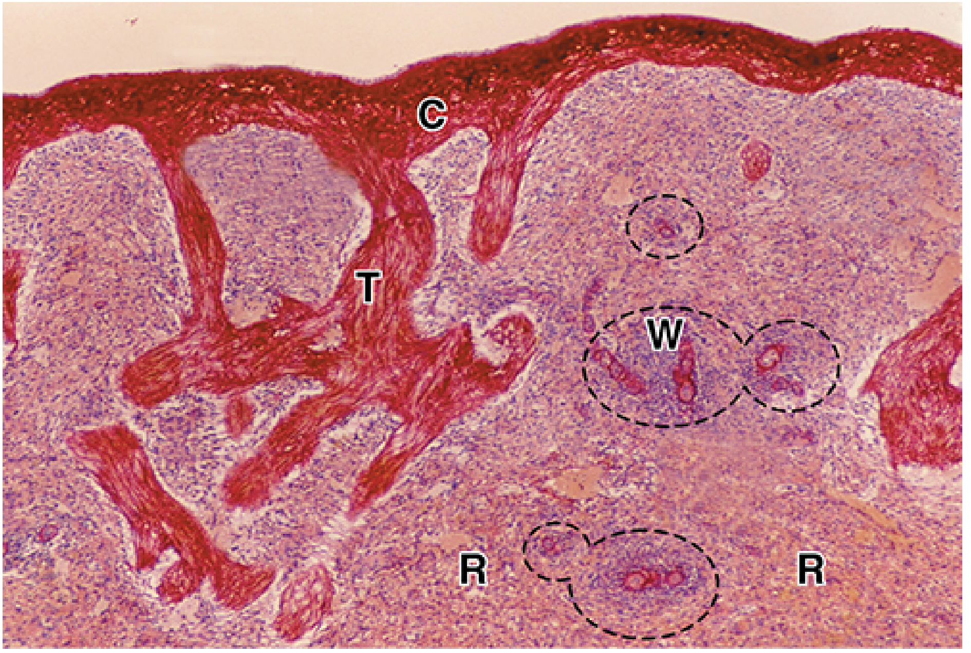
High-yield memory aid
- White pulp = WBC immunity
- Red pulp = RBC removal
The white pulp consists of lymphoid nodules and T-cell-rich PALS, whereas the red pulp is composed of blood-filled sinusoids and splenic cords. Junqueira's Basic Histology - Text and Atlas, 17e, p. 714.
Microscopy difference
Reading File
Finding Sources
Searching the Web
spleen histology microscopy red pulp white pulp features H&E central arteriole sinusoids site:ncbi.nlm.nih.gov
Searching PubMed
spleen histology red pulp white pulp
Microscopic differences on H&E section
| Feature | White pulp | Red pulp |
|---|---|---|
| Overall look | Round or oval dark-blue/purple lymphoid islands | Surrounding pink-red, more diffuse tissue |
| Reason for staining | Densely packed lymphocyte nuclei are basophilic | Numerous erythrocytes plus vascular spaces produce red/pink appearance |
| Landmark | Central arteriole within the lymphoid tissue | No central arteriole as a defining feature |
| Central arteriole position | Often eccentric within a lymphoid follicle | Not applicable |
| Cell arrangement | Dense lymphocytes forming: PALS (T-cell zone) and follicles (B-cell zone) | Loose network of reticular tissue containing RBCs, macrophages, lymphocytes, and plasma cells |
| Vascular spaces | No prominent sinusoids | Many irregular, elongated venous sinusoids |
| Special structures | PALS around arteriole; lymphoid follicle may show a pale germinal center | Splenic cords of Billroth between sinusoids |
| Boundary | Marginal zone at the edge of white pulp | Continues beyond marginal zone |
How to identify in a slide
- Locate a dark, circular lymphocyte-rich area.
- Find the small artery inside it: this is white pulp.
- The broad, pink-red tissue around it with slit-like vascular channels is red pulp.
- Slit-like spaces are sinusoids and intervening cellular tissue is splenic cord.
One-line viva answer:
White pulp is dark-staining lymphoid tissue arranged around a central arteriole, whereas red pulp is a pink-red meshwork of splenic cords and blood-filled sinusoids.
White pulp is dark-staining lymphoid tissue arranged around a central arteriole, whereas red pulp is a pink-red meshwork of splenic cords and blood-filled sinusoids.
Junqueira's Basic Histology - Text and Atlas, 17e, pp. 714-716.
This is a shared conversation. Sign in to Orris to start your own chat.