
Give a brief about alopecia with its types
Reading File
Now let me search the medical library and web in parallel.
Finding Sources
Searching the Web
alopecia types classification overview
Good results. Let me now read the key textbook sections from Harrison's and Andrews' Dermatology simultaneously.
Reading File
Reading File
Now let me read the Andrews' Dermatology section on androgenetic alopecia and also search for more alopecia types in the library.
Reading File
Finding Sources
Let me read the Harrison's classification table and the Goldman-Cecil section for a complete structured overview.
Reading File
Reading File
I now have comprehensive information from the textbooks. Let me compile a thorough answer.
Alopecia - Overview and Types
Alopecia refers to the partial or complete loss of hair from the scalp or any hair-bearing area of the body. It ranges from mild thinning to total scalp or body hair loss, and may be temporary or permanent depending on whether the hair follicles are destroyed.
The fundamental classification divides alopecia into two major categories: scarring (cicatricial) and non-scarring.
- Harrison's Principles of Internal Medicine 22E, p. 437
The Two Major Categories
| Feature | Non-Scarring | Scarring (Cicatricial) |
|---|---|---|
| Follicle status | Preserved (follicles intact) | Destroyed and replaced by fibrosis |
| Reversibility | Potentially reversible | Permanent hair loss |
| Clinical look | Hair shafts absent or miniaturized; follicular openings visible | Smooth shiny scalp, reduced/absent follicular openings |
| Pathology | No fibrosis | Inflammation + fibrosis on biopsy |
NON-SCARRING ALOPECIAS
1. Androgenetic Alopecia (Pattern Alopecia)
The most common form of hair loss, affecting both males and females. It results from the action of dihydrotestosterone (DHT) on genetically susceptible hair follicles, causing progressive follicular miniaturization - terminal hairs are gradually replaced by vellus hairs.
-
Male-pattern: Begins in teens/twenties with frontotemporal recession ("professor angles") and vertex thinning. The parietal and occipital areas are typically spared.
-
Female-pattern: Diffuse thinning over the crown with preservation of the frontal hairline (Ludwig classification). An elevation in circulating androgens from ovarian/adrenal pathology may be found.
-
Mechanism: 5α-reductase converts testosterone to DHT in the scalp. The anagen phase progressively shortens, telogen hairs increase, and the kenogen lag phase lengthens.
-
Histology: Follicular miniaturization, increased telogen:anagen ratio, fibromucinous tract remnants below miniaturized follicles.
-
Andrews' Diseases of the Skin, p. 825
2. Telogen Effluvium
Diffuse shedding caused by a large proportion of follicles being pushed prematurely into the telogen (resting) phase. Hair loss begins ~3 months after the triggering event (the duration of the telogen phase).
-
Acute telogen effluvium: Triggered by systemic illness, high fever, surgery, delivery (postpartum), rapid weight loss, iron deficiency, severe emotional stress, or drugs. Up to 100-200 hairs shed daily. Usually self-limiting once the trigger is removed - spontaneous recovery in a few months.
-
Chronic telogen effluvium: Mostly affects middle-aged women; mild daily shedding (<100 hairs/day) with temporal thinning and decreased hair volume. Often unexplained; no effective treatment. Chronic course with exacerbations.
-
Post-COVID note: Severe telogen effluvium is common 2-3 months after SARS-CoV-2 infection, sometimes with scalp paresthesia (trichodynia).
-
Goldman-Cecil Medicine, p. 4333
3. Anagen Effluvium
Acute, severe diffuse hair loss due to disruption of the anagen (growth) phase of the hair cycle.
- Caused by cancer chemotherapy (anthracyclines, taxanes, busulfan) and scalp radiation.
- Shedding starts 4-6 weeks after drug intake; up to 1000 hairs/day may be lost, including eyebrows and eyelashes.
- Hair regrows after discontinuation, though color and texture may differ. Permanent alopecia can occur with high-dose radiation or certain regimens (busulfan, docetaxel).
- Scalp hypothermia (cold cap) can reduce or prevent chemotherapy-induced hair loss.
4. Alopecia Areata
An autoimmune disorder targeting hair follicles, causing well-demarcated patchy hair loss. It can affect the scalp, beard, or any hair-bearing site.
Subtypes (named by extent):
- Alopecia barbae - patchy loss confined to the beard
- Alopecia ophiasis - band-shaped loss along the scalp margins
- Alopecia totalis - complete loss of all scalp hair
- Alopecia universalis - complete loss of all scalp AND body hair
Hair may regrow spontaneously within a year in some cases, but episodes can recur throughout life. Newer treatments (JAK inhibitors) have shown efficacy in cases unresponsive to older therapies.
5. Traumatic Alopecia
Includes several subtypes caused by physical injury to the hair shaft or follicle:
- Traction alopecia: Chronic tension from tight hairstyles (braids, ponytails) causes hair loss at the hairline and temples. Usually reversible if caught early.
- Trichotillomania: Compulsive hair-pulling behavior; creates irregular patchy areas with broken hairs of varying lengths.
- Pressure alopecia (pressure-induced): Hair loss from prolonged pressure on the scalp (e.g., during surgery).
Early phases are non-scarring and reversible; chronic cases can progress to scarring.
6. Tinea Capitis
Fungal infection of the scalp (usually Trichophyton or Microsporum species) causing patchy, scaly hair loss in children predominantly. The "black dot" pattern (broken hair shafts at the follicular ostium) is characteristic. Treated with systemic antifungals.
7. Psoriasiform / Drug-Induced Alopecia
Alopecia associated with psoriasis-like scalp inflammation, including cases induced by TNF inhibitor biologics. Generally non-scarring and responsive to treatment.
SCARRING (CICATRICIAL) ALOPECIAS
These involve irreversible destruction of hair follicles replaced by fibrotic tissue. Clinically, follicular openings are absent, the scalp is smooth and shiny, and there is often associated inflammation at active lesion margins.
| Condition | Key Features |
|---|---|
| Discoid (Chronic Cutaneous) Lupus | Erythematous scaly plaques; active areas show scale + erythema; healed areas show hypopigmentation rimmed by hyperpigmentation; may occur in systemic LE |
| Lichen Planopilaris (LPP) | Perifollicular violet macules at the periphery; oral involvement possible; frontal fibrosing alopecia is a variant with progressive recession of the frontal hairline |
| Frontal Fibrosing Alopecia (FFA) | A subtype of LPP; band-like recession of the frontal/temporal hairline; also affects eyebrows and eyelashes |
| Central Centrifugal Cicatricial Alopecia (CCCA) | Starts at the vertex, expands centrifugally; most common in Black women; associated with hair care practices |
| Folliculitis Decalvans | Follicular pustules at active margins; can cause reactive arthritis; staph-associated |
| Dissecting Cellulitis | Fluctuant boggy nodules and interconnected sinuses on the scalp; part of the follicular occlusion tetrad |
| Linear Morphea | Localized scleroderma causing linear fibrotic scalp plaques |
| Sarcoidosis | Multiple morphologies; noncaseating granulomas on biopsy; nearly always in Black/African American women with systemic involvement |
Drug-induced scarring alopecia is less common but can result from certain chemotherapeutic agents (taxanes, busulfan).
Quick Summary Diagram
ALOPECIA
├── NON-SCARRING (follicles preserved, potentially reversible)
│   ├── Androgenetic alopecia (male/female pattern)
│   ├── Telogen effluvium (acute / chronic)
│   ├── Anagen effluvium (chemo/radiation)
│   ├── Alopecia areata (autoimmune; subtypes: barbae, ophiasis, totalis, universalis)
│   ├── Traumatic (traction, trichotillomania, pressure)
│   ├── Tinea capitis (fungal)
│   └── Drug-induced / systemic disease (lupus, syphilis, thyroid disorders)
│
└── SCARRING / CICATRICIAL (follicles destroyed, permanent)
    ├── Discoid lupus erythematosus
    ├── Lichen planopilaris / Frontal fibrosing alopecia
    ├── Central centrifugal cicatricial alopecia
    ├── Folliculitis decalvans
    ├── Dissecting cellulitis
    ├── Linear morphea
    └── Sarcoidosis / metastases / chemotherapy agents
Sources:
- Harrison's Principles of Internal Medicine 22E (2025), Chapter 61 - Alopecia classification tables
- Goldman-Cecil Medicine, Chapter 409 - Telogen effluvium and anagen effluvium
- Andrews' Diseases of the Skin: Clinical Dermatology - Pattern alopecia and androgenetic alopecia
- AAD: Alopecia Areata Overview
Vitiligo’s diffrential diagnosis
Searching sources...
Finding Sources
Searching the Web
vitiligo differential diagnosis clinical features
Finding Sources
Reading File
Reading File
Finding Sources
Excellent - found the key sections. Let me read them all in parallel.
Reading File
Reading File
Reading File
I have rich, comprehensive data from multiple authoritative textbooks. Let me also fetch the vitiligo image from Goldman-Cecil and read more of the Fitzpatrick differential table.
Reading File
Loading Image
I now have comprehensive, authoritative content from three major dermatology textbooks. Here is the full differential diagnosis of vitiligo:
Differential Diagnosis of Vitiligo
Vitiligo is characterized by well-defined, milk-white (chalky) depigmented macules and patches due to autoimmune T-cell-mediated destruction of melanocytes. Because many other conditions produce hypo- or depigmentation, distinguishing vitiligo from its mimics is essential for correct prognosis and treatment.
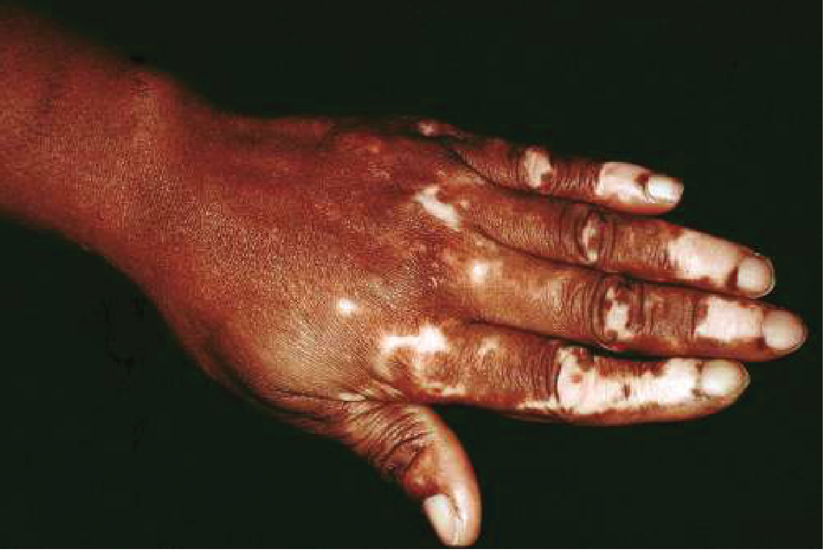
The first helpful step is to determine whether depigmentation is congenital (present at/shortly after birth) vs. acquired. Fair-skinned patients may not manifest congenital lesions until after their first sun exposure.
- Fitzpatrick's Dermatology, Vol. 1-2, Ch. 76
1. INHERITED / CONGENITAL HYPOMELANOSES
These are the most commonly misdiagnosed as vitiligo in neonates and young children.
| Condition | Key Distinguishing Features |
|---|---|
| Piebaldism (KIT gene mutation) | White forelock + anterior body midline depigmentation + bilateral shin depigmentation. Present at birth. Stable, non-progressive. Absence of melanocytes on biopsy. Autosomal dominant |
| Waardenburg Syndrome (PAX3 / MITF mutations) | White forelock, white skin macules, hypertelorism, heterochromic or hypoplastic blue irides, congenital sensorineural deafness, broad nasal root, early greying. Shah-Waardenburg variant includes Hirschsprung disease |
| Tuberous Sclerosis (TSC1 / TSC2 mutations) | Hypomelanotic "ash-leaf" macules in the first years of life + angiofibromas (age 3-4 years) + ungual fibromas + shagreen patches + seizures + confetti-like hypopigmented macules in childhood. Autosomal dominant |
| Hypomelanosis of Ito | Hypopigmented macules/patches along Blaschko lines in a linear/whorled pattern, usually within first 2 years of life. Associated chromosomal or genetic mosaicism |
| Nevus Depigmentosus | Stable, unilateral, often segmental hypopigmented (not fully depigmented) birthmark. Partial decrease in melanin, not absence |
| Oculocutaneous Albinism (OCA) | Diffuse generalized depigmentation of skin, hair, eyes. Nystagmus, photophobia, decreased visual acuity. Total absence of tyrosinase activity in type IA. Differentiated from vitiligo by congenital onset and diffuse involvement |
2. INFECTIOUS DISORDERS
| Condition | Key Distinguishing Features |
|---|---|
| Tinea (Pityriasis) Versicolor | Well-demarcated finely scaling macules/patches in sebaceous areas (chest, back, shoulders). Yellow-green fluorescence on Wood's lamp (due to Malassezia). KOH shows hyphae + spores ("spaghetti and meatballs"). Lesions are hypopigmented, not fully depigmented |
| Leprosy (tuberculoid / borderline forms) | Mainly hypopigmented (not chalk-white) patches with localized anesthesia (loss of sensation), decreased sweating, thickened peripheral nerves. Endemic area exposure. AFB on biopsy or slit-skin smear |
| Secondary Syphilis (leukoderma syphiliticum) | "Necklace of Venus" - postinflammatory hypopigmented/depigmented patches on neck, trunk, limbs. Positive serologic testing (VDRL, TPHA). History of primary chancre |
3. POSTINFLAMMATORY HYPOPIGMENTATION
Loss of pigment follows prior skin inflammation. Distinguished by history of pre-existing inflammatory condition.
- Atopic dermatitis - most common cause; hypopigmented patches in flexural areas, often with itch history
- Discoid (cutaneous) lupus erythematosus - atrophic, scarred patches with follicular plugging; positive ANA, anti-dsDNA; immunofluorescence
- Lichen sclerosis et atrophicus - ivory-white atrophic plaques, often perigenital/perianal; "figure-of-eight" pattern; biopsy shows epidermal atrophy
- Psoriasis - resolving plaques leave hypopigmentation; silvery scales, Auspitz sign in active lesions
- Scleroderma (systemic sclerosis) - leukoderma with retention of perifollicular pigmentation ("salt-and-pepper" pattern), skin tightening, Raynaud's, sclerodactyly
4. CHEMICAL / DRUG-INDUCED LEUKODERMA
| Condition | Key Distinguishing Features |
|---|---|
| Chemical leukoderma | Exposure to cytotoxic melanocyte compounds - monobenzyl ether of hydroquinone, catechols, phenols (industrial exposure). Occupational history critical. Can mimic vitiligo closely and may actually trigger true vitiligo via Koebner phenomenon |
| Drug-induced leukoderma | Imatinib (tyrosine kinase inhibitor), interferon, checkpoint inhibitors. Drug exposure history |
| Leukoderma of melanoma | Depigmented patches around or within a melanoma lesion due to T-cell cross-reactivity to melanocytes. Good prognostic sign with immunotherapy; exclusion of metastases required if spontaneous. Histology and dermoscopy differentiate |
5. PARAMALIGNANT / NEOPLASTIC HYPOMELANOSES
| Condition | Key Distinguishing Features |
|---|---|
| Mycosis Fungoides (Hypopigmented type) | Scarred, irregular hypopigmented patches on non-sun-exposed areas. Atrophic epidermal surface. May have concurrent plaques or tumors. Biopsy shows epidermotropism of atypical lymphocytes (hallmark) |
| Cutaneous Melanoma | Dyschromic lesion with pigmented areas + surrounding depigmentation. Irregular borders, dermoscopic features of malignancy |
6. IDIOPATHIC ACQUIRED HYPOMELANOSES
| Condition | Key Distinguishing Features |
|---|---|
| Idiopathic Guttate Hypomelanosis | Small (2-4 mm), well-demarcated, hypopigmented (not depigmented) macules on photo-exposed areas - shins, extensor forearms. Age-related, common. Slowly progressive, non-confluent |
| Progressive Macular Hypomelanosis | Nummular, non-scaly hypopigmented spots on the trunk, often confluent. Young adults. No fluorescence on Wood's lamp (unlike tinea versicolor) |
| Pityriasis Alba | Poorly defined, faintly scaly, hypopigmented (not depigmented) patches predominantly on the face of children/adolescents. Associated with atopic diathesis. Self-limiting |
| Halo Nevus (Sutton's Nevus) | Depigmented white halo surrounding a central pigmented melanocytic nevus. The halo represents autoimmune destruction of nevus melanocytes. Isolated finding; may coexist with vitiligo |
| Nevus Anemicus | Pale patch due to localized vasoconstriction, not loss of pigment. Lesion disappears with diascopy (pressure). Wood's lamp negative (no true depigmentation) |
7. SYSTEMIC DISEASES TO EXCLUDE
Four systemic diseases should be specifically considered when vitiligo-like findings are present:
- Systemic Sclerosis (Scleroderma) - salt-and-pepper depigmentation pattern, skin induration, Raynaud's
- Vogt-Koyanagi-Harada (VKH) Syndrome - vitiligo + uveitis + poliosis + meningitis + hearing loss. Most severe uveitis associated with vitiligo
- Waardenburg Syndrome - (see above)
- Shah-Waardenburg Syndrome - Waardenburg features + Hirschsprung disease
Key Diagnostic Tools
| Tool | Use |
|---|---|
| Wood's Lamp (365 nm UV) | Vitiligo fluoresces bright blue-white (accentuated depigmentation). Tinea versicolor shows yellow-green. Helpful in fair-skinned patients where lesions may not be visible in normal light |
| KOH preparation | Fungal hyphae and spores confirm tinea versicolor |
| Skin biopsy | Absence of melanocytes (S100, Melan-A negative) in vitiligo. Epidermotropic lymphocytes in mycosis fungoides. Granulomas in sarcoidosis |
| Serology | Treponemal tests for syphilis; ANA, anti-dsDNA for lupus; thyroid antibodies (up to 30% of vitiligo patients have associated thyroid disease) |
Summary Table
| Category | Key Conditions |
|---|---|
| Inherited | Piebaldism, Waardenburg syndrome, Tuberous sclerosis, Hypomelanosis of Ito, OCA |
| Infectious | Tinea versicolor, Leprosy, Secondary syphilis |
| Postinflammatory | Atopic dermatitis, Discoid LE, Lichen sclerosis, Psoriasis, Scleroderma |
| Chemical/Drug | Phenol leukoderma, Imatinib, Melanoma-associated leukoderma |
| Neoplastic | Hypopigmented mycosis fungoides, Cutaneous melanoma |
| Idiopathic | Idiopathic guttate hypomelanosis, Pityriasis alba, Halo nevus, Progressive macular hypomelanosis |
| Systemic | Scleroderma, VKH syndrome, Waardenburg, Shah-Waardenburg |
Sources:
- Fitzpatrick's Dermatology, Vol. 1-2, Chapter 76 (Vitiligo - Differential Diagnosis Tables 76-1 and 76-2)
- Harrison's Principles of Internal Medicine 22E, Chapter 61 (Hypopigmentation - Table 61-10)
- Goldman-Cecil Medicine, Chapter 408 (Circumscribed Hypopigmentation)
- Dermatology 2-Volume Set 5e, Chapter 66
This is a shared conversation. Sign in to Orris to start your own chat.