
Cardiac conditions requiring open heart surgery: briefly describe the pathophysiology, clinical presentations and management of the following conditions:
I notice the question lists conditions requiring open heart surgery but the specific conditions weren't included in the message. Let me ask for clarification before proceeding.
Asking for Preferences
Which cardiac conditions would you like covered? Please list them below.
The user wants coverage of Atrial Septal Defect (ASD). Let me read the medical library skill and search for content in parallel.
Reading File
Now let me search the medical library for ASD content in parallel across headings and paragraphs:
Finding Sources
Finding Sources
The best sources are Sabiston (surgery textbook), Robbins & Kumar (pathology), and Goldman-Cecil (medicine). Let me read the most relevant blocks in parallel:
Reading File
Reading File
Reading File
Excellent - I have rich content from all three key sources. Let me now fetch the ASD diagram from Robbins and get the Sabiston content on surgical details/types, and also get a recent PubMed search:
Loading Image
Reading File
Reading File
Searching PubMed
"atrial septal defect" AND management
I now have all the information needed for a thorough, well-cited response. Let me also fetch the surgical closure diagram:
Loading Image
I now have everything needed for a thorough answer. Here is the complete response:
Atrial Septal Defect (ASD)
ASD is one of the most common congenital cardiac lesions, occurring in approximately 13 per 10,000 live births, and is the most common congenital defect diagnosed in adults because - unlike VSDs - ASDs rarely close spontaneously. - Sabiston Textbook of Surgery, p. 2564
Types of ASD
There are three anatomic subtypes, classified by location:
| Type | Frequency | Location | Associated Anomalies |
|---|---|---|---|
| Secundum | ~90% | Fossa ovalis / central septum | Usually isolated |
| Primum | ~5% | Adjacent to AV valves | AV valve abnormalities, VSD |
| Sinus venosus | ~5% | Near SVC or IVC orifice | Anomalous pulmonary venous return |
- Robbins, Cotran & Kumar Pathologic Basis of Disease, p. 504
Pathophysiology
Normal fetal development: In fetal life, the lungs are non-functional and pulmonary pressures exceed systemic pressures, keeping the foramen ovale open (right-to-left). At birth, lung expansion drops pulmonary vascular resistance (PVR), left atrial pressure rises above right atrial pressure, and the foramen ovale closes.
ASD mechanism: When closure is incomplete (especially of the septum primum in secundum ASDs), a persistent communication between the atria remains. Because systemic vascular resistance (SVR) greatly exceeds PVR, and because the right ventricle is far more compliant than the left, blood flows left-to-right across the defect. - Robbins, p. 504

Fig. 12.3 - Common congenital causes of left-to-right shunts. Arrow in (A) shows the direction of ASD flow from LA to RA. (Robbins, Cotran & Kumar)
Consequences of chronic left-to-right shunting:
-
Pulmonary blood flow may be 2-8x normal (Qp/Qs can exceed 3:1 in large defects)
-
Right heart volume overload - right atrial and right ventricular dilation and hypertrophy
-
Increased flow through the pulmonary valve causes a flow murmur
-
Over decades, sustained right-sided pressure and volume overload can lead to pulmonary arterial hypertension - less common in ASDs than VSDs, but possible
-
Paradoxical right-to-left shunting can occur transiently during increased intrathoracic pressure (Valsalva, coughing), risking paradoxical embolism and stroke
-
In advanced disease: Eisenmenger syndrome (reversal of shunt to right-to-left, cyanosis) - though this is far less common than with VSDs
-
Sabiston Textbook of Surgery, p. 2564; Robbins, p. 504-505
Clinical Presentation
In children: Most are asymptomatic or have only mild exercise intolerance and frequent respiratory tract infections. - Sabiston, p. 2564
In adults (symptoms typically emerge in the 3rd-5th decade):
-
Dyspnea on exertion - the most common presenting symptom
-
Fatigue and exercise intolerance
-
Palpitations, atrial fibrillation/flutter - due to atrial dilation and volume overload
-
Syncope or stroke - from atrial arrhythmias or paradoxical embolism
-
Signs of right heart failure: peripheral edema, elevated JVP, hepatomegaly in late disease
-
More than 70% of patients are functionally impaired by the 5th decade if untreated
-
Goldman-Cecil Medicine, p. 980
Physical Examination
| Finding | Mechanism |
|---|---|
| Wide, fixed splitting of S2 | Hallmark sign - increased venous return raises right atrial pressure during inspiration, offsetting normal respiratory variation; delayed pulmonary valve closure from RV overload |
| Soft midsystolic murmur (2nd left interspace) | Increased flow across the pulmonary valve |
| Mid-diastolic murmur (lower left sternal border) | Increased flow across the tricuspid valve (with large shunts) |
| Right ventricular heave | RV volume overload, felt at left parasternal area |
- Goldman-Cecil Medicine, p. 984
Investigations
-
ECG: Incomplete right bundle branch block (rSR' pattern in V1) is characteristic; right axis deviation; prolonged PR interval, atrial fibrillation/flutter with advanced disease
-
Chest X-ray: Pulmonary vascular plethora (bilateral increased lung markings), dilated main pulmonary artery and branches, right atrial and right ventricular enlargement
-
Echocardiography (TTE/TEE): Definitive diagnostic tool - identifies the ASD location and size, quantifies the shunt ratio (Qp/Qs), estimates pulmonary artery pressures, and assesses RV function. Note: sinus venosus ASDs can be missed on TTE and require TEE or MRI
-
Cardiac catheterization: Not routinely needed, but used when pulmonary hypertension needs quantification
-
Goldman-Cecil Medicine, p. 985-986
Management
Indications for Closure
Closure is recommended when there is:
- Qp/Qs ratio ≥ 1.5:1 with evidence of right heart volume overload
- Paradoxical embolism
- Symptomatic patients at any age
Most centers recommend closure before school age (4-5 years) to prevent long-term right heart remodeling. Closure is generally contraindicated once irreversible pulmonary hypertension (Eisenmenger syndrome) has developed.
1. Catheter-Based (Device) Closure
Now the preferred approach for secundum ASDs, accounting for over 60% of ASD interventions worldwide. The Amplatzer Septal Occluder (and similar nitinol mesh devices) is deployed via transfemoral catheter under echocardiographic and fluoroscopic guidance. Requirements:
- Adequate septal rim (≥5 mm on most borders)
- Suitable defect anatomy (secundum type)
- Defect diameter typically ≤38 mm
A 2025 meta-analysis (de Liyis et al., PMID 38597284) comparing surgical vs. transcatheter closure in pediatric secundum ASD confirmed that both are highly effective, with transcatheter closure offering shorter hospital stays and fewer procedural complications.
2. Surgical Closure (Open Heart Surgery)
Indications for surgery over catheter closure:
- Primum and sinus venosus ASDs (not amenable to device closure)
- Inadequate septal rim for device anchorage
- Associated anomalies requiring surgical correction (anomalous pulmonary veins, AV valve repair)
- Failed device closure
Procedure: Performed under cardiopulmonary bypass via median sternotomy or minimally invasive right thoracotomy. A right atriotomy is made, and the defect is closed by:
- Direct suture closure - for small defects
- Patch closure - autologous pericardium or prosthetic material (Dacron/Gore-Tex) for larger defects
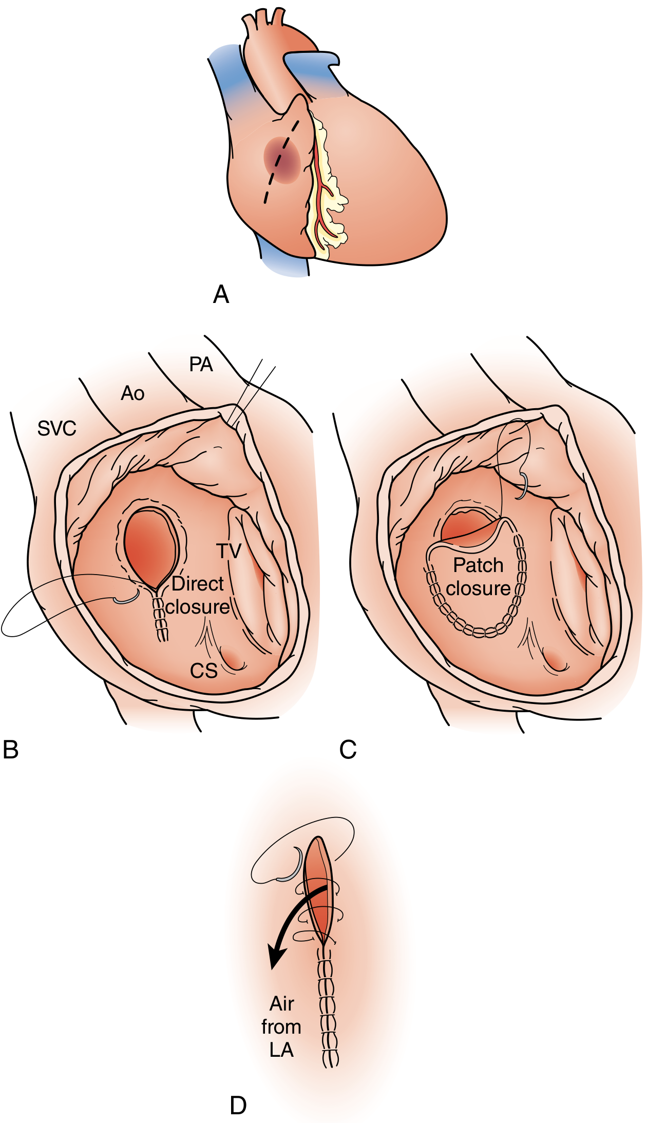
FIGURE 113.10 - Surgical closure for ASD: (A) Right atriotomy, (B) Direct suture closure, (C) Patch closure, (D) De-airing the LA. (Sabiston Textbook of Surgery)
For sinus venosus ASDs with partial anomalous pulmonary venous return, more complex repairs are required:
-
Superior type: intracardiac patch baffle, or the Warden procedure (SVC translocation) when anomalous veins drain high
-
Inferior type (scimitar syndrome): patch baffle in the IVC, potentially requiring hypothermic circulatory arrest
-
Sabiston Textbook of Surgery, p. 2564-2397
Outcomes: Surgical closure carries a very low perioperative mortality, with virtually no risk of residual defects. Post-operative survival is comparable to the general population when repair is done before the development of significant pulmonary hypertension. A 2025 systematic review (El-Andari et al., PMID 40517112) confirmed that minimally invasive approaches to surgical ASD repair have equivalent outcomes to conventional sternotomy with improved cosmesis and recovery.
3. Medical Management
- There is no medical therapy that closes the defect
- Antiplatelet therapy (aspirin) may be prescribed post-device closure for 3-6 months
- Rate/rhythm control and anticoagulation for atrial arrhythmias
- Diuretics and heart failure therapy if right heart failure is present
Summary Table
| Feature | Detail |
|---|---|
| Incidence | 13/10,000 live births |
| Most common type | Secundum (90%) |
| Shunt direction | Left-to-right (initially) |
| Hallmark sign | Wide, fixed splitting of S2 |
| ECG finding | Incomplete RBBB (rSR' in V1) |
| Echo finding | RV dilation, interatrial shunt on Doppler |
| Primary treatment | Device closure (most secundum ASDs) or surgery |
| Surgery needed for | Primum, sinus venosus, complex anatomy |
| Bypass technique | Cardiopulmonary bypass via right atriotomy |
| Prognosis (repaired) | Normal life expectancy if closed before PH develops |
Exam answer detailed
Asking for Preferences
Which conditions would you like detailed exam answers for? · What exam level is this for?
Searching sources...
Let me pull additional depth from the textbooks on embryology, complications, Eisenmenger, and anesthetic considerations for an MBBS finals-level answer.
Finding Sources
Finding Sources
Finding Sources
Now let me pull more depth from Braunwald's and the Maternal-Fetal Medicine book on ASD-specific clinical details:
Reading File
Finding Sources
Finding Sources
Let me also pull the Harrison's 22E content on ASD and the Braunwald's clinical features section for maximum exam-level depth:
Reading File
Reading File
I also need the Braunwald's section on ASD clinical features and management indications for completeness:
Reading File
I now have comprehensive, exam-level content from multiple authoritative sources (Braunwald's, Harrison's, Sabiston, Robbins, Goldman-Cecil). Here is the full, structured MBBS finals exam answer:
ATRIAL SEPTAL DEFECT (ASD)
Detailed MBBS Finals Exam Answer
DEFINITION
An Atrial Septal Defect (ASD) is an abnormal, fixed communication between the left and right atria through the interatrial septum. It is one of the most common congenital cardiac defects, occurring in 13 per 10,000 live births, and is the most frequent congenital heart lesion diagnosed in adults (since most ASDs - unlike VSDs - do not close spontaneously). - Robbins & Kumar, Pathologic Basis of Disease, p. 503
EMBRYOLOGY & CLASSIFICATION
Understanding ASD classification requires knowing normal atrial septum development:
Normal septal development:
- The septum primum grows down as a crescent-shaped membrane to partition the atria; the anterior gap beneath it is the ostium primum (allows fetal right-to-left flow)
- Before the ostium primum closes, perforations develop posteriorly forming the ostium secundum
- A second ingrowth - the septum secundum - grows from the right side to cover the ostium secundum, leaving a residual channel: the foramen ovale
- In fetal life, pulmonary pressure > systemic pressure, so the foramen ovale stays open (right-to-left)
- At birth, lung expansion drops pulmonary vascular resistance (PVR), left atrial pressure exceeds right atrial pressure, and the foramen ovale closes
ASD develops when this closure is incomplete or the septum is deficient. - Robbins, p. 503-504
Types of ASD (Classified by Location)
| Type | Frequency | Location | Key Associations |
|---|---|---|---|
| Secundum ASD | 90% | Fossa ovalis / central septum | Deficient septum primum; usually isolated; can be single, multiple, or fenestrated |
| Primum ASD | 5% | Adjacent to AV valves (AV canal region) | Always with AV valve abnormalities (mitral cleft); often + VSD; associated with Down syndrome |
| Sinus venosus ASD | 5% | Near SVC or IVC junction | Associated with partial anomalous pulmonary venous return (PAPVR); NOT amenable to device closure |
| Coronary sinus defect | Rare | Between coronary sinus and LA | Rare; requires surgery |
- Harrison's Principles of Internal Medicine 22E, p. 2114; Robbins, p. 504
Exam tip: Primum ASD + mitral cleft + VSD = Atrioventricular Septal Defect (AVSD) / Endocardial cushion defect - seen in Down syndrome (trisomy 21).
Do NOT confuse ASD with Patent Foramen Ovale (PFO): PFO is persistence of the flap valve of the fossa ovalis without true septal deficiency. It causes no right heart dilation and is present in ~25% of adults. It is detected by bubble contrast echo (agitated saline) with Valsalva. - Harrison's, p. 2114
PATHOPHYSIOLOGY
Direction of Shunt
In a normal heart after birth, systemic vascular resistance (SVR) greatly exceeds pulmonary vascular resistance (PVR), and the right ventricle is far more compliant (distensible) than the left ventricle. Through the ASD, blood flows left-to-right, from the high-pressure/low-compliance left atrium to the lower-pressure/high-compliance right side. - Robbins, p. 505
Hemodynamic Consequences
Left atrium → (through ASD) → Right atrium
↓
Increased RV volume load
↓
Increased pulmonary blood flow (Qp/Qs may be 2-8x normal, up to >3:1)
↓
Right heart dilation (RA + RV enlargement)
↓
Dilated pulmonary artery trunk and branches
- Robbins, p. 505; Sabiston Textbook of Surgery, p. 2564
Progression over Time
| Stage | Mechanism | Clinical Consequence |
|---|---|---|
| Early (childhood) | Compensated RV volume overload | Often asymptomatic |
| Middle (3rd-4th decade) | Progressive RA/RV dilation | Exercise intolerance, palpitations, SVT, AF |
| Late (4th-5th decade) | RV failure, PA dilation | Dyspnea at rest, right heart failure |
| End-stage | Sustained pulmonary hypertension | Eisenmenger syndrome (shunt reversal to R→L, cyanosis) |
Eisenmenger Syndrome: Chronic high pulmonary blood flow causes intimal hyperplasia and medial hypertrophy of pulmonary arterioles. PVR rises progressively until it exceeds SVR, reversing the shunt to right-to-left. The patient becomes cyanotic. At this stage, ASD closure is contraindicated (would acutely overload the left heart). - Braunwald's Heart Disease, p. 879
Exam tip: Eisenmenger is less common in ASDs than in VSDs or PDAs because the lower-pressure atrial shunt causes less vascular trauma to pulmonary arterioles.
Paradoxical Embolism
Even before Eisenmenger, patients are at risk of transient right-to-left shunting during moments of raised intrathoracic pressure (Valsalva, coughing, sneezing). A venous thrombus (even a small DVT) can cross the defect and enter the arterial circulation, causing cryptogenic stroke or systemic embolism. - Braunwald's Heart Disease, p. 969
CLINICAL FEATURES
In Children
Most children are asymptomatic or have mild:
- Recurrent respiratory tract infections (due to increased pulmonary blood flow)
- Mild exercise intolerance
In Adults (symptoms typically emerge after the 3rd decade)
>70% of patients are functionally impaired by the 5th decade if untreated. - Goldman-Cecil Medicine, p. 980
Symptoms:
- Dyspnea on exertion (most common presenting symptom)
- Fatigue and reduced exercise tolerance
- Palpitations - from supraventricular arrhythmias (SVT, atrial flutter, AF)
- Syncope - from arrhythmias or reduced cardiac output
- Stroke - from paradoxical embolism or AF
- Signs of right heart failure in advanced disease: peripheral edema, elevated JVP, hepatomegaly
Physical Examination Findings
| Sign | Mechanism | Significance |
|---|---|---|
| Wide, FIXED splitting of S2 | Hallmark sign. RV ejection is prolonged by volume overload. Inspiration normally increases right-sided return and delays P2, but in ASD the increased venous return also decreases left-to-right shunting, offsetting the change - so splitting is constant regardless of respiration | Pathognomonic for ASD |
| Soft midsystolic ejection murmur (2nd left interspace) | Increased flow through pulmonary valve (flow murmur, NOT the ASD itself) | Heard in majority |
| Mid-diastolic murmur (lower left sternal border) | Increased tricuspid flow with large shunts | Indicates significant shunt |
| RV heave / parasternal lift | Right ventricular hypertrophy and dilation | Left parasternal area |
| Pulmonary artery pulsation | Dilated pulmonary trunk | 2nd left intercostal space |
| Cyanosis + clubbing | Late - Eisenmenger syndrome (shunt reversal) | Poor prognostic sign |
- Goldman-Cecil Medicine, p. 984; Braunwald's, p. 878
INVESTIGATIONS
1. ECG
- Incomplete right bundle branch block (rSR' pattern in V1) - most characteristic finding
- Right axis deviation
- Right atrial enlargement (tall peaked P waves)
- Prolonged PR interval (1st degree heart block)
- Atrial fibrillation / flutter in older patients
- In primum ASD: left axis deviation (due to involvement of the AV node region)
Exam tip: Left axis deviation on ECG + ASD = Primum ASD (down syndrome link). Incomplete RBBB = Secundum ASD.
2. Chest X-Ray
- Pulmonary plethora - increased bilateral vascular markings (increased pulmonary blood flow)
- Dilated main pulmonary artery and its branches
- Right atrial and right ventricular enlargement
- Normal or small aortic knuckle (relatively reduced systemic flow)
- In Eisenmenger: oligaemic lung fields peripherally + dilated central pulmonary arteries ("pruning")
3. Echocardiography (Definitive Investigation)
- TTE (transthoracic echo): Identifies primum and secundum ASDs, measures defect size, shows right heart dilation. Doppler estimates Qp/Qs and pulmonary artery pressure
- TEE (transoesophageal echo): Required for sinus venosus ASDs (often missed on TTE) and for pre-procedural sizing before device closure. Also used intra-operatively during device deployment
- Bubble contrast echo (agitated saline): Detects right-to-left shunting (bubbles in LA/LV = positive); needed to detect PFO during Valsalva
- Findings: drop-out of interatrial septum signal, RV volume overload (RV dilation, paradoxical septal motion), LA-to-RA color Doppler jet
4. Cardiac MRI
- Excellent for sinus venosus defects, anomalous pulmonary venous anatomy, and pre-surgical planning
- Accurately quantifies Qp/Qs
5. Cardiac Catheterization
- Not routinely needed for diagnosis
- Indicated when: pulmonary hypertension suspected, conflicting echo results, pre-operative assessment of PVR
- Finding: step-up in oxygen saturation at the right atrial level (due to mixing of oxygenated left atrial blood)
- Qp/Qs calculated from oxygen saturations
6. Pulse Oximetry
- At rest and on exercise - to detect right-to-left shunting (desaturation) in patients with suspected Eisenmenger syndrome - Braunwald's, p. 878
MANAGEMENT
General Principles
- Closure is recommended when Qp/Qs ≥ 1.5:1 with RV volume overload, regardless of symptoms
- Optimal timing: before school age (4-5 years); adult repair still beneficial if done before irreversible PH develops
- Contraindication to closure: Eisenmenger syndrome (fixed severe pulmonary hypertension with resting right-to-left shunt)
-
60% of ASD interventions worldwide are now catheter-based - Sabiston, p. 2565
1. Catheter-Based (Percutaneous) Device Closure
Indications: Secundum ASD with adequate septal rim (≥5 mm on most sides) and defect diameter typically ≤38 mm
Procedure:
- Transfemoral venous access; catheter guided across the defect
- Amplatzer Septal Occluder (most widely used) - a self-centering, double-disc nitinol mesh device deployed under TEE + fluoroscopic guidance
- Device endothelialises within 3-6 months
Advantages: No thoracotomy, shorter hospital stay, faster recovery, excellent cosmesis
Post-procedure complications:
- Atrial arrhythmias
- Complete heart block (rare)
- Device thrombus (aspirin prescribed for 3-6 months)
- Device erosion (~1 in 1000 cases) - can cause haemopericardium; any chest pain post-procedure warrants urgent echo - Braunwald's, p. 878-879
A 2025 meta-analysis (de Liyis et al., PMID 38597284) comparing surgical vs. transcatheter closure in pediatric secundum ASD confirmed both are highly effective, with transcatheter offering fewer complications and shorter hospital stays.
2. Surgical Closure (Open Heart Surgery)
Indications for surgery over device closure:
- Primum ASD (always requires surgery; device not possible)
- Sinus venosus ASD (always requires surgery; cannot be catheter-closed)
- Coronary sinus defect
- Secundum ASD with inadequate rims for device anchorage
- Very large secundum defects
- Associated anomalies requiring surgical repair (AV valve repair, PAPVR repair)
- Failed or complicated device closure
Procedure:
- General anaesthesia; median sternotomy (or right mini-thoracotomy for minimally invasive approach)
- Cardiopulmonary bypass established (aortic + venous cannulation)
- Aortic cross-clamp + cardioplegia for myocardial protection
- Right atriotomy - direct visualisation of the defect
- Closure:
- Direct suture closure - for small defects (primary closure with continuous suture)
- Patch closure - autologous pericardium or synthetic patch (Dacron / Gore-Tex) for larger defects
- De-airing of the left atrium before closure
- Termination of bypass; closure
Sinus venosus ASD (superior type) with PAPVR to SVC:
- Intracardiac patch baffle redirecting anomalous pulmonary veins to LA
- Warden procedure: SVC is divided, the cardiac end oversewn, and the cephalic SVC end is anastomosed to the right atrial appendage; a patch baffles the anomalous veins to the LA
Inferior sinus venosus ASD (Scimitar syndrome):
- Complex repair; may require patch baffle within the intrahepatic IVC
- May need hypothermic circulatory arrest - Sabiston, p. 2396
A 2025 systematic review (El-Andari et al., PMID 40517112) confirmed that minimally invasive surgical approaches (right mini-thoracotomy, robotic) produce equivalent outcomes to sternotomy with better cosmesis and shorter recovery.
Outcomes: Perioperative mortality is very low (<1%). Post-operative survival equals that of the normal population when repair is performed before development of significant pulmonary hypertension. - Robbins, p. 505
Residual issues post-repair:
- Atrial arrhythmias may persist (especially AF) - surgery does not reliably reduce arrhythmia burden in older adults - Braunwald's, p. 878
- Follow-up TTE at 1 week, 1 month, then annually for at least 5 years (device closure)
- Patients with elevated PVR at closure need ongoing monitoring as PAH can progress
3. Medical Management
- No pharmacological therapy closes the defect
- Antiplatelet therapy (aspirin 75-150 mg/day) for 3-6 months post-device closure (prophylaxis against device thrombus)
- Rate/rhythm control + anticoagulation (warfarin or NOAC) for atrial fibrillation
- Diuretics + ACE inhibitors for right heart failure symptoms
- Pulmonary arterial hypertension (PAH) therapy (e.g., sildenafil, bosentan, prostacyclins) for Eisenmenger syndrome - treats symptoms but does not reverse the shunt
- Endocarditis prophylaxis: NOT routinely required for isolated secundum ASD (low risk); required for 6 months post-device/surgical repair
COMPLICATIONS OF UNTREATED ASD
| Complication | Mechanism |
|---|---|
| Right heart failure | Chronic RV volume overload |
| Atrial fibrillation / flutter | RA dilation causing electrical remodelling |
| Pulmonary arterial hypertension | Chronic high-flow pulmonary circulation |
| Eisenmenger syndrome | Reversal of shunt due to fixed PVR > SVR |
| Paradoxical embolism / stroke | Venous thrombus crossing atrial defect |
| Infective endocarditis | Rare in secundum ASD; more common with primum |
QUICK COMPARISON: ASD vs PFO
| Feature | ASD | PFO |
|---|---|---|
| Definition | True deficiency of septal tissue | Incomplete fusion of flap valve |
| Prevalence | 13/10,000 live births | ~25% of adults |
| Right heart dilation | Yes (if large) | No |
| Detectable on TTE | Usually | No (needs bubble echo + TEE) |
| Spontaneous closure | Rarely | Often remains patent |
| Clinical significance | Shunting, RV overload, arrhythmias | Paradoxical embolism risk |
| Treatment | Closure (device or surgery) | Device closure if cryptogenic stroke |
SUMMARY - KEY EXAM POINTS
| Feature | Answer |
|---|---|
| Incidence | 13 per 10,000 live births |
| Most common type | Secundum (90%) |
| Most common CHD in adults | ASD (due to lack of spontaneous closure) |
| Shunt direction | Left-to-right initially |
| Hallmark auscultatory sign | Wide, fixed splitting of S2 |
| ECG in secundum ASD | Incomplete RBBB (rSR' in V1), right axis deviation |
| ECG in primum ASD | Incomplete RBBB + left axis deviation |
| CXR findings | Pulmonary plethora, dilated PA, RA+RV enlargement |
| Definitive investigation | Echocardiography (TTE ± TEE) |
| Step-up in O2 saturation at catheter | At right atrial level |
| Qp/Qs for closure | ≥ 1.5:1 with RV overload |
| Preferred treatment (secundum) | Amplatzer device (catheter-based) |
| Types requiring surgery | Primum, sinus venosus, coronary sinus defects |
| Surgical approach | Cardiopulmonary bypass, right atriotomy, suture or patch |
| Contraindication to closure | Eisenmenger syndrome |
| Down syndrome association | Primum ASD (AVSD / endocardial cushion defect) |
| Prognosis after timely repair | Normal life expectancy |
Next ventricular septal defect tetralogy of Fallot, double outlet right ventricle. transposition of great vessels, AV canal defect.
Searching sources... Running all 5 condition searches in parallel.
Finding Sources
Finding Sources
Finding Sources
Finding Sources
Finding Sources
Excellent results. Now reading the richest blocks for each condition in parallel - Schwartz's for VSD, Robbins for TOF, Sabiston for TGA & AVSD, Schwartz's for DORV:
Reading File
Reading File
Reading File
Reading File
Reading File
Now fetching VSD management details from Schwartz's (continued), TOF clinical & surgical details from Sabiston, TGA surgical continuation, and AVSD surgical details:
Reading File
Finding Sources
Reading File
Reading File
Now let me get TOF surgical details from Schwartz's, TGA arterial switch details from Sabiston, and fetch key diagrams in parallel:
Reading File
Reading File
Loading Image
Loading Image
Now fetching the TOF anatomy diagram and the AVSD two-patch diagram:
Loading Image
Loading Image
Reading File
I have comprehensive content from all sources. Here is the full MBBS Finals exam answer:
CONGENITAL HEART DEFECTS REQUIRING OPEN HEART SURGERY
Detailed MBBS Finals Exam Answers
1. VENTRICULAR SEPTAL DEFECT (VSD)
Definition
A VSD is a pathologic communication (hole) between the left and right ventricles through the interventricular septum. It is the most common form of congenital heart disease, comprising 20-30% of all CHD cases. - Schwartz's Principles of Surgery, p. 814
Classification (by Location)
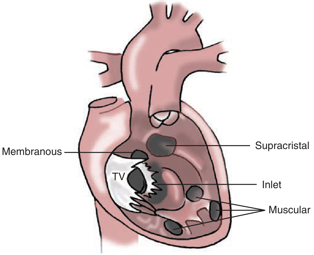
| Type | Frequency | Location | Key Features |
|---|---|---|---|
| Perimembranous (membranous) | ~80% | Membranous septum, near tricuspid valve | Most common surgical type; includes malalignment VSDs in TOF |
| Muscular | Most common overall | Trabecular muscular septum | May be multiple ("Swiss-cheese septum"); can close spontaneously |
| AV canal (inlet) | ~5% | Below tricuspid annulus, no muscle between defect and AV valve | Part of AVSD spectrum |
| Supracristal (outlet) | ~5% | Infundibular septum, below pulmonary valve | Risk of aortic valve prolapse into defect |
Pathophysiology
The LV systolic pressure greatly exceeds RV pressure under normal conditions, so blood flows left-to-right through the VSD.
-
Small (restrictive) VSDs: Offer high resistance to flow; RV pressure remains normal or near-normal; Qp/Qs rarely exceeds 1.5. The high-velocity jet of blood creates turbulence but causes no significant volume overload. Risk: endocarditis from endocardial damage.
-
Large (non-restrictive) VSDs: Equalize LV and RV pressures; Qp/Qs can be very high. Causes massive pulmonary overcirculation and LV volume overload. If untreated, sustained high pulmonary blood flow causes pulmonary vascular remodeling, rising PVR, and ultimately Eisenmenger syndrome (shunt reversal to R→L, cyanosis). VSDs carry a higher risk of Eisenmenger than ASDs.
-
Schwartz's Surgery, p. 814-815
Exam tip: ~50% of small/moderate muscular VSDs close spontaneously in the first 2 years of life. Perimembranous VSDs also may close but less often. Outlet VSDs virtually never close spontaneously.
Clinical Presentation
Symptoms
- Small VSD: Asymptomatic (discovered incidentally on auscultation)
- Large VSD: In infancy - congestive heart failure (tachypnoea, poor feeding, failure to thrive, recurrent chest infections, diaphoresis with feeding); exercise intolerance; growth retardation
Signs
| Sign | Mechanism |
|---|---|
| Harsh pansystolic murmur (lower left sternal border) | High-velocity L→R jet through VSD; loudest at 3rd-4th left intercostal space |
| Loud P2 | Pulmonary hypertension (increased flow) |
| Apical mid-diastolic rumble | Increased flow across mitral valve (Qp/Qs ≥2) |
| Thrill | Often palpable in large VSDs |
| RV heave | RV pressure/volume overload |
| Cyanosis + clubbing | Late sign - Eisenmenger syndrome (shunt reversal) |
Exam tip: A very small VSD makes a loud murmur (high-velocity jet, maladie de Roger). A very large VSD may make a softer murmur (pressures equalize, less turbulence) but causes heart failure.
Investigations
- ECG: Left ventricular hypertrophy (or biventricular in large shunts); left atrial enlargement
- CXR: Cardiomegaly; pulmonary plethora (increased vascular markings); enlarged LA, LV; dilated pulmonary artery
- Echocardiography (TTE): Definitive - shows defect, measures shunt, estimates PA pressure
- Cardiac catheterisation: Step-up in O2 saturation at right ventricular level; used to measure PVR when Eisenmenger is suspected
Management
Indications for Closure
- Qp/Qs ≥ 2:1 with symptoms or evidence of LV volume overload
- Any VSD causing heart failure, failure to thrive
- Outlet VSDs (risk of aortic regurgitation from valve prolapse)
- History of infective endocarditis
- Contraindication: Eisenmenger syndrome (fixed PVR; closure would acutely right-heart overload)
1. Surgical Closure (Primary treatment)
- Cardiopulmonary bypass, right ventriculotomy or right atriotomy (preferred - avoids RV incision)
- Patch closure with Dacron or autologous pericardium using interrupted pledgeted sutures (Fig. 20-60 - intraoperative interrupted suture technique)
- Device (catheter-based) closure: available for selected muscular and perimembranous VSDs in centres with expertise
- Pulmonary artery banding: Palliative procedure to reduce pulmonary blood flow in small infants not suitable for primary repair; debanded when definitive repair is done
2. Medical (pre-operative)
- Diuretics (furosemide), ACE inhibitors (captopril) for heart failure
- High-calorie feeds / nasogastric feeding for growth failure
- Endocarditis prophylaxis
Outcomes
Excellent - operative mortality <1-2%. Eisenmenger patients: not candidates for repair; managed with PAH therapies, oxygen; may be considered for heart-lung transplantation.
2. TETRALOGY OF FALLOT (TOF)
Definition
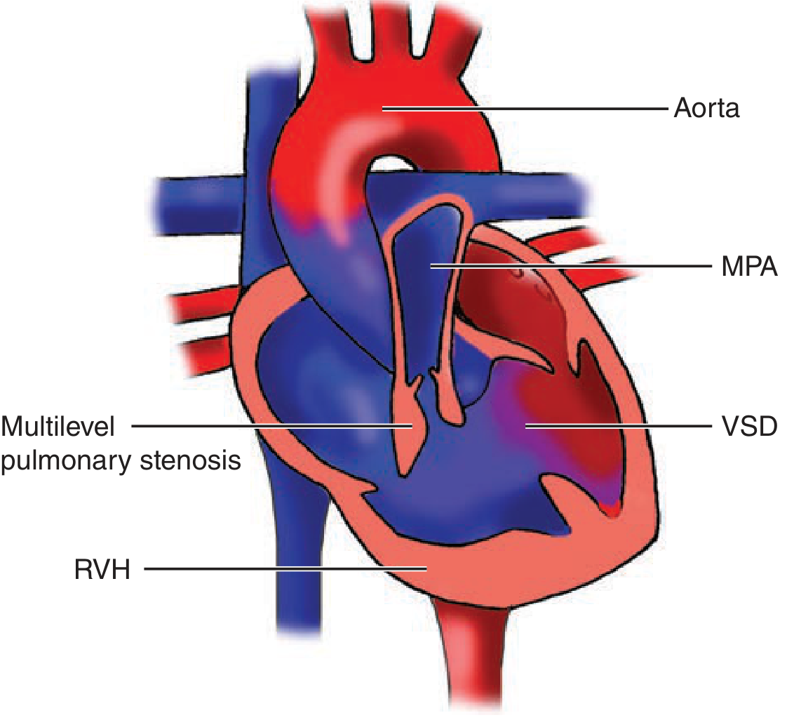
TOF is the most common cyanotic congenital heart disease, accounting for ~5% of all CHD. It is defined by four anatomical features, all caused by a single embryological defect: anterosuperior displacement of the infundibular (outlet) septum. - Robbins & Kumar Basic Pathology, p. 271
The Four Features (Mnemonic: PROVE)
- Pulmonary stenosis (RVOT obstruction - subvalvular infundibular stenosis ± valvular stenosis ± PA hypoplasia)
- Right ventricular hypertrophy (secondary to RVOT obstruction)
- Overriding aorta (aorta straddles the VSD, receiving blood from both ventricles)
- VSD (large, perimembranous, non-restrictive - the only "hole")
"Monology of Fallot" - Van Praagh: all four features result from the single malalignment of the infundibular septum. - Schwartz's Surgery, p. 812
Pathophysiology
The key determinant of clinical severity is the degree of RVOT obstruction:
RVOT obstruction → RV pressure rises
↓
Blood preferentially shunts RIGHT-TO-LEFT through VSD (deoxygenated blood → aorta)
↓
Desaturated blood enters systemic circulation → CYANOSIS
↓
Decreased pulmonary blood flow → Less oxygen exchange → Worsening hypoxaemia
↓
Polycythaemia (compensatory) → Thrombotic risk
-
If RVOT obstruction is mild: shunt may be L→R initially (acyanotic or "pink TOF")
-
If RVOT obstruction is severe: dominant R→L shunt, early cyanosis
-
Heart is enlarged, "boot-shaped" (coeur en sabot) on CXR due to RVH + concave pulmonary bay
-
The proximal aorta is dilated; pulmonary trunk is hypoplastic
-
In severe cases (pulmonary atresia): PDA or bronchial collaterals are the only source of pulmonary blood flow
-
Robbins, p. 288-290
Coronary anomaly: In ~3-5% of TOF, the left anterior descending (LAD) arises from the right coronary artery and crosses the RVOT - a surgically critical finding that alters the approach to RVOT relief.
Clinical Presentation
Symptoms
- Cyanosis - present at birth or develops in first months of life
- Clubbing of fingers and toes (chronic hypoxaemia)
- Polycythaemia (compensatory erythropoiesis)
- Squatting - characteristic posture in children after exertion; squatting increases SVR (compresses femoral arteries), decreasing R→L shunt and temporarily improving saturation
- Failure to thrive, exercise intolerance
Tet Spells (Hypercyanotic Episodes)
A medical emergency - peak incidence 2-4 months of age:
- Sudden infundibular spasm → acute decrease in pulmonary blood flow → severe hypoxaemia → crying/agitation → further spasm → vicious cycle
- Triggers: Crying, feeding, defecation, fever, tachycardia
- Features: Sudden deepening cyanosis, hyperpnoea, limpness, syncope, possible seizure/death
Management of tet spell:
- Knee-chest position (simulates squatting - increases SVR)
- Oxygen (100%)
- Morphine (0.1 mg/kg IV/SC) - reduces infundibular spasm, decreases tachycardia
- IV fluid bolus (increases preload)
- IV propranolol - reduces RVOT spasm
- IV phenylephrine - increases SVR, reduces R→L shunt
- Sodium bicarbonate if metabolic acidosis
- Urgent surgical repair if spells are refractory
Physical Signs
| Sign | Detail |
|---|---|
| Cyanosis and clubbing | Central cyanosis from birth or early infancy |
| Ejection systolic murmur | 2nd-3rd left intercostal space - from RVOT obstruction (NOT the VSD) |
| Soft or absent P2 | Reduced pulmonary blood flow |
| Absent/single S2 | Aortic closure only |
| RV heave | RVH |
| Absent VSD murmur | Large, non-restrictive VSD - no significant pressure gradient across it |
Exam tip: In TOF the murmur comes from the pulmonary stenosis, not the VSD. The louder the murmur, the less severe the TOF (more blood going through the RVOT). A quiet murmur = very severe obstruction = worse cyanosis.
Investigations
- ECG: Right axis deviation; right ventricular hypertrophy (tall R in V1, deep S in V5-V6)
- CXR: Boot-shaped heart (coeur en sabot) - RVH + upturned apex; decreased pulmonary vascular markings; right aortic arch in 25%
- Echocardiography: Definitive diagnosis; defines RVOT anatomy, VSD position, coronary origins, PA size
- Cardiac catheterisation: Rarely needed; can precipitate a tet spell; reserved for defining coronary anatomy or PA size if echo inconclusive
- MRI: Used in repaired TOF follow-up to quantify pulmonary regurgitation and RV volumes
Management
Pre-operative palliation
- Beta-blockers (propranolol) to reduce frequency of tet spells
- Modified Blalock-Taussig (BT) shunt: In small neonates not suitable for primary repair - subclavian artery to ipsilateral pulmonary artery (via Gore-Tex graft), increasing pulmonary blood flow; staged approach to definitive repair
Definitive Surgical Repair (Intracardiac repair)
Performed under cardiopulmonary bypass, usually at 3-6 months of age (or earlier if tet spells):
- VSD closure: Patch placed through right atrium (or right ventriculotomy), directing LV outflow to aorta
- RVOT relief: Resection of infundibular muscle; pulmonary valvotomy; transannular patch (pericardium or synthetic) if pulmonary annulus is hypoplastic
- Pulmonary artery reconstruction if necessary
Transannular patching (widening RVOT across the pulmonary valve annulus) relieves obstruction but creates pulmonary regurgitation - the major long-term complication requiring pulmonary valve replacement in adulthood.
Long-term Follow-up After Repair
- Pulmonary regurgitation - progressive RV dilation → RV failure → need pulmonary valve replacement (surgical or transcatheter Melody valve)
- Residual RVOT obstruction
- Ventricular arrhythmias (VT) - from RV fibrosis at ventriculotomy scar; risk of sudden cardiac death
- Right bundle branch block (RBBB) - common post-repair
- QRS duration >180ms on ECG = risk factor for VT and sudden death
3. DOUBLE OUTLET RIGHT VENTRICLE (DORV)
Definition
DORV is a spectrum of cardiac malformations in which both the aorta and pulmonary artery arise wholly or in large part from the right ventricle. It accounts for ~5% of CHD. The vast majority have a concomitant VSD, which is the only outlet for the LV. - Schwartz's Surgery, p. 812
Classification (by VSD Position)
The physiology and management of DORV are dictated primarily by the relationship of the VSD to the great vessels:
| VSD Type | Frequency | Physiology | Clinical Analogy |
|---|---|---|---|
| Subaortic VSD | 47% | LV blood → VSD → aorta; oxygenated | Large isolated VSD / congestive heart failure |
| Doubly committed VSD | 4% | Beneath both great vessels | Large VSD with pulmonary overcirculation |
| Non-committed VSD | 26% | Remote from both great vessels | Complex; variable |
| Subpulmonic VSD (Taussig-Bing anomaly) | 23% | LV blood → VSD → pulmonary artery; deoxygenated blood → aorta | Functionally like D-TGA; cyanosis |
Pathophysiology
The critical factors determining presentation:
- Size and location of VSD relative to great vessels
- Presence or absence of RVOT (pulmonary stenosis) obstruction
- Associated anomalies (arch hypoplasia, coarctation)
- Subaortic VSD without PS → pulmonary overcirculation, CHF (like large VSD)
- Subaortic VSD + PS → cyanosis (like TOF)
- Subpulmonic VSD (Taussig-Bing) → oxygenated LV blood streams into pulmonary artery; deoxygenated RV blood into aorta → cyanosis (like TGA)
Clinical Presentation
Three clinical scenarios depending on anatomy:
- Pulmonary overcirculation/CHF (subaortic or doubly committed VSD without PS) - presents in infancy with tachypnoea, failure to thrive, recurrent infections
- Cyanosis (subaortic VSD + PS) - resembles TOF
- Cyanosis with CHF (Taussig-Bing / subpulmonic VSD) - resembles TGA
Investigations: Echocardiography is the mainstay. MRI / cardiac catheterisation used for surgical planning. Key questions: coronary anatomy, additional VSDs, tricuspid-to-pulmonary valve distance.
Management
All DORV requires open heart surgery. The specific repair depends on anatomy:
| VSD Type | Surgical Strategy |
|---|---|
| Subaortic VSD (without PS) | Intraventricular tunnel connecting LV → VSD → aorta (biventricular repair) |
| Subaortic VSD + PS | Intracardiac tunnel + RVOT reconstruction (like TOF repair + VSD closure) |
| Subpulmonic VSD (Taussig-Bing) | Arterial Switch Operation (like TGA repair) + VSD closure |
| Non-committed VSD | Complex tunnel repair; or single-ventricle pathway if biventricular repair not feasible |
- Rastelli procedure: Intraventricular LV-to-aorta tunnel + RV-to-PA conduit (for VSD + PS cases)
- Arterial Switch Operation (ASO): For Taussig-Bing anatomy
- Palliative PA banding: If complex anatomy defers definitive repair
Reintervention is required in ~37% of patients at 15 years, mainly for RVOT reconstruction or conduit replacement. - Schwartz's Surgery, p. 813
4. TRANSPOSITION OF THE GREAT ARTERIES (TGA / D-TGA)
Definition
TGA is a discordant ventriculoarterial connection - the aorta arises from the morphologic RV and the pulmonary artery arises from the morphologic LV - while the atrioventricular connections are concordant (normal). It occurs in 2-3 per 10,000 live births and is the most common cause of cyanotic CHD presenting in the neonatal period. - Sabiston Textbook of Surgery, p. 2575
Exam tip - Concordance rules:
- TGA (D-TGA): AV concordant (RA→RV, LA→LV) + VA discordant (RV→Ao, LV→PA) = Complete TGA
- Congenitally Corrected TGA (ccTGA): AV discordant + VA discordant = physiologically corrected but with morphologic RV as systemic pump
Embryology
Failure of the truncal and aortopulmonary septa to spiral normally during development, so the aorta stays anterior and rightward, arising from the RV. - Robbins, p. 320
Pathophysiology
Systemic venous (deoxygenated) blood:
IVC/SVC → RA → RV → AORTA → systemic circulation → (never reaches lungs)
Pulmonary venous (oxygenated) blood:
Pulmonary veins → LA → LV → PULMONARY ARTERY → lungs → (never reaches systemic circulation)
The result is two parallel, non-communicating circulations - incompatible with postnatal life. Survival depends entirely on mixing at one or more levels:
- ASD / patent foramen ovale
- VSD (present in ~30% of cases - "TGA-VSD")
- PDA (patent ductus arteriosus)
Without adequate mixing, severe hypoxaemia and death occur within days of birth.
- Robbins, p. 320-322; Sabiston, p. 2575
Variants
- TGA-IVS (intact ventricular septum): Most common (~65%); presents with profound cyanosis at birth when PDA closes
- TGA-VSD (~35%): Sufficient mixing via VSD; may present with less cyanosis but with pulmonary overcirculation and CHF
- TGA-VSD with PS: Reduced pulmonary blood flow + cyanosis
Clinical Presentation
Symptoms
- Severe central cyanosis from birth (in TGA-IVS) - does NOT respond to oxygen administration (pathognomonic)
- Tachypnoea without respiratory distress (the "happy but blue" neonate)
- Metabolic acidosis from tissue hypoxia
- RV hypertrophy (RV acts as systemic ventricle): LV is small/hypoplastic (low-pressure pulmonary circuit)
Physical Signs
| Sign | Detail |
|---|---|
| Profound cyanosis | Present from birth |
| Single loud S2 | Anterior aortic valve closure (PA posterior, P2 inaudible) |
| No/soft murmur | In TGA-IVS; murmur present with VSD or PS |
| RV heave | Systemic RV |
Investigations
- CXR: Classic "egg on a string" (egg-shaped heart with narrow superior mediastinum due to parallel great vessels lying on top of each other, no thymic shadow); increased pulmonary vascular markings
- ECG: Right axis deviation; RVH
- Echocardiography: Definitive - shows AV concordance, VA discordance, coronary origins, VSD, size of LA/LV (important for ASO planning)
- Arterial blood gas: Profound hypoxaemia with metabolic acidosis; hyperoxia test - PaO2 fails to rise above ~150 mmHg on 100% O2 (unlike respiratory causes)
Management - This is a Neonatal Emergency
Immediate Stabilisation
- IV Prostaglandin E1 (PGE1) - maintains/reopens the ductus arteriosus, allowing mixing and improving systemic saturation (first-line emergency treatment)
- Balloon Atrial Septostomy (Rashkind procedure) - a balloon catheter is passed via the femoral/umbilical vein across the foramen ovale; the balloon is inflated in the LA and forcefully withdrawn to tear the atrial septum, creating an open ASD to allow atrial-level mixing (Fig. 113.27). Achieves SaO2 of 70-80%, buying time for definitive surgery
- Metabolic correction (bicarbonate if acidotic)
Definitive Surgery - Arterial Switch Operation (ASO / Jatene procedure)
The ASO is the gold-standard repair, providing anatomical correction. It must be done within the first 2-3 weeks of life (before the LV regresses from the low-pressure pulmonary circuit and loses its ability to support systemic work).
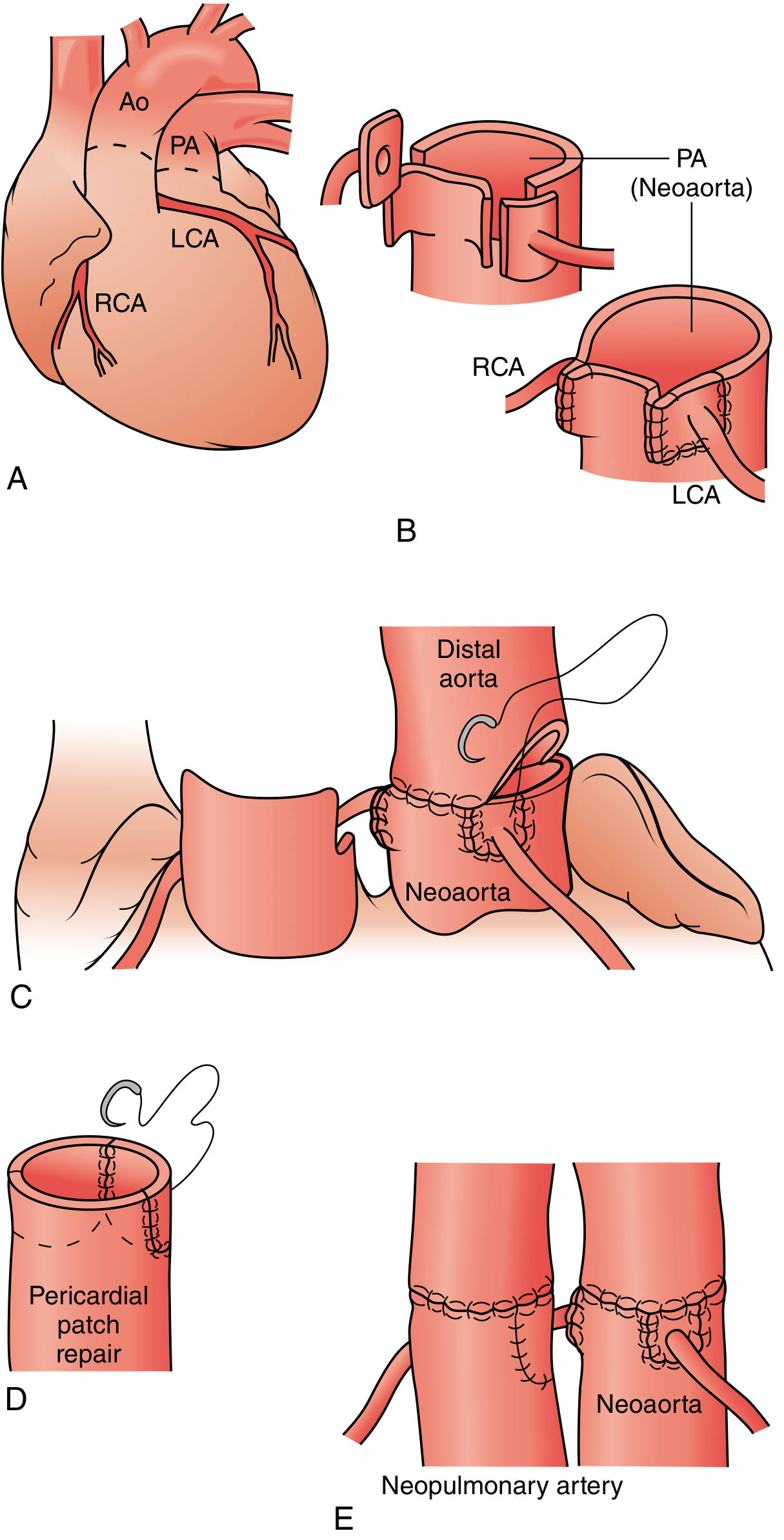
Steps of the ASO:
- Cardiopulmonary bypass + deep hypothermic circulatory arrest (neonates)
- Transect both great vessels above the sinuses of Valsalva
- Excise coronary arteries with buttons of aortic wall from the aortic root (now neoaortic/pulmonary root)
- Lecompte maneuver: The distal pulmonary artery bifurcation is brought anterior to the reconstructed neoaorta to prevent compression
- Coronary reimplantation into the pulmonary root (now neoaorta) - the most technically challenging step
- Distal aorta anastomosed to neoaorta (the original pulmonary root)
- Distal pulmonary artery (neopulmonary artery) anastomosed to what was the aortic root
- Pericardial patches to reconstruct the donor coronary sites on the original aorta (now neopulmonary artery)
- VSD closure (if present) via right atrium
Result: Morphologic LV now ejects to the aorta (systemic); morphologic RV now ejects to pulmonary artery. Anatomically and physiologically corrected.
Long-term complications of ASO:
- Neopulmonary artery stenosis (most common - at anastomotic sites)
- Coronary artery complications (kinking, stenosis, occlusion)
- Neoaortic valve insufficiency
- Excellent overall survival: >95% at 20 years in experienced centres
Historical (now obsolete): Atrial Switch Operations
- Mustard procedure (pericardial baffle) and Senning procedure (native atrial tissue baffle): Redirected systemic venous return to LV→PA and pulmonary venous return to RV→Ao = physiologic but not anatomic correction
- Problem: The morphologic RV remained the systemic ventricle and failed progressively → systemic RV failure, baffle obstruction/leaks, atrial arrhythmias
- Still seen in adults who were repaired before the ASO era
Congenitally Corrected TGA (ccTGA)
- Both AV and VA connections discordant: RA→morphologic LV→PA and LA→morphologic RV→Ao
- Physiologically corrected but RV acts as systemic ventricle
- May be asymptomatic for decades; progressive systemic RV failure; high incidence of complete heart block
- Surgical option: Double switch (atrial switch + ASO) to make morphologic LV the systemic ventricle; requires prior LV retraining if LV has been decompressed - Sabiston, p. 2578-2780
5. ATRIOVENTRICULAR CANAL DEFECT (AV CANAL / AVSD)
Definition
AV canal defect (AVSD / atrioventricular septal defect / endocardial cushion defect) is a complex spectrum of lesions involving:
- Deficiency of the atrial septum (primum ASD)
- Deficiency of the ventricular septum (inlet VSD)
- Abnormal AV valves (single common AV valve or cleft mitral valve)
It occurs in approximately 1 in 2,100 live births and results from failure of fusion of the endocardial cushions during embryogenesis. - Sabiston Textbook of Surgery, p. 2567
Key association: ~60-70% of complete AVSDs occur in patients with Trisomy 21 (Down syndrome). AVSD is the most common cardiac defect in Down syndrome.
Classification
| Type | ASD | VSD | AV Valves |
|---|---|---|---|
| Partial AVSD | Primum ASD | Absent | Cleft in anterior mitral leaflet; two separate AV valve orifices |
| Transitional AVSD | Primum ASD | Small restrictive inlet VSD | Two separate AV valve orifices |
| Complete AVSD | Primum ASD | Large non-restrictive inlet VSD | Common AV valve with 5 leaflets (most severe) |
Rastelli Classification (for complete AVSD - based on attachment of the superior bridging leaflet of the common AV valve):
- Type A (most common): Superior bridging leaflet committed to LV side; chordae attach to crest of VSD
- Type B: Bridging leaflet chordae attach to an anomalous papillary muscle in RV (straddling)
- Type C: "Free-floating" superior bridging leaflet; no chordal attachment to septum
Pathophysiology
In complete AVSD, there are shunts at both atrial and ventricular levels (L→R), plus AV valve regurgitation:
Primum ASD → L→R atrial shunt
+
Inlet VSD → L→R ventricular shunt (large, non-restrictive)
+
Common AV valve regurgitation → volume overload of both atria and ventricles
= Massive pulmonary overcirculation + biventricular volume overload
= CHF in infancy + early development of pulmonary vascular disease
Anatomical features of surgical importance:
- AV node and bundle of His are displaced inferiorly to the rim of the primum ASD and along the inferior edge of the VSD - must be protected during repair to avoid complete heart block
- "Goose-neck" deformity of LVOT on angiography - elongated LV outflow tract due to anterior displacement of the aortic valve
- Risk of early Eisenmenger syndrome (especially in Down syndrome patients, who have particularly reactive pulmonary vasculature)
Clinical Presentation
Partial AVSD
- May be asymptomatic until adulthood (behaves like an ASD with mitral regurgitation)
- Exercise intolerance, dyspnoea
- Signs: wide fixed S2 splitting + pansystolic murmur of MR (left axillary radiation)
- Left axis deviation on ECG is characteristic (due to displacement of conduction system)
Complete AVSD
- Presents in infancy (2-6 weeks) with:
- Congestive heart failure: tachypnoea, poor feeding, diaphoresis, failure to thrive
- Recurrent respiratory infections
- Cardiomegaly
- Physical signs: tachycardia, gallop rhythm, hepatomegaly, pansystolic murmur (MR/TR and VSD), wide fixed S2 splitting
Physical Signs Summary
| Sign | Detail |
|---|---|
| Pansystolic murmur | AV valve regurgitation and/or VSD shunting |
| Wide fixed S2 splitting | Due to primum ASD component |
| Signs of CHF | Hepatomegaly, oedema, crepitations |
| Cyanosis (late) | Eisenmenger; or in unbalanced forms |
Investigations
- ECG: Left axis deviation (characteristic - due to posteriorly displaced conduction system); superior QRS axis; incomplete RBBB; biventricular hypertrophy; prolonged PR interval
- CXR: Cardiomegaly; pulmonary plethora; dilated main pulmonary artery
- Echocardiography: Definitive - shows primum ASD, inlet VSD, common AV valve, valve regurgitation, Rastelli type, ventricular balance, PA pressure
- Cardiac catheterisation: In older patients to assess PVR (step-up at both RA and RV level)
- Karyotype/genetic testing: Given high association with Down syndrome (trisomy 21)
Exam tip - ECG axis: Partial AVSD = left axis deviation. Complete AVSD = superior axis (extreme left axis). This contrasts with secundum ASD (right axis deviation). Left axis deviation in a child with a murmur = think AVSD.
Management
Timing
- Complete AVSD: Surgical repair recommended by 3-6 months of age (before irreversible pulmonary vascular disease develops; Down syndrome patients may need even earlier repair due to more reactive pulmonary vasculature)
- Partial AVSD: May be deferred to 2-4 years of age if hemodynamically stable
Medical (Pre-operative)
- Diuretics, ACE inhibitors for CHF
- High-calorie feeds / NG feeds for growth
- Pulmonary vasodilators if pulmonary hypertension present
Surgical Repair - Open Heart Surgery
All AVSD requires surgical repair under cardiopulmonary bypass:
Goals:
- Close primum ASD
- Close inlet VSD
- Divide common AV valve into two separate (left and right) competent valves
- Protect the displaced AV node and bundle of His to avoid complete heart block
Techniques:
- Single-patch technique: One large patch used to close both ASD and VSD; common AV valve sutured to the patch
- Two-patch technique (preferred): Separate patches for the VSD (pericardial or Dacron) and ASD; AV valve divided and sutured to patches independently. Preserves more AV valve tissue and is associated with better valve function - Sabiston, p. 2567
Mitral valve (left AV valve): The cleft in the anterior mitral leaflet is sutured closed. Residual MR may require annuloplasty.
Critical intraoperative concern: The conduction tissue runs along the inferior rim of the VSD and the posterior-inferior border of the primum ASD - sutures must be placed away from this to avoid complete AV block.
Post-operative Considerations
- Pulmonary hypertensive crisis - may occur in the early post-operative period (ICU management: oxygen, hyperventilation, sedation, inhaled nitric oxide)
- Long-term risk: recurrent left AV valve regurgitation (most common reason for re-operation), LVOT obstruction (goose-neck deformity), arrhythmias, heart block
Outcomes
In experienced centres, operative mortality for complete AVSD repair is 2-5%. Freedom from reoperation at 10 years is ~85-90%. Patients with Down syndrome have similar outcomes to those without. - Braunwald's Heart Disease
MASTER COMPARISON TABLE
| Feature | VSD | TOF | DORV | TGA | AVSD |
|---|---|---|---|---|---|
| Shunt | L→R | R→L | Variable | Parallel circuits | L→R (both levels) |
| Cyanosis | Late (Eisenmenger) | Yes (early) | Variable | Yes (profound, at birth) | Late (Eisenmenger) |
| Most common type | Perimembranous | Classic | Subaortic VSD | TGA-IVS | Partial/Complete |
| Association | Isolated | DiGeorge (22q11) | Heterotaxy | None specific | Down syndrome (Trisomy 21) |
| ECG hallmark | LVH or BVH | RVH + RAD | Variable | RVH | Left/superior axis deviation |
| CXR hallmark | Cardiomegaly, plethora | Boot-shaped heart | Variable | Egg on a string | Cardiomegaly, plethora |
| Murmur | Pansystolic (LLSB) | ESM (RVOT) | Variable | Absent/minimal | PSM + wide fixed S2 |
| Emergency Rx | Medical CHF Rx | Tet spell Rx | PGE1 if needed | PGE1 + BAS (Rashkind) | Medical CHF Rx |
| Definitive surgery | Patch closure | VSD patch + RVOT relief | Intraventricular tunnel | Arterial Switch Op (ASO) | 2-patch repair + AV valve reconstruction |
| Timing of repair | 3-6 months (large VSD) | 3-6 months | Variable | Within 2-3 weeks of life | 3-6 months |
| Key surgical risk | Heart block | Residual PS, PR | Reoperation (37%) | Coronary transfer | Complete heart block |
Sources: Schwartz's Principles of Surgery 11e; Sabiston Textbook of Surgery; Robbins & Kumar Pathologic Basis of Disease 10e; Robbins Basic Pathology; Goldman-Cecil Medicine; Braunwald's Heart Disease 12e; Harrison's Principles of Internal Medicine 22eHere is the complete MBBS Finals exam-level answer for all five conditions. Here's a quick orientation guide:
Quick Navigation
| Condition | Key Exam Points |
|---|---|
| VSD | Most common CHD; L→R shunt; pansystolic murmur at LLSB; patch closure via right atriotomy |
| TOF | Most common cyanotic CHD; 4 features from one malalignment; boot-shaped heart; tet spells; repair = VSD patch + RVOT relief |
| DORV | Both vessels from RV; physiology depends on VSD position; Taussig-Bing = TGA-like; repair strategy varies by anatomy |
| TGA | Aorta from RV; parallel circuits; cyanosis at birth; emergency PGE1 + Rashkind BAS; definitive = Arterial Switch Operation within 2-3 weeks |
| AVSD | ASD + VSD + AV valve defect; endocardial cushion failure; Down syndrome; left axis deviation on ECG; 2-patch repair protecting AV node |
The single most important distinguishing exam fact for each:
- VSD: loudest murmur in small defects (maladie de Roger)
- TOF: murmur from RVOT stenosis, NOT the VSD; squatting relieves tet spells
- DORV: Taussig-Bing (subpulmonic VSD) is physiologically TGA
- TGA: the only condition where cyanosis does NOT respond to oxygen; single loud S2; egg-on-a-string CXR
- AVSD: left/superior axis deviation on ECG + Down syndrome = pathognomonic combination
Focus more on surgery part and resend
Searching sources... Pulling deeper surgical detail from Sabiston, Schwartz's, and Mulholland for all 5 conditions in parallel.
Reading File
Reading File
Reading File
Reading File
Now reading TOF surgical repair in detail from Schwartz's, DORV repair, and TGA Rastelli/atrial switch from Sabiston:
Reading File
Reading File
Reading File
Reading File
Now reading TOF intracardiac repair from Sabiston, TGA atrial switch operations, and fetching key surgical diagrams:
Reading File
Reading File
Loading Image
Loading Image
I now have all surgical detail needed. Here is the surgery-focused MBBS Finals exam answer:
CONGENITAL HEART DEFECTS: SURGICAL MANAGEMENT
Surgery-Focused MBBS Finals Exam Answer
For each condition the answer is structured: Brief recap → Surgical indications → Pre-operative preparation → Operative steps (detailed) → Post-operative issues & outcomes
1. VENTRICULAR SEPTAL DEFECT (VSD)
Brief Recap
VSD is the most common CHD. Blood shunts left-to-right through a hole in the interventricular septum. The four types (perimembranous 80%, muscular, inlet, supracristal/outlet) have different surgical implications.
Indications for Surgical Closure
| Indication | Detail |
|---|---|
| Large VSD with CHF or failure to thrive | Especially in infancy - proceed regardless of age or weight |
| Qp/Qs ≥ 2:1 with LV volume overload | Even if asymptomatic |
| Subarterial (supracristal/outlet) VSD | Always close - progressive right coronary cusp prolapse → aortic regurgitation; does not close spontaneously |
| VSD after infective endocarditis | Remove vegetation + close defect |
| Moderate VSD with cardiomegaly | Even without severe symptoms |
| Small VSD with prior endocarditis | Controversial; some advocate prophylactic closure given low surgical risk |
| Contraindication: Eisenmenger syndrome | Fixed PVR; closure causes acute right heart failure and death |
~50% of small muscular VSDs close spontaneously by age 2 and may be observed. Perimembranous may also decrease in size. Outlet VSDs never close spontaneously.
Pre-operative Preparation
- Optimise nutritional status (high-calorie feeds / NG feeding)
- Diuretics (furosemide) + ACE inhibitors (captopril) for heart failure
- Echocardiography to define anatomy; cardiac catheterisation only if PVR measurement needed
- Pulmonary artery banding (PA band): Palliative option for tiny infants (<2 kg) with complex or multiple (Swiss-cheese) VSDs not amenable to primary repair. A band is placed around the main PA to restrict pulmonary blood flow and prevent vascular disease. The band is removed when the child is taken for definitive repair.
Surgical Technique
Setup
- General anaesthesia with arterial line, central venous access
- Median sternotomy
- Cardiopulmonary bypass (CPB) established: aortic and bicaval venous cannulation (separate SVC and IVC cannulae to allow right heart work without flooding the field)
- Moderate hypothermia (28-32°C) and cold cardioplegic arrest (antegrade, into aortic root)
Approach
- Right atriotomy - preferred for most perimembranous, inlet, and many muscular VSDs. The tricuspid valve leaflets are retracted to expose the VSD through the right atrium.
- Right ventriculotomy - for apical muscular VSDs and certain complex cases (produces RV scar, so avoided if possible)
- Pulmonary arteriotomy or infundibular RV incision - for supracristal/outlet VSDs
Closure
- VSD is inspected carefully; relationship to the tricuspid valve, aortic valve, and His bundle noted
- Patch closure is standard for all but the smallest defects:
- Dacron (polyester) or Gore-Tex (PTFE) synthetic patch, or
- Autologous pericardium (treated with glutaraldehyde to stiffen it)
- Patch is sutured with interrupted pledgeted sutures or running suture technique (interrupted preferred near conduction tissue - allows sutures to loosen if oedema causes tension post-op)
Critical Anatomic Hazard: The Conduction System
The His bundle and AV node run along the posteroinferior rim of perimembranous VSDs (between the defect and the coronary sinus). All sutures in this zone must be placed in the right ventricular muscle away from the septum, or on the right side of the septum, to avoid complete heart block - the most feared surgical complication.
- For the anterosuperior rim: sutures can safely be placed on the septum
- For the posteroinferior rim: sutures placed 3-5 mm away from the rim into RV myocardium
Supracristal VSD (special case)
The right coronary aortic cusp may prolapse into the defect. At repair, any valve cusp prolapse must be assessed; in advanced cases, aortic valve repair or resuspension of the prolapsed cusp is required in addition to VSD closure. Approach via pulmonary arteriotomy or subpulmonary incision.
Swiss-cheese (multiple muscular) VSDs
- Very challenging: some advocate PA banding in infancy + staged closure later
- Modern techniques: oversized patch, fibrin glue, combined intraoperative device closure (Amplatzer muscular VSD occluder) deployed through a purse-string in the RV with echocardiographic guidance, transatrial repair techniques
Percutaneous Device Closure
- Amplatzer Muscular VSD Occluder: for muscular VSDs
- Amplatzer Membranous VSD Occluder: for perimembranous VSDs - risk of heart block because the device sits adjacent to the His bundle; less commonly used than for muscular VSDs
- Also used for residual VSDs after surgery, and per-ventricular (hybrid) approach via mini-sternotomy
Post-operative Considerations & Outcomes
| Issue | Detail |
|---|---|
| Heart block | Most feared complication; may be transient oedema-related (recovers in days) or permanent (requires pacemaker) |
| Residual VSD | Assess with intraoperative TEE before coming off bypass; a small residual jet (Qp/Qs <1.5) may be observed; larger residuals may require re-bypass and re-repair |
| Right bundle branch block | Common post-ventriculotomy; usually benign |
| Pulmonary hypertensive crisis | Particularly in patients with pre-existing elevated PVR; managed with inhaled NO, hyperventilation, sedation |
| Hospital mortality | <1% for isolated VSD repair in experienced centres, even in very small infants; rises with complex associated lesions |
- Schwartz's Principles of Surgery, p. 814-816; Sabiston Textbook of Surgery, p. 2566
2. TETRALOGY OF FALLOT (TOF)
Brief Recap
TOF = malalignment VSD + RVOT obstruction + overriding aorta + RVH. All caused by anterior malalignment of the infundibular septum. Most common cyanotic CHD (1 in 2,500 live births).
Indications for Surgery
Surgery is the mainstay of therapy - TOF is a surgical disease. Medical therapy only temporises. The natural history of untreated TOF is death from progressive cyanosis before age 10 in most patients. - Sabiston, p. 2573
- Primary intracardiac repair at 3-6 months of age (standard current approach at most centres)
- Neonatal repair if profound cyanosis or severe desaturation
- Staged approach (palliative shunt first, then repair) in:
- Neonates <3 months who are haemodynamically unstable
- Requirement for an extracardiac conduit (due to anomalous LAD crossing RVOT)
- Severe branch PA hypoplasia (shunt promotes PA growth before repair)
- Associated major aortopulmonary collaterals (MAPCAs) - unifocalisation surgery needed
Pre-operative Preparation
- Echocardiography - define RVOT anatomy, VSD, PA size, branch PA anatomy, coronary origins
- CT/MRI angiography - PA anatomy, aortic arch, coronary origins if anomalous
- Cardiac catheterisation - rarely needed; can precipitate tet spell (RVOT spasm); used only if coronary anatomy unclear
- Beta-blockers (propranolol) - reduce tet spells pre-operatively
- PGE1 - in severe neonatal cases to maintain ductal patency and pulmonary blood flow
Palliative Shunts (Staged Approach)
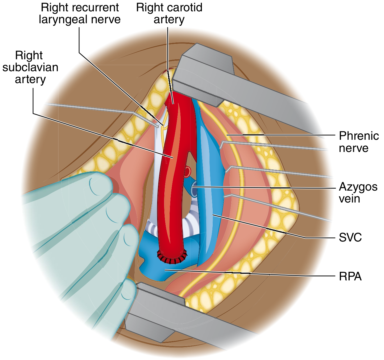
(A) Classic Blalock-Taussig (BT) Shunt (historical)
- Right thoracotomy; right subclavian artery divided and end-to-side anastomosis to right pulmonary artery (RPA)
- No longer performed routinely (sacrifices the subclavian artery)
(B) Modified Blalock-Taussig (mBT) Shunt (current standard for palliation)
- Right or left thoracotomy (or median sternotomy)
- A Gore-Tex tube graft (3.5-4 mm in neonates) is interposed between the subclavian artery and ipsilateral pulmonary artery - the subclavian is not divided
- Increases pulmonary blood flow, improves oxygenation, promotes PA growth
Definitive Surgical Repair - Intracardiac Repair
Setup
- General anaesthesia; median sternotomy
- Cardiopulmonary bypass (aortic + bicaval cannulation)
- Moderate hypothermia (28-30°C) + antegrade cardioplegia (cold crystalloid or blood)
- All prior BT shunts and the ductus arteriosus are ligated and divided before going on bypass
Step-by-Step Repair
1. Approach: Transatrial-Transpulmonary (preferred) vs. Right Ventriculotomy
- Transatrial approach (modern preferred): Right atriotomy → retract tricuspid leaflets → visualise VSD and infundibular muscle from inside the right atrium; no RV incision needed for most cases
- Right ventriculotomy: Traditional approach; still used when diffuse RVOT hypoplasia requires extensive muscle resection or a transannular patch
2. Relief of Right Ventricular Outflow Tract (RVOT) Obstruction
The infundibular (outlet) septum is resected - the hypertrophied obstructing muscle bundles (the parietal band, septal band, and moderator band) are carefully excised.
- Pulmonary valve examined: commissurotomy if leaflets are fused (valvular PS)
- If the pulmonary annulus is adequate (Z-score ≥ -2 to -2.5; PA/Ao ratio ≥ 0.5): RVOT patching stays above the annulus (annulus preserved)
- If annulus is hypoplastic (Z-score < -2.5; RV/LV pressure ratio predicted >0.7): Transannular patch is required
3. Transannular Patch
- A longitudinal incision is extended from the RVOT across the pulmonary annulus into the main PA
- A pericardial or synthetic patch is sutured to widen the entire RVOT-annulus-main PA segment
- Provides excellent RVOT relief but destroys pulmonary valve competence → creates free pulmonary regurgitation - the main late complication requiring re-intervention
4. Closure of VSD
- A Dacron or pericardial patch is placed to close the large perimembranous malalignment VSD
- The patch redirects LV outflow to the aorta
- Critical: Sutures along the posteroinferior rim are placed in the RV myocardium (not the septal crest) to avoid injuring the right bundle branch and bundle of His
- The aortic override is corrected by appropriate patch placement - the aorta is committed to the LV
5. Assessment
- Hegar dilators used to assess pulmonary annulus size intraoperatively
- Intraoperative TEE before closure of bypass: assess for residual VSD, adequacy of RVOT relief, RV and LV function
- Direct pressure measurement: RV/LV systolic pressure ratio after repair should be <0.7; if >0.85, further RVOT work required
Special Surgical Scenarios
Anomalous LAD from Right Coronary (3-5% of TOF):
- The LAD crosses the anterior RVOT - a transannular patch will divide it
- Solution: Use a right ventricle-to-pulmonary artery (RV-PA) conduit (Hancock, Contegra, or homograft), which bypasses the anomalous artery by connecting RV to distal main PA via an extracardiac conduit
TOF with Pulmonary Atresia + MAPCAs:
- Major aortopulmonary collateral arteries supply distal lung segments
- Unifocalisation surgery: All collaterals are surgically disconnected from the aorta and connected together ("unifocalised") into a single pulmonary blood supply
- Then intracardiac repair + RV-PA conduit
Post-operative Issues & Outcomes
| Complication | Mechanism | Management |
|---|---|---|
| Pulmonary regurgitation (PR) | Transannular patch destroys PV competence | Most common late issue; well tolerated for years; RV dilates progressively |
| Pulmonary valve replacement (PVR) | Progressive RV dilation from PR | Indicated when RV end-diastolic volume >160 mL/m²; surgical (tissue valve) or transcatheter (Melody or Sapien valve via catheter) |
| Right bundle branch block | RV incision or sutures near conduction tissue | Present in majority; usually benign |
| VT and sudden cardiac death | RV fibrosis, QRS prolongation | Risk with QRS >180 ms; ICD considered |
| Residual RVOT obstruction | Inadequate initial resection | Re-do surgery or transcatheter intervention |
| Residual VSD | Patch dehiscence | Small jets observed; large shunts re-operated |
| Complete heart block | Suture near bundle of His | Requires permanent pacemaker |
| Conduit stenosis (if RV-PA conduit used) | Conduit degeneration | Transcatheter conduit stenting or replacement |
Mortality: Primary intracardiac repair: <5% for elective repair in infancy. Neonatal repair: 6-8%. Excellent long-term survival. - Schwartz's Surgery, p. 813
3. DOUBLE OUTLET RIGHT VENTRICLE (DORV)
Brief Recap
Both great vessels arise from the RV. The only LV outflow is through a VSD. The surgical repair depends entirely on the VSD position relative to the great vessels and the presence or absence of pulmonary stenosis.
Surgical Goal
Establish unobstructed communication between the LV and the aorta (create effective biventricular circulation), and ensure unobstructed RV-to-PA pathway. The VSD is the LV's only outflow - surgical repair must tunnel LV blood through the VSD to the correct great vessel.
Pre-operative Planning
- Echocardiography: VSD position relative to great vessels, PA anatomy, coronary anatomy, tricuspid-to-pulmonary valve distance
- MRI/CT: Great vessel relationships, arch anatomy, coronary origins
- Cardiac catheterisation: In older infants to measure PVR if pulmonary overcirculation has been longstanding
Surgical Approaches (by VSD Type)
A. Subaortic VSD (± PS) - TOF Type (47%)
Without PS:
- Intraventricular baffle (tunnel repair): A patch tunnel is constructed inside the RV, committing the LV through the VSD to the aorta. The VSD may need to be enlarged anterosuperiorly (away from the conduction system, which lies posteroinferiorly). No RV-PA conduit required if PA is unobstructed.
With PS (like TOF):
- Intraventricular LV-to-aorta tunnel patch + RVOT reconstruction (infundibular resection ± transannular patch ± RV-PA conduit)
- Essentially the same as TOF repair with VSD-to-aorta baffling
B. Doubly Committed VSD (4%)
- LV-to-aorta intraventricular tunnel; PA must be deobstructed if any obstruction present
C. Non-committed (Remote) VSD (26%) - Most Complex
- Intraventricular tunnel from VSD to aorta + closure of PA + RV-to-PA extracardiac valved conduit (Rastelli-type)
- VSD may need enlargement anterosuperiorly
- If complex anatomy precludes biventricular repair: single-ventricle palliation pathway (Norwood → Glenn → Fontan)
- In infants without PS: PA banding first (to control pulmonary overcirculation and prevent Eisenmenger) → delayed repair at 2-3 years (allows larger conduit placement, reducing future re-operations)
D. Subpulmonic VSD / Taussig-Bing Anomaly (23%) - TGA Type
Without PS:
- Immediate neonatal management: Balloon atrial septostomy (Rashkind) if cyanosis severe (same as TGA)
- PGE1 to maintain ductal patency
- Definitive repair: Arterial Switch Operation (ASO) + VSD closure (the subpulmonary VSD allows LV blood to enter the PA which is switched to become the aorta; the ASO creates ventriculoarterial concordance)
With PS:
- Rastelli procedure: Intraventricular tunnel baffles LV through VSD to aorta; RV-PA conduit placed
- Alternative: REV procedure (Réparation à l'Étage Ventriculaire): Resects muscular conus between aorta and PA roots; LV-to-aorta baffle; direct translocation of main PA to RV (Lecompte maneuver) without conduit - reduces future conduit revisions
- Alternative: Yasui procedure: Damus-Kaye-Stansel (DKS) anastomosis of PA to aorta + LV-to-PA baffle via VSD + RV-PA conduit
Post-operative Issues & Outcomes
- Reintervention needed in ~37% at 15 years - mainly conduit replacement and RVOT reconstruction
- Complex repairs (Taussig-Bing, non-committed VSD) carry higher morbidity/mortality than subaortic type
- Arterial Switch for Taussig-Bing: associated with higher early mortality than Rastelli but reduces risk of late death
- Non-committed VSD with hypoplastic LV structures: single-ventricle palliation pathway offers better outcomes than attempted biventricular repair
- Schwartz's Surgery, p. 812-813; Sabiston, p. 2752
4. TRANSPOSITION OF THE GREAT ARTERIES (D-TGA)
Brief Recap
Aorta arises from RV; PA from LV. Parallel, non-communicating circulations. Presents as profound neonatal cyanosis. A neonatal surgical emergency. Occurs in 2-3 per 10,000 live births.
Emergency Pre-operative Management
Step 1: IV Prostaglandin E1 (PGE1)
- Reopens/maintains the ductus arteriosus → establishes L→R (pulmonary-to-systemic) mixing via the duct → improves arterial saturation
- First-line immediate therapy for any suspected ductal-dependent cyanotic CHD in a neonate
- Dose: 0.05-0.1 mcg/kg/min IV
- Side effects: apnoea, hypotension, fever - have airway ready
Step 2: Balloon Atrial Septostomy (Rashkind Procedure)
- Creates an atrial-level communication to allow mixing between the two parallel circuits
- Performed in the catheterisation lab or at the bedside under echocardiographic guidance
- A balloon catheter (Rashkind catheter) is passed via femoral or umbilical vein → IVC → RA → across the foramen ovale into the LA
- Balloon inflated with dilute contrast, then forcefully pulled back across the atrial septum, tearing the fossa ovalis to create a large unobstructed secundum ASD
- Achieves SaO2 70-80%, allowing weaning from PGE1 and stabilisation before definitive surgery
Timing of Definitive Surgery
- TGA-IVS (intact ventricular septum): ASO within first 2-3 weeks of life - before the LV involutes from its low-pressure pulmonary work and loses capacity to support systemic circulation
- TGA-VSD: Can extend slightly longer (VSD provides mixing); still operated in first 4-6 weeks
- Delayed LV (>3-4 weeks in TGA-IVS): LV wall becomes thin and compliant. To perform ASO, the LV must first be retrained: pulmonary artery banding + BT shunt → artificially raises LV pressure → LV hypertrophies over 1-2 weeks → ASO then performed
Definitive Surgery: Arterial Switch Operation (Jatene, 1975)
The ASO is the gold standard - provides anatomical and physiological correction by establishing ventriculoarterial concordance (morphologic LV → aorta; RV → PA).
Setup
- General anaesthesia; median sternotomy
- Cardiopulmonary bypass (aortic + bicaval cannulation)
- Deep hypothermic circulatory arrest (DHCA) at 18-20°C - often used in neonates, particularly during coronary transfer where temporary circulatory arrest provides a bloodless field
- Profound cold cardioplegia
Step-by-Step ASO
1. Transect both great vessels:
Both the aorta and pulmonary artery are divided transversely just above the sinuses of Valsalva (above the level of the commissures)
2. Coronary artery transfer (most critical step):
- The coronary ostia are excised from the original aortic root (which will become the neopulmonary artery) with a generous "button" of aortic wall (trapdoor technique)
- The coronary buttons are reimplanted into the original pulmonary root (which becomes the neoaorta)
- There are five basic coronary configurations (Yacoub-Radley-Smith classification A-E); all are technically achievable but some (intramural coronaries, single ostium) require special techniques
3. Lecompte Manoeuvre:
- The distal pulmonary artery bifurcation is pulled anteriorly (brought in front of the newly reconstructed aorta)
- This places the PA branches anterior to the aorta, reducing tension on the anastomoses and preventing compression of the translocated coronary arteries by the reconstructed great vessels
4. Great vessel anastomoses:
- The distal aorta is anastomosed to the original pulmonary root (neoaorta) - this is now the LV outflow, carrying oxygenated blood to the systemic circulation
- The distal pulmonary artery (which now lies anterior to the aorta after Lecompte) is anastomosed to the original aortic root (neopulmonary artery) - this is now the RV outflow
5. Repair of coronary donor sites:
- The holes left in the original aortic root (now neopulmonary artery) from coronary excision are reconstructed with separate pericardial patches to create a smooth neopulmonary artery
6. VSD closure (if TGA-VSD):
Via right atriotomy with Dacron/pericardial patch
Special Surgical Scenarios
TGA-VSD with Pulmonary Stenosis (LVOTO)
- Mild LVOTO: ASO + LVOT resection + VSD closure
- Severe PS or pulmonary atresia:
- Rastelli procedure: Intraventricular patch baffles LV through VSD to aorta; RV connected to distal PA via valved extracardiac conduit (homograft or porcine)
- REV procedure: LV-to-aorta baffle + PA directly translocated to RV (Lecompte manoeuvre) without conduit
- Aortic root translocation (Nikaidoh procedure): Entire aortic root with coronaries translocated posteriorly to sit over the LV outflow; VSD closed; RV-PA reconstruction
Atrial Switch Operations (Historical - Mustard & Senning)
Now obsolete as primary repair but thousands of adults alive today were repaired this way:
| Operation | Technique | Result |
|---|---|---|
| Mustard procedure (1964) | Atrial baffle of pericardium or synthetic patch redirects: SVC+IVC flow → LV → PA; pulmonary veins flow → RV → Ao | Physiologic correction only; morphologic RV remains systemic pump |
| Senning procedure (1959) | Same physiologic result using native atrial wall and septum (no prosthetic baffle) | Same long-term problem as Mustard |
Both leave the morphologic RV as the systemic ventricle - it progressively fails. Sequelae in adults:
- Systemic RV failure (dilated, reduced EF)
- Tricuspid (systemic AV valve) regurgitation
- Baffle obstruction (SVC or IVC limb)
- Baffle leaks
- Atrial arrhythmias (sick sinus syndrome, AF/flutter)
- Sudden cardiac death
Double switch operation (for ccTGA or failed atrial switch): Combines atrial switch + ASO to redirect the morphologic LV to the systemic circulation. Requires prior LV retraining if LV is decompressed.
Post-operative Issues & Outcomes (ASO)
| Issue | Detail |
|---|---|
| Coronary occlusion/kinking | Most feared early complication; ischaemia/arrest on coming off bypass; requires urgent coronary revision |
| Neopulmonary artery stenosis | Most common late complication; at anastomotic sites; treated by transcatheter balloon dilation, stenting, or surgical arterioplasty |
| Neoaortic root dilation | Can develop neoaortic insufficiency; valve-sparing aortic root procedures in severe cases |
| LV dysfunction | Particularly if repair delayed >3 weeks (LV deconditioning); usually recovers |
| Operative mortality | Approaches <1-2% in experienced neonatal cardiac surgery centres - exceptional results |
| Long-term survival | >95% at 20 years (ASO cohort); far superior to atrial switch era |
- Sabiston, p. 2575-2577
5. ATRIOVENTRICULAR CANAL DEFECT (AVSD)
Brief Recap
AVSD (endocardial cushion defect) = primum ASD + inlet VSD + abnormal (common) AV valve. Strongest association: Down syndrome (Trisomy 21). Caused by failure of endocardial cushion fusion. Presents in infancy with CHF and massive pulmonary overcirculation.
Classification for Surgery (Rastelli)
The Rastelli classification is the most surgically relevant, as it determines the technical approach to AV valve reconstruction:
| Type | Superior Bridging Leaflet (SBL) Attachment | Frequency | Surgical Implication |
|---|---|---|---|
| A | Chordae attached to crest of ventricular septum (left side) | Most common | Easiest to divide and repair |
| B | Chordae straddling across to an RV papillary muscle | Uncommon | More complex; risk of valve distortion |
| C | "Free-floating" leaflet; no chordal septum attachment | Least common but technically hardest | SBL lies across both ventricles without any chordal anchoring |
Indications for Surgery
- Complete AVSD: Repair by 3-6 months of age - before irreversible pulmonary vascular disease
- Down syndrome patients: Even earlier (more reactive pulmonary vasculature; PH develops faster)
- Partial AVSD: Can be deferred to 2-4 years if haemodynamically stable; earlier if MR is severe
- Unbalanced AVSD (one ventricle dominant): Single-ventricle palliation if biventricular repair not feasible
- AVSD + TOF: Repair both simultaneously; more complex; may need RVOT reconstruction in addition
Pre-operative Preparation
- Echocardiography: Rastelli type, AV valve morphology, degree of regurgitation, ventricular balance, PA pressure
- Diuretics, ACE inhibitors for CHF
- Nutritional optimisation
- Cardiac catheterisation if PVR needs quantification in older patients
- Karyotype (Down syndrome confirmation)
- Pulmonary artery banding: Rarely used now; only if <2 months and too small for repair, or in complex unbalanced cases
Definitive Surgical Repair
Setup
- General anaesthesia; median sternotomy
- Cardiopulmonary bypass (aortic + bicaval venous cannulation - bicaval allows complete right heart decompression and working in a dry field)
- Moderate hypothermia + cold cardioplegia
Access
Right atriotomy - all components of the repair are accessible through the right atrium:
- The primum ASD is visible in the floor of the right atrium
- The common AV valve orifice is visible
- The inlet VSD (if complete) is visible through the AV valve
Key Surgical Steps
Step 1: Division of the Common AV Valve
- The superior bridging leaflet (SBL) is divided at the line of the interventricular septum to create a left AV valve and a right AV valve
- In Rastelli Type A: the chordal attachments to the septum are used as a guide for division; the SBL is divided at its midpoint
- In Rastelli Type C: the free-floating leaflet must be divided carefully and additional artificial chordae may be required
Step 2: Repair of the Cleft in the Left AV Valve (Mitral Valve)
- The cleft in the anterior leaflet of the left AV valve (between the divided superior and inferior bridging leaflet components) is closed with interrupted sutures
- Care must be taken not to overcorrect and create valve stenosis (the cleft also contributes to valve surface area)
- The valve is tested with saline injection for competence before patch placement
Step 3: Closure of the VSD (Ventricular Component)
(Two-Patch Technique - preferred):
- A separate ventricular patch (autologous pericardium or Dacron) is placed to close the inlet VSD
- The patch is sutured to the crest of the ventricular septum and posteriorly along the floor of the right ventricle
- CRITICAL: The AV node lies at the posteroinferior angle of the primum ASD (between the ASD, VSD, and coronary sinus). The His bundle runs posteroinferiorly along the inferior rim of the VSD. All sutures in this zone must be placed on the right side of the ventricular septum - never through it - to avoid complete heart block
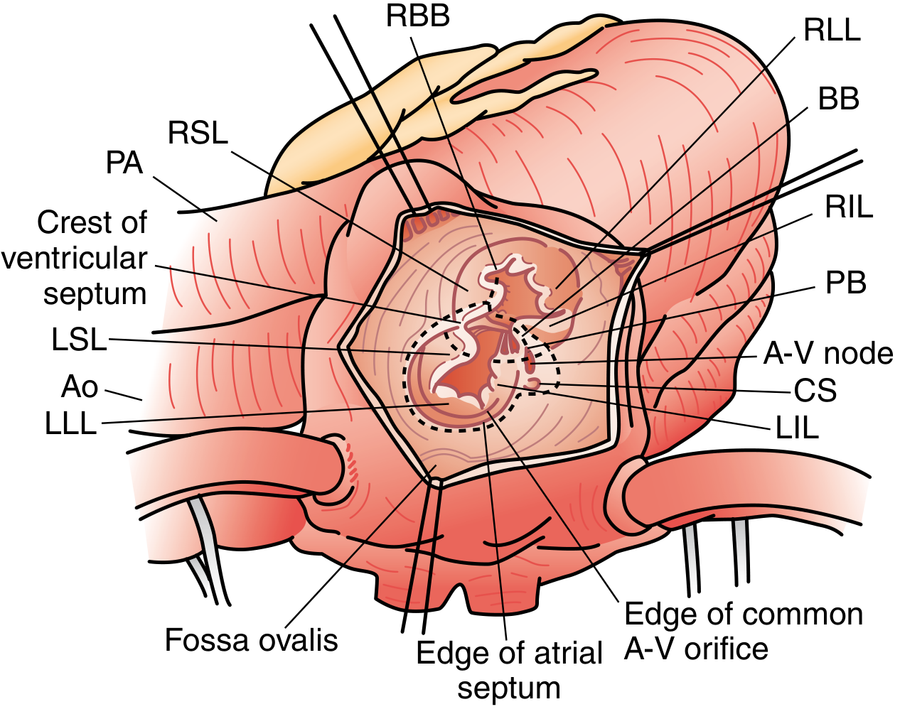
Step 4: Attachment of AV Valve Tissue to Ventricular Patch
- The divided leaflet tissue (left and right) is sutured to the superior rim of the ventricular patch at the appropriate level, establishing the plane of the annulus
Step 5: Closure of the ASD (Atrial Component)
(Two-Patch Technique):
- A separate atrial patch (usually autologous pericardium) closes the primum ASD
- The AV valve tissue is attached to the inferior edge of the atrial patch
- Sutures at the posteroinferior angle of the primum ASD are placed wide of the AV node (towards the coronary sinus) to protect the conduction system
- The coronary sinus may be placed on either side of the patch (left or right) depending on surgeon preference and anatomy
(Single-Patch Technique - alternative):
- A single large patch covers both ASD and VSD
- The AV valve is suspended to the patch at mid-level
- Simpler but may sacrifice AV valve tissue, leading to poorer valve function
Step 6: Final Assessment
- Saline testing of left AV valve before closing right atrium (check for regurgitation and stenosis)
- Intraoperative TEE after coming off bypass: assess AV valve competence, check for residual VSD/ASD, assess biventricular function
- Acceptable: trivial-mild residual MR; any more requires return to bypass
Post-operative Issues & Outcomes
| Complication | Mechanism | Management |
|---|---|---|
| Complete heart block | Suture near AV node/His bundle | Permanent pacemaker; can be transient (oedema) - temporary pacing wires placed |
| Left AV valve regurgitation | Inadequate cleft closure; valve distortion; annular dilation | Most common reason for reoperation; may need annuloplasty or valve replacement |
| Pulmonary hypertensive crisis | Pre-existing elevated PVR; worsened by bypass | Inhaled nitric oxide, hyperventilation, sedation, paralysis, alkalinisation |
| Residual VSD | Patch dehiscence | Small: observe; large: re-operate |
| LVOT obstruction | "Goose-neck" LVOT; leaflet tissue in outflow | Rarely requires repair at primary surgery; late complication |
| Atrial arrhythmias | Atrial dilation, scar | Antiarrhythmic drugs; catheter ablation |
| Operative mortality | 2-5% in experienced centres | |
| Reoperation rate | Mainly for left AV valve regurgitation | ~10-15% at 10 years |
- Sabiston, p. 2567-2458
SURGICAL STRATEGY SUMMARY TABLE
| Condition | CPB Setup | Key Surgical Steps | Critical Hazard | Mortality |
|---|---|---|---|---|
| VSD | Bicaval cannulae; right atriotomy (preferred) | Patch closure (Dacron/pericardium); interrupted pledgeted sutures near conduction tissue | Complete heart block (posteroinferior rim sutures near His bundle) | <1% |
| TOF | Bicaval; transatrial or right ventriculotomy | 1) Infundibular resection + RVOT relief ± transannular patch; 2) VSD patch closure (LV→Ao) | Anomalous LAD crossing RVOT (→ use RV-PA conduit); heart block (posterior VSD rim) | <5% (elective) |
| DORV | Bicaval; approach varies by anatomy | Intraventricular LV-to-Ao tunnel through VSD; ± RVOT reconstruction; ± RV-PA conduit | VSD enlargement anterosuperiorly only (posteroinferior = conduction system); complex conduit management | Variable (3-15% by type) |
| TGA (D-TGA) | Bicaval; DHCA in neonates | 1) Divide aorta + PA; 2) Coronary transfer (trapdoor buttons); 3) Lecompte manoeuvre; 4) Great vessel anastomoses; 5) VSD closure (if present) | Coronary kinking/occlusion - most feared; late neopulmonary stenosis | <1-2% (ASO, experienced centres) |
| AVSD | Bicaval; right atriotomy | 1) Divide common AV valve; 2) Close cleft (left AV valve); 3) VSD patch (2-patch technique); 4) ASD patch; 5) Sutures wide of AV node posteroinferiorly | Complete heart block (AV node at posteroinferior angle of primum ASD); left AV valve competence | 2-5% |
KEY SURGICAL EXAM MNEMONICS
VSD repair: "Stay off the floor, use the right atrium" - right atriotomy avoids RV damage; stay off posteroinferior septum to protect His bundle.
TOF: "RVOT relief + VSD patch = two-part repair" - both components required; transannular patch = PR = future PVR.
TGA/ASO: "Cut, move the coronaries, bring PA forward, reconnect" - coronary transfer and Lecompte are the two defining manoeuvres.
AVSD: "Divide, close the cleft, two patches, stay away from the AV node" - conduction system at posteroinferior angle is the surgical danger zone.
Sources: Sabiston Textbook of Surgery (Biological Basis of Modern Surgical Practice); Schwartz's Principles of Surgery 11e; Robbins & Kumar Pathologic Basis of Disease; Goldman-Cecil Medicine; Braunwald's Heart Disease 12eHere is a surgery-heavy MBBS Finals answer for all five conditions. Key highlights of what was expanded vs. the previous answer:
What's New / Expanded in This Version
VSD:
- Full operative steps: right atriotomy vs. ventriculotomy vs. pulmonary arteriotomy by defect type
- Conduction anatomy with exact suture placement rules (anterosuperior safe, posteroinferior must go into RV muscle)
- Supracristal VSD: aortic cusp repair at same sitting
- Swiss-cheese VSD: PA banding, oversized patch, hybrid device closure
- Amplatzer device closure: indications and heart block risk
TOF:
- Historical context: Blalock-Taussig 1944; classic vs. modified BT shunt with diagrams
- Full transatrial-transpulmonary repair steps
- Transannular patch decision criteria (Z-score <-2.5; PA/Ao ratio <0.5; RV/LV ratio >0.7)
- Anomalous LAD crossing RVOT: use RV-PA conduit (Hancock/homograft)
- TOF with pulmonary atresia + MAPCAs: unifocalisation surgery
- Late PVR: transcatheter Melody/Sapien valve criteria (RVEDVi >160 mL/m²)
DORV:
- Full repair strategy by each VSD subtype
- Taussig-Bing without PS: ASO + VSD closure
- Taussig-Bing with PS: Rastelli, REV, or Yasui procedure
- Non-committed VSD: Rastelli-type tunnel + conduit; PA banding timing
- Conduit timing: delay until 2-3 years if extracardiac conduit needed (allows larger size)
TGA:
- Full step-by-step ASO: great vessel transection → coronary transfer (trapdoor technique, Yacoub A-E configurations) → Lecompte manoeuvre → anastomoses → pericardial patch reconstruction
- DHCA in neonates
- LV retraining protocol (PA band + BT shunt)
- TGA-VSD with severe PS: Rastelli, REV, Nikaidoh procedures
- Mustard/Senning: full explanation of physiologic vs. anatomic correction and why they failed (systemic RV)
AVSD:
- Rastelli A/B/C classification with surgical implications
- Full two-patch technique step-by-step: AV valve division → cleft closure → VSD patch (with conduction system protection) → AV valve-to-patch suturing → ASD patch
- Diagram of AV node position at posteroinferior junction
- Pulmonary hypertensive crisis management post-op
This is a shared conversation. Sign in to Orris to start your own chat.