
Cubital fossa
Reading File
The cubital fossa is a triangular depression on the anterior aspect of the elbow, bounded by the brachioradialis (laterally), pronator teres (medially), and a line joining the epicondyles (superiorly). Its floor is the brachialis and supinator, and its roof is the deep fascia reinforced by the bicipital aponeurosis. Contents (lateral to medial): biceps tendon, brachial artery, median nerve (mnemonic: TAN - Tendon, Artery, Nerve).
Searching sources...
Finding Sources
Searching the Web
cubital fossa anatomy boundaries contents clinical importance
Reading File
Finding Sources
Reading File
Loading Image
Loading Image
Here is a complete overview of the cubital fossa from Gray's Anatomy for Students and the Thieme Atlas of Anatomy.
Cubital Fossa
The cubital fossa (also called the antecubital fossa) is a triangular depression on the anterior aspect of the elbow joint, serving as the key transition zone between the arm and the forearm. All major neurovascular structures pass through or in close relation to it.
Shape and Borders
The fossa is triangular in shape, with:
| Boundary | Structure |
|---|---|
| Base (superior) | Imaginary horizontal line connecting the medial and lateral epicondyles of the humerus |
| Medial border | Pronator teres (arising from the medial epicondyle) |
| Lateral border | Brachioradialis (arising from the lateral supraepicondylar ridge) |
| Apex | Where the two muscles meet distally |
Roof and Floor
- Roof: Skin and superficial fascia, reinforced by the bicipital aponeurosis (a flat connective tissue membrane passing from the medial side of the biceps tendon to the deep fascia of the forearm). The sharp medial margin of the bicipital aponeurosis can often be felt clinically.
- Floor: Mainly the brachialis muscle proximally, and the supinator muscle distally.
Contents (Lateral to Medial: mnemonic TAN)
| Structure | Notes |
|---|---|
| T - Tendon of biceps brachii | Passes to attach to the radial tuberosity |
| A - Brachial artery | Bifurcates into radial and ulnar arteries at the apex of the fossa |
| N - Median nerve | Lies just medial to the brachial artery; exits by passing between the ulnar and humeral heads of pronator teres |
Additionally, the radial nerve lies just deep to the lateral lip of the brachioradialis (outside the classic "contents" but within the fossa region). Here it divides into:
- Superficial branch - continues deep to brachioradialis into the forearm
- Deep branch - passes between the two heads of the supinator to reach the posterior compartment
Important negative: The ulnar nerve does NOT pass through the cubital fossa. It passes posterior to the medial epicondyle in the cubital tunnel.
Diagrams from Gray's Anatomy for Students
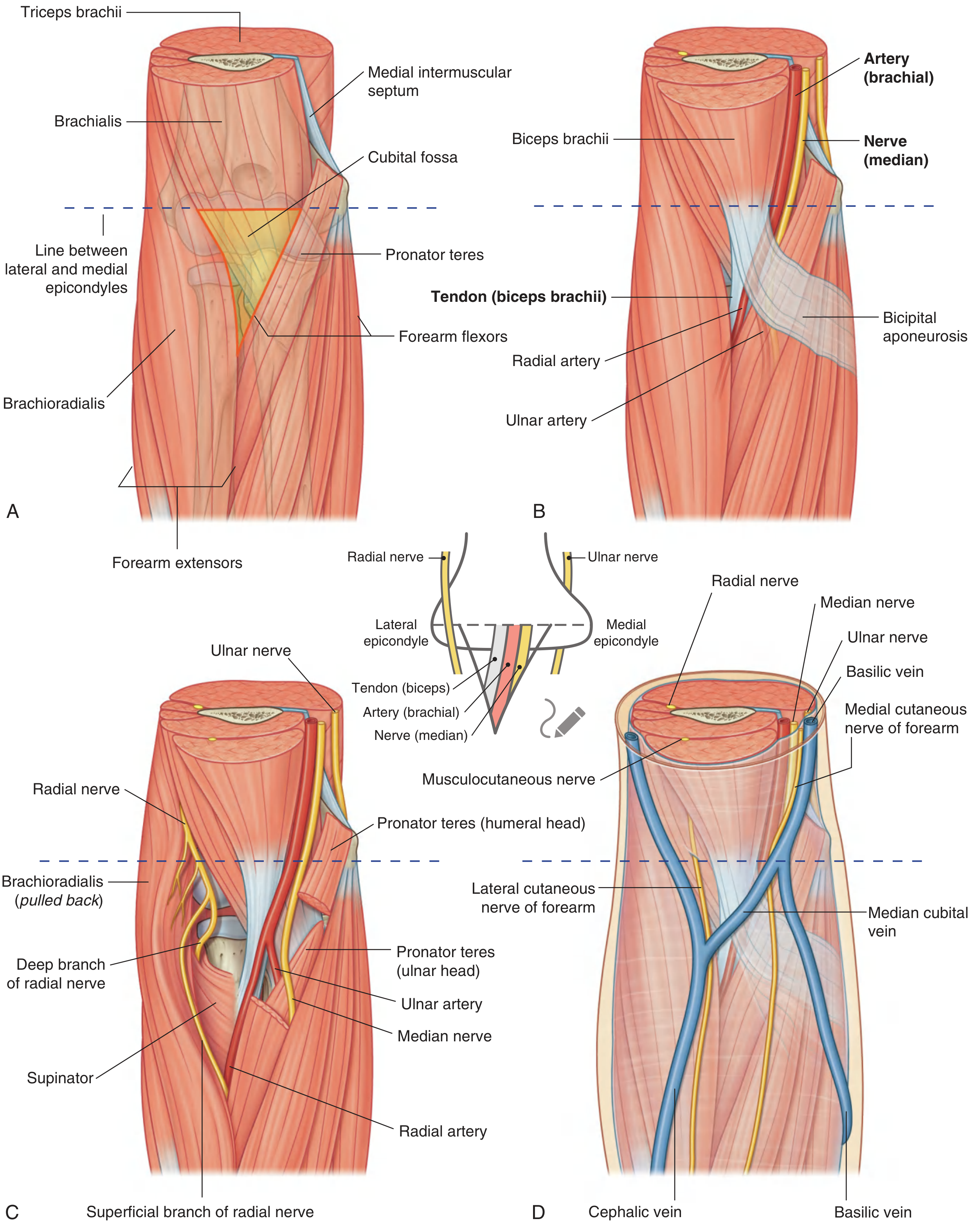
Fig. 7.77 - (A) Margins, (B) Contents, (C) Radial nerve position, (D) Superficial structures - Gray's Anatomy for Students
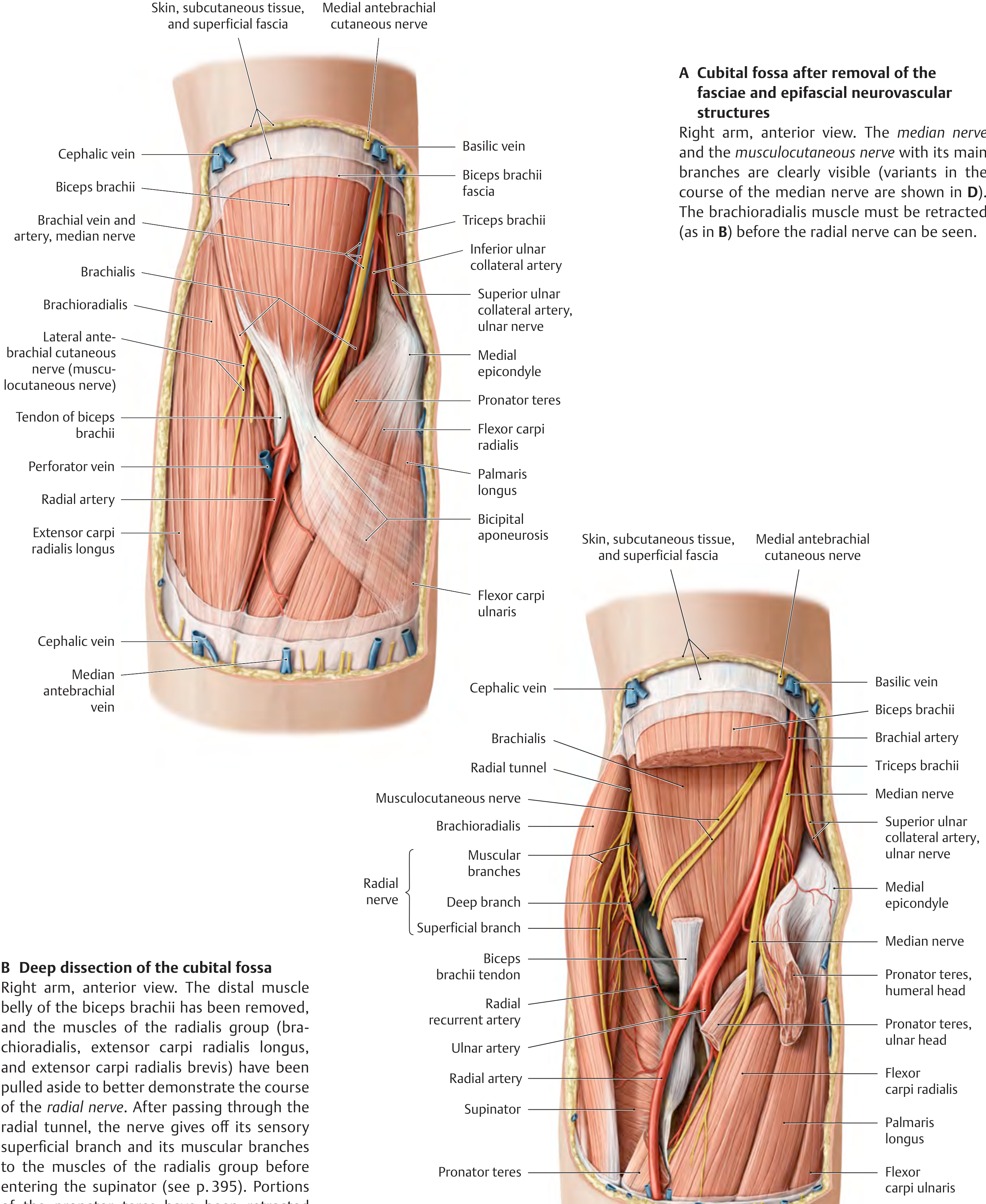
Deep dissection showing the cubital fossa - Thieme Atlas of Anatomy
Structures in the Roof (Superficial / Epifascial)
These lie in the superficial fascia above the bicipital aponeurosis:
- Median cubital vein - the most important; crosses diagonally from the cephalic to basilic vein
- Lateral cutaneous nerve of the forearm (terminal branch of the musculocutaneous nerve)
- Medial cutaneous nerve of the forearm
Clinical Significance
| Application | Details |
|---|---|
| Venipuncture / IV access | The median cubital vein in the roof is the most common site for blood draws and IV cannulation. It is protected from the brachial artery beneath by the bicipital aponeurosis. |
| Blood pressure measurement | The stethoscope diaphragm is placed over the cubital fossa to auscultate the brachial artery (Korotkoff sounds). |
| Brachial artery pulse | Palpable medial to the biceps tendon; used in BP measurement and for arterial access. |
| Nerve block / injury | The median nerve's position makes it vulnerable to laceration, compression, or iatrogenic injury in this region. |
| Dialysis fistula | The brachial artery is anastomosed to the cephalic vein at the elbow (brachiocephalic fistula) to create high-flow access for hemodialysis. |
| Brachial artery bifurcation | Normally occurs at the apex of the fossa, but the bifurcation can occur much higher - even in the axilla. This is important during arterial procedures. |
| Drug injection | Repeated injections into the cubital fossa veins (e.g., IV drug use) can cause thrombosed veins, abscesses, and pigmented "rail-road track" scars. |
Sources: Gray's Anatomy for Students, p. 889-890 | Thieme Atlas of Anatomy (General Anatomy and Musculoskeletal System)
This is a shared conversation. Sign in to Orris to start your own chat.