
tinea corporis
I'll read the medical library skill first, then search for tinea corporis content in parallel.
Reading File
Now let me search the library and PubMed in parallel.
Finding Sources
Searching PubMed
"tinea corporis"[MeSH Terms] AND treatment
Good. Now let me read the most comprehensive sources in parallel - Dermatology 5e (most detailed) and Fitzpatrick's, plus Andrews'.
Reading File
Reading File
Reading File
Let me read more from Dermatology 5e (which had the most detailed entry) and Fitzpatrick's treatment section.
Reading File
Reading File
Now let me get the Dermatology 5e detailed section on clinical features, pathogenesis, and treatment, and also fetch a key image.
Reading File
Loading Image
I now have comprehensive information. Let me compile the full answer.
Tinea Corporis
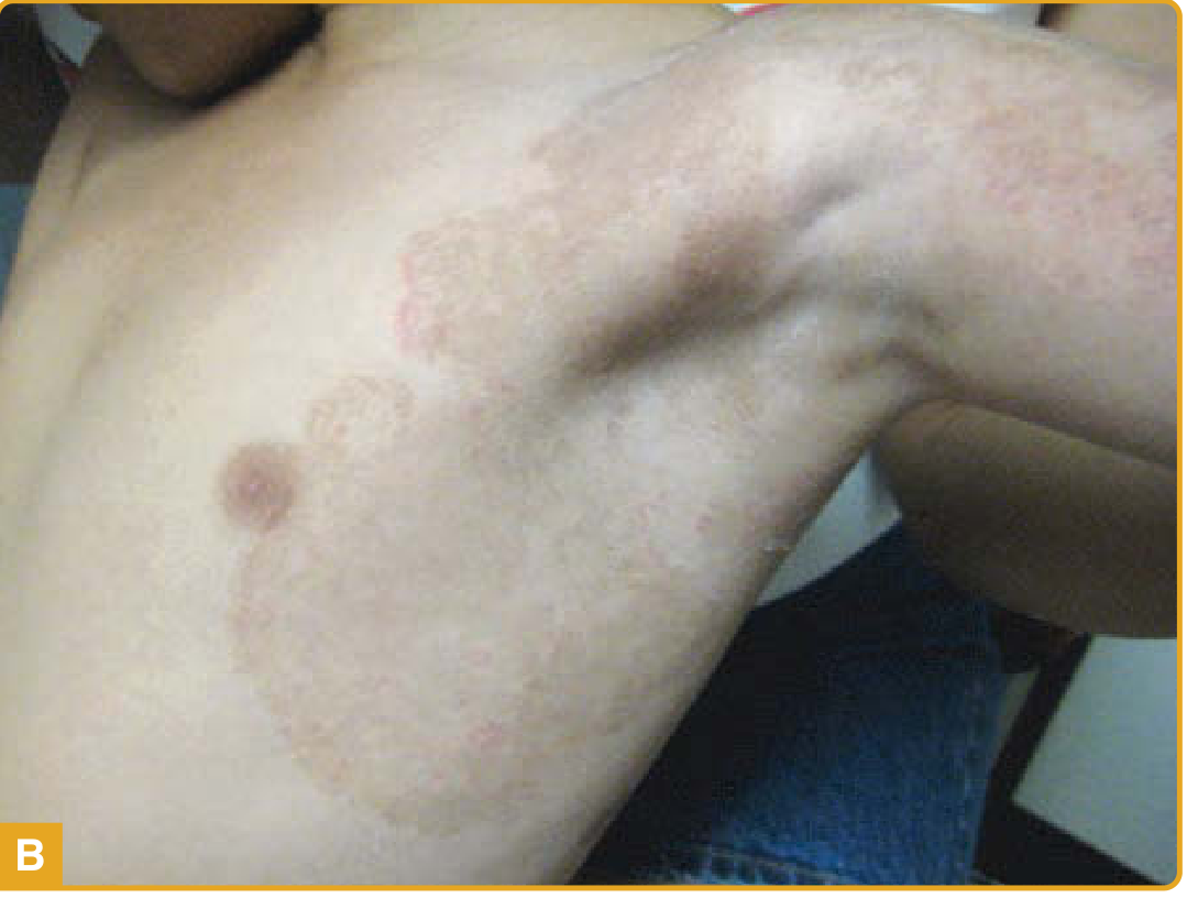
Tinea corporis (ringworm) is a superficial dermatophyte infection of the glabrous skin of the trunk and extremities, excluding the scalp, beard, face, palms, soles, and groin. The infection is confined to the stratum corneum.
Etiology
Any dermatophyte can cause tinea corporis. The most common pathogens are:
| Organism | Notes |
|---|---|
| Trichophyton rubrum | Most common overall; responsible for chronic, widespread, and follicular forms |
| T. mentagrophytes / T. interdigitale | Common; zoophilic strains cause inflammatory lesions |
| T. indotineae (formerly T. mentagrophytes var. Vill) | Emerging resistant strain, especially from South Asia |
| Microsporum canis | Common in children; acquired from dogs and cats |
| T. tonsurans | Cause of tinea corporis gladiatorum in wrestlers |
| Epidermophyton floccosum | Common in humid climates; no microconidia |
| T. concentricum | Causes tinea imbricata; restricted to the Far East, South Pacific, Central/South America |
- Dermatology 5e, p. 1604; Fitzpatrick's Dermatology, p. 2974
Epidemiology & Transmission
- Transmitted by direct contact with infected humans (anthropophilic), animals (zoophilic), or soil (geophilic), as well as via fomites or autoinoculation from other body sites (feet are a common reservoir).
- Children are more prone to zoophilic infections, especially M. canis from pets.
- Risk factors: humid climates, occlusive clothing, skin-to-skin contact, minor skin trauma, immunosuppression.
- Tinea corporis gladiatorum: caused by T. tonsurans in wrestlers; head, neck, and arms most affected due to mat contact and skin abrasion.
Clinical Features
Classic presentation:
- One or more annular ("ringworm") plaques with a raised, erythematous, scaly border that advances centrifugally
- Central clearing (or residual scale) as the active border spreads outward
- The border may be vesicular; pruritus is common
Morphologic variants:
| Variant | Features |
|---|---|
| Annular | Classic ring; raised scaly border, central clearing |
| Polycyclic | Multiple coalescing rings, large confluent plaques |
| Psoriasiform | Thick scaly plaques mimicking psoriasis; common in immunosuppressed |
| Tinea imbricata | Concentric scaly rings; minimal vesiculation; caused by T. concentricum |
| Tinea incognito | Modified by topical steroids - border blurred, scale minimal, widespread hyphae on KOH |
| Majocchi granuloma | Deep follicular involvement; scaly follicular papules/nodules in annular arrangement; most common on shaved legs of women |
- Fitzpatrick's Dermatology, p. 2973; Andrews' Diseases of the Skin, p. 347
Diagnosis
1. KOH preparation (potassium hydroxide)
- Scrape from the active advancing border (not the center)
- Branching, septate hyphae confirm fungal infection
- Pitfall: "mosaic false hyphae" from cell wall junctions of normal keratinocytes is the most common artifact mimicking positive KOH
- Cotton/synthetic fibers from clothing can also mimic hyphae
2. Fungal culture
- Growth visible in 1-2 weeks; identifiable to genus level by gross and microscopic morphology
3. Skin biopsy
- Periodic acid-Schiff (PAS) or Grocott methenamine silver (GMS) stain
- Useful in chronic refractory cases (tinea incognita)
- Hyphae found in the stratum corneum
4. Wood's lamp
- M. canis and select Microsporum spp. fluoresce green; Trichophyton spp. do not
Differential Diagnosis
| Category | Conditions |
|---|---|
| Most likely | Erythema annulare centrifugum, nummular eczema, psoriasis, tinea versicolor, subacute cutaneous lupus erythematosus, cutaneous candidiasis |
| Consider | Contact dermatitis, atopic dermatitis, pityriasis rosea, seborrheic dermatitis |
| Rule out | Mycosis fungoides, parapsoriasis, secondary syphilis |
- Fitzpatrick's Dermatology, p. 2974
Treatment
Topical (first-line for localized disease)
Apply to lesion plus 2 cm beyond the active border. Duration: 2-4 weeks (or 1 week for terbinafine).
| Drug Class | Examples | Frequency |
|---|---|---|
| Allylamines (fungicidal, preferred) | Terbinafine (Lamisil), naftifine (Naftin), butenafine (Mentax) | Once daily; terbinafine can be 1-week course |
| Imidazoles | Clotrimazole, miconazole, econazole, ketoconazole, oxiconazole, sulconazole | Once or twice daily x 2-4 weeks |
| Benzylamine | Butenafine | Once daily |
| Hydroxypyridone | Ciclopirox olamine (Loprox) | Twice daily |
| Thiocarbamate | Tolnaftate | Twice daily |
- Allylamines generally require a shorter treatment course than azoles due to fungicidal vs. fungistatic activity.
- Avoid combination steroid-antifungal products (e.g., clotrimazole/betamethasone dipropionate) - these frequently produce widespread tinea and fungal folliculitis.
Oral (for extensive, recurrent, or follicular disease)
| Drug | Dose | Duration |
|---|---|---|
| Terbinafine | 250 mg/day | 2-4 weeks |
| Itraconazole | 200 mg/day or 100 mg/day | 1 week or 2 weeks |
| Fluconazole | 150-200 mg/week | 4-6 weeks |
| Griseofulvin | 500 mg/day (microsize) | 4-8 weeks |
- Oral therapy is indicated for Majocchi granuloma, tinea incognito, immunocompromised patients, and widespread/recalcitrant disease.
- Andrews' Diseases of the Skin, p. 347; Fitzpatrick's Dermatology, p. 2974
Important note - T. indotineae
This emerging species has high-level terbinafine resistance (due to SQLE gene mutations). Itraconazole or voriconazole may be required. It has been reported increasingly from the Indian subcontinent and in travelers globally.
Special Situations
- Immunocompromised patients: More extensive, atypical presentations (polycyclic, psoriasiform); oral therapy usually required.
- Tinea corporis gladiatorum: Oral terbinafine preferred; prophylaxis with topical antifungals during wrestling season.
- Majocchi granuloma: Oral antifungal required (topical therapy insufficient due to deep follicular involvement); terbinafine 250 mg/day x 4-8 weeks is standard.
- Pregnancy: Topical agents preferred; clotrimazole and miconazole are safest; systemic azoles are teratogenic (Pregnancy Category C/D).
Sources: Dermatology 2-Volume Set 5e, p. 1604-1606 | Fitzpatrick's Dermatology 9e, p. 2973-2974 | Andrews' Diseases of the Skin, p. 347
This is a shared conversation. Sign in to Orris to start your own chat.