
Nasopharynx easy and detailed explanation in the book of bdc,grays anatomy
Reading File
Now let me search the library for nasopharynx content in BDC (Chaurasia) and Gray's Anatomy simultaneously.
Finding Sources
Finding Sources
I can see Gray's Anatomy for Students has a Nasopharynx section. Let me also search for BDC (BD Chaurasia's Human Anatomy) and read both simultaneously.
Finding Sources
Reading File
I have Gray's Anatomy for Students content. BDC (BD Chaurasia) is not in the library, but let me read more of Gray's content and also search for more anatomy content on nasopharynx. Let me also fetch the diagram image and search for more nasopharynx anatomy content.
Reading File
Loading Image
Searching Images
nasopharynx anatomy diagram lateral wall pharyngotympanic tube
Excellent! I have good content from Gray's Anatomy for Students. Now let me also search for nasopharynx paragraphs with more detail (walls, blood supply, nerve supply) and check if there's a separate "Gray's Anatomy" (the full edition) vs "Gray's Anatomy for Students":
Finding Sources
Finding Sources
Only "Gray's Anatomy for Students" is available (not the full Gray's Anatomy). BDC (BD Chaurasia) is not in the library. Let me read more of the Gray's content including the TOC block that has walls/vessels/nerves info, and also get the Scott-Brown's applied anatomy content which covers nasopharynx walls in detail:
Reading File
Reading File
Now let me read the pharyngeal wall section and vessels/nerves from Gray's Anatomy for Students to complete the picture:
Finding Sources
Finding Sources
Now let me read the arteries, lymphatics, and nerves sections:
Reading File
Reading File
I now have comprehensive content. Let me read the nerves section fully and also fetch the venous/lymphatic diagram:
Reading File
I now have all the content I need for a comprehensive answer. Let me compile everything into a detailed, well-structured explanation.
Nasopharynx - Detailed Explanation (Gray's Anatomy for Students)
Note on BDC: BD Chaurasia's Human Anatomy is not available in the library. The explanation below is drawn from Gray's Anatomy for Students (the standard reference), supplemented with Scott-Brown's Otorhinolaryngology for applied anatomy. The content directly parallels what BDC covers in its pharynx chapters.
1. What is the Nasopharynx?
The nasopharynx is the uppermost part of the pharynx - the part that lies purely in the respiratory pathway (not the digestive tract). It is the region:
- Behind the posterior apertures (choanae) of the nasal cavities
- Above the level of the soft palate
Unlike the oropharynx and laryngopharynx below it, the nasopharynx is always open - it does not collapse during breathing.
- Gray's Anatomy for Students, p. 1031
2. Boundaries / Walls
Roof and Posterior Wall
The ceiling (roof) is formed by the sloping base of the skull:
- The posterior part of the body of the sphenoid bone (floor of sphenoid sinus)
- The basal part of the occipital bone (clivus)
The roof slopes down posteriorly to form the posterior wall, which is the clivus bone. Together, the ceiling and lateral walls form a domed vault at the top of the pharynx.
Anterior Wall
Communicates with the two nasal cavities through the choanae (posterior nasal apertures).
Inferior boundary - the Pharyngeal Isthmus
Below, the nasopharynx is continuous with the oropharynx at the pharyngeal isthmus. This isthmus is marked on the pharyngeal wall by a mucosal fold caused by the underlying palatopharyngeal sphincter (part of the superior constrictor muscle).
- During swallowing and phonation: elevation of the soft palate + constriction of the palatopharyngeal sphincter closes the pharyngeal isthmus, separating nasopharynx from oropharynx
- This prevents food/liquid from regurgitating into the nose
3. Lateral Walls - Key Features
Each lateral wall has two prominent features:
A. Pharyngeal Opening of the Pharyngotympanic (Eustachian) Tube
- Located posterior to and slightly above the level of the hard palate, lateral to the top of the soft palate
- The tube projects into the nasopharynx from a posterolateral direction
- The posterior rim of the tube forms a bulge on the pharyngeal wall called the torus tubarius (tubal elevation)
- Behind the torus tubarius is a deep recess called the pharyngeal recess (also known as the fossa of Rosenmüller - clinically important as the most common site for nasopharyngeal carcinoma)
B. Mucosal Elevations and Folds
Two important folds are associated with the pharyngotympanic tube:
- Salpingopharyngeal fold - a small vertical fold descending from the tubal elevation, overlying the salpingopharyngeus muscle
- Torus levatorius - a broad elevation emerging from just under the tube opening, overlying the levator veli palatini muscle as it enters the soft palate
- Gray's Anatomy for Students, p. 1031-1032
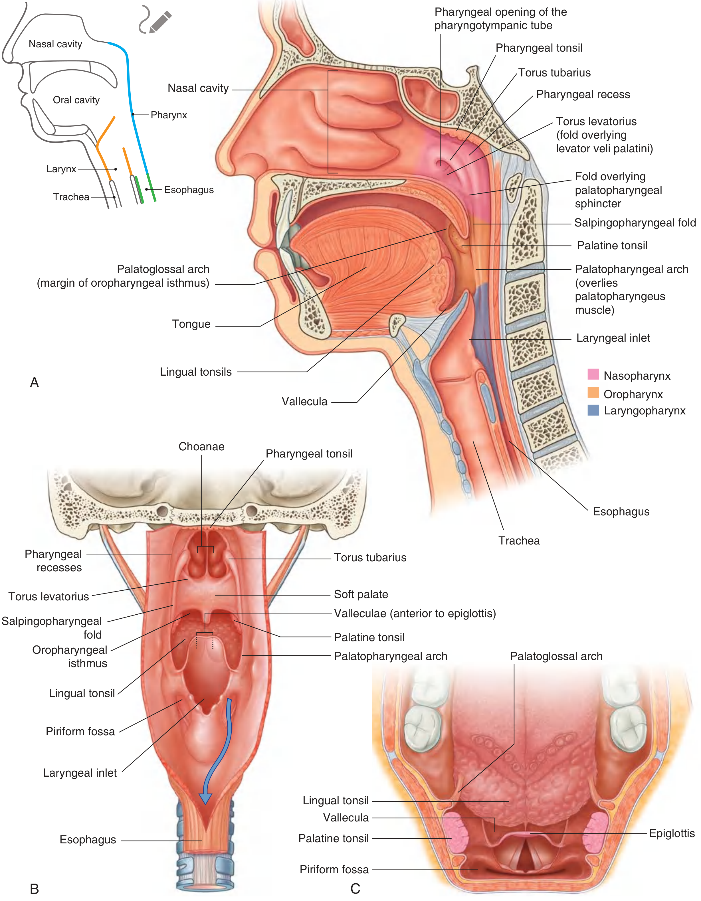
Fig. 8.208 - Mucosal Features of the Pharynx. (A) Lateral view showing nasopharynx (pink), oropharynx (orange), and laryngopharynx (blue). (B) Posterior view. (C) Superior view. [Gray's Anatomy for Students]
4. Pharyngeal Tonsil (Adenoids)
The roof and posterior wall of the nasopharynx contains a large collection of lymphoid tissue - the pharyngeal tonsil (part of Waldeyer's tonsillar ring).
- When enlarged (especially in children), it is called adenoids
- Enlarged adenoids can occlude the nasopharynx, forcing the child to breathe only through the mouth
- This is one of the most common causes of chronic nasal obstruction in children
5. Fascia of the Nasopharynx
The pharyngeal wall has two layers of fascia:
| Fascia | Location | Role |
|---|---|---|
| Buccopharyngeal fascia | Outside the muscular wall | Part of pretracheal cervical fascia |
| Pharyngobasilar fascia | Lines the inner surface (thick) | Reinforces the wall where muscle is deficient |
At the upper part (roof and posterior wall), the superior constrictor muscle is absent. Here the pharyngeal wall consists almost entirely of the pharyngobasilar fascia, reinforced externally by the levator and tensor veli palatini muscles of the soft palate. Anteriorly, this fascia attaches to the medial pterygoid plates on both sides.
6. Blood Supply
Arterial supply to the nasopharynx (upper pharynx) comes from branches of the external carotid artery:
- Ascending pharyngeal artery (main supply to the nasopharynx)
- Ascending palatine artery and tonsillar branches of the facial artery
- Numerous branches of the maxillary artery and lingual artery
Venous drainage: Pharyngeal veins form a venous plexus that drains:
-
Superiorly → pterygoid plexus (in the infratemporal fossa)
-
Inferiorly → facial and internal jugular veins
-
Gray's Anatomy for Students, p. 1034
7. Lymphatic Drainage
Lymphatics from the nasopharynx drain into:
- Retropharyngeal lymph nodes (nodes of Rouvière) - the first-echelon nodes, lying between the nasopharynx and the vertebral column
- Then to upper deep cervical (jugulodigastric) nodes
- Then further down in a step-wise fashion to lower neck nodes
This bilateral lymphatic drainage explains why nasopharyngeal carcinoma frequently presents with bilateral cervical lymphadenopathy.
8. Nerve Supply
Motor innervation
All muscles of the pharynx are supplied by the vagus nerve [X] through the pharyngeal plexus, EXCEPT the stylopharyngeus, which is innervated directly by the glossopharyngeal nerve [IX].
The pharyngeal plexus is formed by:
- Pharyngeal branch of vagus [X] (major motor nerve)
- Branches from the external laryngeal nerve (from vagus [X])
- Pharyngeal branches of the glossopharyngeal nerve [IX]
Sensory innervation (important!)
Each part of the pharynx has different sensory innervation:
| Region | Sensory Nerve |
|---|---|
| Nasopharynx | Pharyngeal branch of maxillary nerve [V2] - originates in pterygopalatine fossa, travels through the palatovaginal canal in the sphenoid bone to reach the roof |
| Oropharynx | Glossopharyngeal nerve [IX] |
| Laryngopharynx | Vagus [X] via internal branch of superior laryngeal nerve |
- Gray's Anatomy for Students, p. 1036
9. Epithelium / Histology
The nasopharynx is covered with pseudostratified ciliated columnar (respiratory) epithelium - similar to the mucosa of the nasal cavities and paranasal sinuses. This is in contrast to the oropharynx and laryngopharynx which are lined by stratified squamous epithelium.
10. Clinical Significance
| Condition | Mechanism |
|---|---|
| Adenoids (enlarged pharyngeal tonsil) | Blocks choanae → nasal obstruction, mouth breathing, snoring |
| Otitis media with effusion (glue ear) | Adenoid enlargement blocks/infects the pharyngotympanic tube opening |
| Nasopharyngeal carcinoma (NPC) | Most commonly arises from the fossa of Rosenmüller; spreads to parapharyngeal space, invades skull base via sphenoid; associated with EBV infection |
| Eustachian tube dysfunction | Inflammation of the nasopharyngeal mucosa (e.g., in URTI) obstructs tube opening → ear pain, hearing loss |
| Regurgitation through nose | Damaged soft palate/pharyngeal isthmus incompetence → food/liquid enters nasopharynx |
Summary: Key Points for Exams
- Location: Behind choanae, above soft palate - purely respiratory part of pharynx
- Roof: Sphenoid body + basal occipital (clivus) - always open (not collapsed)
- Inferior boundary: Pharyngeal isthmus (closed by soft palate during swallowing)
- Lateral wall landmark: Torus tubarius (around Eustachian tube opening) + pharyngeal recess (fossa of Rosenmüller)
- Folds: Salpingopharyngeal fold (salpingopharyngeus) + Torus levatorius (levator palatini)
- Lymphoid tissue: Pharyngeal tonsil / adenoids
- Arterial supply: Ascending pharyngeal artery (main) - branch of external carotid
- Sensory nerve: Maxillary nerve V2 (pharyngeal branch via palatovaginal canal)
- Motor nerve: Vagus nerve [X] via pharyngeal plexus
- Lymphatics: → Retropharyngeal nodes (Rouvière) → upper deep cervical nodes
- Epithelium: Pseudostratified ciliated columnar (respiratory type)
Source: Gray's Anatomy for Students, Chapter 8 (Head and Neck), pp. 1031-1036; Scott-Brown's Otorhinolaryngology Head & Neck Surgery, Applied Anatomy of the Nasopharynx.
Regarding BDC (BD Chaurasia): BD Chaurasia's Human Anatomy is not available in this library. However, the coverage above directly corresponds to the standard BDC pharynx chapter (Vol. 3 - Head, Neck & Brain), which covers the same structures: boundaries, lateral wall features, pharyngotympanic tube, pharyngeal tonsil, blood supply, lymphatics, and nerve supply in the same sequence. The Gray's Anatomy for Students content is more detailed and clinically oriented than BDC, making it an excellent complement.
This is a shared conversation. Sign in to Orris to start your own chat.