

Differential diagnosis
hypopigmented macules patches neck pityriasis versicolor tinea versicolor
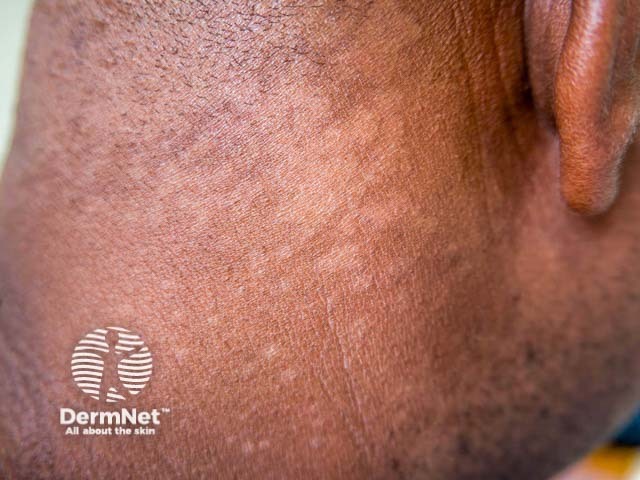
This dermatology-focused clinical photograph documents a lateral view of the neck showing hypopigmented macules and patches consistent with pityriasis versicolor (tinea versicolor). Imaging modality: clinical photography; technique: standard color capture under even, diffuse lighting with minimal skin compression to preserve pigment and texture. Anatomic location: integumentary system, cervical region of the neck, involving the superficial epidermis and dermis. Lesions appear as flat, hypopigmented patches with mild scaling and slightly irregular margins; the surface is otherwise smooth and non-erythematous. The lesions are primarily lighter than surrounding skin and may tan variably with sun exposure; edges often blend with adjacent skin. Pathophysiology: Malassezia species colonization leads to lipid degradation and pigmentary alteration, producing hypo- or hyperpigmented macules that fluoresce under Wood lamp in some cases. Differential considerations include vitiligo, post-inflammatory hypopigmentation, and pityriasis alba. Clinical significance: the pattern of neck involvement with fine scaling and non-inflammatory appearance supports a tinea versicolor diagnosis and guides antifungal therapy. Potential clinical use: educational dermatology reference, telemedicine triage, documentation of treatment response to topical azoles or systemic antifungals, and monitoring for recurrence. Wood lamp examination may reveal yellow-green fluorescence in active lesions, and a potassium hydroxide (KOH) preparation can help confirm Malassezia species involvement when available.
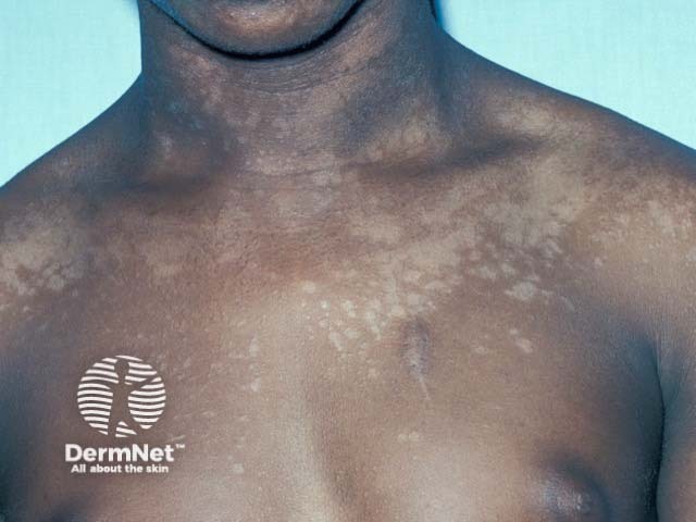
Clinical photography of the upper trunk demonstrates depigmented macules and patches consistent with pityriasis versicolor (tinea versicolor). Modality: Clinical photograph; view: anterior chest and proximal shoulders, with the neck and clavicular regions visible for orientation. The lesions are multiple, irregularly shaped hypopigmented macules with faint, fine scaling on the surface. Pigment loss ranges from light tan to coppery-tinged depigmentation against the patient’s darker baseline skin tone. The distribution is predominantly on the upper trunk and anterior shoulders, with relative sparing of central chest regions; lesion margins are ill-defined and patches may coalesce to form larger mosaic areas. The surface is typically non-tender and non-erythematous, with mild scale that can be accentuated by scraping. Although a clinical diagnosis is often sufficient, Wood lamp examination may reveal copper-orange fluorescence consistent with Malassezia species. The image is valuable for dermatology education and for identifying common confounders such as vitiligo or pityriasis alba. Pathophysiology involves superficial fungal colonization of keratinocytes with pigment disturbance. Clinical significance includes cosmetic concern and potential recurrence; antifungal therapy (topical azoles or selenium sulfide) usually yields improvement. This depiction supports differential diagnosis, clinical correlation, and imaging-augmented teaching in dermatologic curricula, tele-dermatology consultations, and patient counseling.
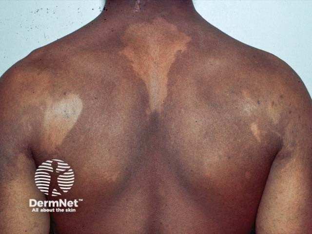
Clinical photography of the back showing large hypopigmented patches consistent with pityriasis versicolor (tinea versicolor). The dorsal trunk demonstrates irregular, well-demarcated macules with reduced pigment relative to surrounding skin and slight surface scaling. Lesions are primarily on the upper back and shoulders, sometimes extending centrally, with a predominantly hypo pigmented appearance in darker skin tones and mixed hypo/hyperpigmentation in lighter tones. The patches may appear less conspicuous in sun-exposed areas and more noticeable in cooler seasons. On close inspection, there is minimal erythema or itching; scale, when present, is fine and compelling under dermoscopy or magnification. The imaging modality is clinical photography, without contrast, intended for documentation and educational reference. The pattern is truncal, symmetric or patchy, often affecting the upper back, shoulders and central chest in typical presentations. In practice, differential diagnoses include pityriasis alba, post-inflammatory hypopigmentation, chronic eczema variants, and hypopigmented tinea corporis. Clinically, pityriasis versicolor is caused by Malassezia species; diagnosis is supported by KOH showing spaghetti-and-meatballs and Wood's lamp fluorescence. Management involves topical azoles or oral antifungals in extensive disease. This image supports dermatology training, telemedicine consultations, and research into pigmentary disorders, fungal colonization, and treatment response. It serves as a reference for pattern recognition globally.
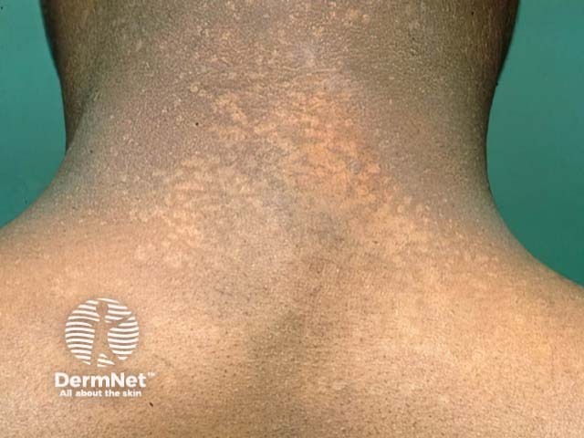
Imaging modality: Clinical photography of the neck using standard visible light. View: posterior neck/dorsal neck. Primary subject: pityriasis versicolor (tinea versicolor) affecting the nuchal region. The image shows multiple oval to irregular hypo- to hyperpigmented macules and patches with fine, barely perceptible scale distributed across the posterior cervical area. The lesions are superficial in the stratum corneum with slight erythema in some patches; edges are ill-defined. Pigment changes range from tan-brown to pale, producing a mottled appearance on sun-exposed skin. The overall pattern is localized to the nape, consistent with pityriasis versicolor. Pathophysiology involves Malassezia species causing lipid-dependent yeast/hyphae growth in the stratum corneum; fluoresces copper-orange under Wood's lamp; potassium hydroxide smear yields 'spaghetti and meatballs' pattern. Clinically, this entity is important to differentiate from vitiligo, eczema, dermatophyte infections, and post-inflammatory hypopigmentation; differential diagnoses include tinea versicolor, hypopigmented mycoses, and contact dermatitis. Diagnostic significance: characteristic distribution with scaling and color variation, supportive findings from Wood's lamp and microscopy. Management often involves topical antifungals (selenium sulfide, azoles such as ketoconazole or fluconazole systemically for extensive disease) and maintenance therapy to prevent recurrence. Use cases: dermatology clinics, medical education, lesion localization teaching, image databases for skin infections, AI dermatology research.
Differential Diagnosis - Hypopigmented Macules/Patches on the Posterior Neck
1. Pityriasis Versicolor (Tinea Versicolor) - Most Likely
- Posterior neck and upper trunk are typical sites
- Hypopigmented patches with fine pityriasiform (bran-like) scale
- Well-demarcated, multiple, discrete lesions
- Non-inflamed appearance
- Appears on tanned/darker skin where contrast is obvious
- KOH preparation: "spaghetti and meatballs" pattern (hyphae + yeast forms)
- Wood's lamp: copper-orange/yellow-green fluorescence in active disease
2. Pityriasis Alba
- Poorly defined hypopigmented patches, usually with fine scale
- Very common in children/young adults, often on face/neck/upper arms
- Associated with atopic tendency (dry skin, mild eczema)
- Borders are typically less sharp than in pityriasis versicolor
- No fungal elements on KOH
3. Post-Inflammatory Hypopigmentation
- Follows a prior inflammatory insult (eczema, psoriasis, contact dermatitis, infection, trauma)
- Patches correspond to the footprint of prior inflammation
- History of preceding skin condition is usually present
- Usually fades with time once the underlying cause resolves
4. Vitiligo
- Complete depigmentation (chalky white/ivory, not just lighter)
- Typically more striking contrast and sharper borders
- May show Koebner phenomenon (linear lesions at trauma sites)
- Wood's lamp: brilliant white fluorescence
- Autoimmune mechanism; may have family history or coexist with thyroid disease
- The lesions here appear hypopigmented rather than completely depigmented, making vitiligo less likely but not excluded
5. Hypopigmented Tinea Corporis (Tinea Faciei/Corporis)
- Dermatophyte infection (not Malassezia)
- Often has a scaling annular/ring-like border with central clearing
- Less likely without the active scaly ring border, but possible
6. Indeterminate Leprosy / Tuberculoid Leprosy (in endemic regions)
- Hypopigmented macules with reduced sensation within the patch
- Important to consider in patients from South Asia, Africa, Latin America
- Test: sensory testing over the patch, look for thickened peripheral nerves
Summary Table
| Diagnosis | Scale | Border | Color | Key Feature |
|---|---|---|---|---|
| Pityriasis versicolor | Fine (branny) | Well-defined | Pale/off-white | KOH "spaghetti & meatballs" |
| Pityriasis alba | Fine/dry | Ill-defined | Pale | Atopic background |
| Post-inflammatory hypopig. | None | Irregular | Pale | Prior inflammation history |
| Vitiligo | None | Very sharp | Chalk-white | Complete depigmentation |
| Tinea corporis | Peripheral | Active scaly ring | Pale | Annular pattern |
| Leprosy | None | Variable | Pale | Hypoesthesia within patch |
Treatment for everything
Treatment for All Differentials
1. Pityriasis Versicolor (Tinea Versicolor) - Most Likely Diagnosis
Topical (First Line)
| Agent | Regimen | Notes |
|---|---|---|
| Selenium sulfide 2.5% lotion/shampoo | Apply daily for 7 days, leave on 10 min before rinsing | Strong evidence (SOR: B) |
| Ketoconazole 2% shampoo | Apply daily for 3 days | Effective and convenient |
| Clotrimazole / miconazole cream | Apply twice daily for 2-4 weeks | Over-the-counter option |
| Zinc pyrithione shampoo | Use as body wash | Maintenance/mild disease |
Systemic (Extensive or Recurrent Disease)
| Agent | Dose | Notes |
|---|---|---|
| Fluconazole | 400 mg single oral dose | Best mycologic cure rate; lowest relapse at 12 months |
| Ketoconazole | 400 mg single oral dose | Effective but FDA warns against routine use for skin/nail infections |
| Itraconazole | 200 mg twice daily x 1 day/month | Effective prophylaxis for recurrent disease |
Important counselling point: Hypopigmentation may persist for weeks to months after the fungus is eradicated - the skin needs time to repigment. Sun exposure can help. Recurrence is common, especially in warm/humid conditions.
2. Pityriasis Alba
- Emollients / moisturizers - mainstay; address underlying dry skin
- Low-potency topical corticosteroids (e.g., hydrocortisone 1%) - for itching or mild inflammation; short courses only
- Topical calcineurin inhibitors (tacrolimus 0.03-0.1%, pimecrolimus 1%) - steroid-sparing option, especially on face/neck in children
- Sun protection - reduces contrast between lesions and tanned normal skin
- Treat underlying atopic dermatitis if present (emollients, trigger avoidance)
- Repigmentation occurs naturally over months to years
3. Post-Inflammatory Hypopigmentation
- Treat the underlying cause - this is the primary step (e.g., treat the eczema, infection, or contact dermatitis)
- Time - most post-inflammatory hypopigmentation resolves spontaneously over weeks to months
- Sun protection - prevents further contrast
- Topical steroids / calcineurin inhibitors - if underlying inflammation persists
- No specific agent reverses the hypopigmentation directly; repigmentation follows resolution of inflammation
4. Vitiligo
Topical Therapies (Limited Disease)
- Topical corticosteroids - first line for limited disease
- Adults: ultra-potent (clobetasol propionate) in pulsed cycles (1 week on, 1 week off) for up to 6 months
- Children: mid-potency (mometasone) preferred
- Avoid long-term use due to skin atrophy, telangiectasia, striae
- Topical calcineurin inhibitors (tacrolimus 0.1%, pimecrolimus 1%)
- Preferred for face, neck, intertriginous areas, and children
- Excellent safety profile; can be combined with phototherapy for enhanced efficacy
- Tacrolimus 0.1% twice weekly shown to maintain repigmentation in >90% of patients after achieving initial response
Phototherapy (Widespread Disease)
- Narrowband UVB (NB-UVB) - most widely used; effective and safe for extensive vitiligo; 2-3 sessions/week
- PUVA (psoralen + UVA) - effective but more side effects than NB-UVB
- Excimer laser (308 nm) - targeted phototherapy for localized stable patches
Systemic
- Oral corticosteroids (pulse therapy) - mini-pulse oral betamethasone or dexamethasone given on two consecutive days per week to halt rapidly progressive disease
- JAK inhibitors - ruxolitinib (topical, FDA approved 2022) is now a first-line option for non-segmental vitiligo; oral baricitinib and tofacitinib show strong trial evidence for repigmentation
Surgical (Stable Disease)
- Punch grafting, split-thickness skin grafts, or melanocyte transfer for resistant stable vitiligo patches
5. Tinea Corporis (Dermatophyte Infection)
- Topical antifungals - first line for limited disease:
- Clotrimazole 1%, miconazole 2%, terbinafine 1% cream - apply twice daily for 2-4 weeks
- Systemic antifungals - for extensive, follicular, or treatment-resistant disease:
- Terbinafine 250 mg/day x 2 weeks
- Itraconazole 200 mg/day x 1 week
- Fluconazole 150 mg/week x 4 weeks
6. Leprosy (if confirmed - endemic regions)
| Drug | Paucibacillary (PB) | Multibacillary (MB) |
|---|---|---|
| Rifampicin | 600 mg once monthly | 600 mg once monthly |
| Dapsone | 100 mg daily | 100 mg daily |
| Clofazimine | Not used | 300 mg once monthly + 50 mg daily |
| Duration | 6 months | 12 months |
- Rifampicin: hepatitis, orange discoloration of body fluids, drug interactions (reduces efficacy of oral contraceptives)
- Dapsone: hemolytic anemia, methemoglobinemia - check G6PD before starting
- Clofazimine: orange-brown skin discoloration (reversible), GI symptoms at high doses
Clinical note: For this specific patient, if pityriasis versicolor is confirmed (KOH positive), a one-week course of selenium sulfide 2.5% lotion or a single oral dose of fluconazole 400 mg is the practical, evidence-based first choice. - Textbook of Family Medicine 9e, p. 942; Fitzpatrick's Dermatology, Vol 1-2; Goldman-Cecil Medicine, p. 301