
Stroke management
Reading File
Finding Sources
Searching PubMed
"ischemic stroke" AND "management"
Reading File
Reading File
Finding Sources
Finding Sources
Finding Sources
Reading File
Reading File
Reading File
Finding Sources
Finding Sources
Reading File
Reading File
Reading File
Finding Sources
Reading File
Reading File
I now have comprehensive information from multiple authoritative sources. Let me compile the full response.
Stroke Management
Stroke is a medical emergency defined as a sudden focal neurological deficit caused by cerebrovascular disease. Management depends on stroke type (ischemic vs. hemorrhagic), time of onset, and the patient's clinical profile. Below is a structured, evidence-based overview.
1. Initial Assessment and Triage
Act FAST - Face drooping, Arm weakness, Speech difficulty, Time to call emergency services.
All patients with suspected stroke or TIA presenting within 72 hours of symptom onset should be admitted immediately - preferably to a dedicated stroke unit or ICU, which is associated with lower mortality, shorter hospital stay, and less discharge to nursing facilities. - Bradley and Daroff's Neurology in Clinical Practice
Immediate Priorities (ABCs)
- Airway: Protect from obstruction, hypoventilation, and aspiration. Use pulse oximetry; add supplemental O2 if SpO2 < 94%.
- Breathing: Avoid hyperthermia (worsens ischemic outcome); treat fever aggressively.
- Circulation: Cardiac monitoring for at least 48 hours (high frequency of arrhythmias, including new-onset atrial fibrillation). Obtain 12-lead ECG and troponin level immediately.
- Disability (Neurologic): Assess with the NIHSS (NIH Stroke Scale) to quantify deficits.
Emergency Imaging
- Non-contrast CT head - first-line; performed immediately to differentiate ischemic from hemorrhagic stroke. Diagnostic for SAH in ~90% of cases within 24 hours.
- CT Angiography (CTA) ± CT Perfusion - now standard in all patients with suspected large vessel occlusion (LVO) to screen for endovascular thrombectomy eligibility.
- MRI (DWI) - most sensitive for early ischemic change; not always needed if clinical diagnosis is clear. - Washington Manual of Medical Therapeutics
2. Ischemic Stroke
A. Intravenous Thrombolysis (IV tPA / Alteplase)
Dose: 0.9 mg/kg IV (maximum 90 mg) over 60 minutes, with 10% given as a bolus over the first minute. - ROSEN's Emergency Medicine (AHA/ASA 2019 Guidelines)
| Time Window | Recommendation |
|---|---|
| 0-3 hours | Recommended for all eligible patients ≥18 years, regardless of age (≤80 or >80), including severe stroke and mild disabling stroke |
| 3-4.5 hours | Recommended for patients ≤80 years, no history of both DM + prior stroke, NIHSS ≤25, not on oral anticoagulants, no ischemic injury >1/3 MCA territory |
Pre-treatment BP requirement: Must be < 185/110 mmHg (or safely lowered to this level before starting).
Key exclusions (0-3 h window):
- Mild non-disabling stroke (NIHSS 0-5)
- Intracranial hemorrhage, mass, or mass effect
- Active bleeding or coagulopathy (thrombocytopenia <30, INR >3)
- Active anticoagulation with elevated aPTT
- SBP >185 or DBP >110 despite treatment
Important: Aspirin and anticoagulants should be held for the first 24 hours after tPA.
B. Mechanical Thrombectomy (Endovascular)
This represents the most significant advance in acute ischemic stroke therapy since IV tPA in 1996. - Washington Manual
Indication: Acute ischemic stroke with large artery occlusion (LVO) in the anterior circulation, within 24 hours of last known well, regardless of whether IV alteplase was given. - ROSEN's Emergency Medicine
0-6 hours criteria (AHA/ASA):
- Pre-stroke mRS ≤1 (no significant disability)
- Causative occlusion of ICA or M1 segment of MCA
- NIHSS ≥6
- ASPECTS ≥6
Extended windows:
- 6-16 hours: Select patients using DAWN or DEFUSE-3 trial criteria (CT perfusion-guided mismatch selection)
- 16-24 hours: DAWN trial criteria apply
Every 1-hour delay to reperfusion is associated with less functional independence. In pooled data from MR CLEAN, ESCAPE, SWIFT PRIME, EXTEND-IA, REVASCAT, and THRACE trials, benefit became nonsignificant after 7 hours without perfusion imaging. - ROSEN's
Key 2025 update - A 2025 meta-analysis (PMID 40245349) confirmed benefit of endovascular thrombectomy even in patients with large ischemic core stroke, expanding eligibility criteria beyond traditional ASPECTS ≥6 thresholds.
C. Blood Pressure Management (Ischemic Stroke)
- BP often transiently elevated after stroke and tends to fall spontaneously within days.
- Do NOT aggressively lower BP unless: active MI/CHF, hypertensive emergency with end-organ damage, SBP >220 mmHg, or DBP >120 mmHg.
- For patients receiving IV tPA: target BP < 180/105 mmHg after treatment.
- A 15% reduction in the first 24 hours is a reasonable goal if lowering is needed.
- If urgent lowering is needed: IV labetalol (10 mg over 1-2 min, repeat as needed). - Bradley and Daroff's; Washington Manual
D. Antiplatelet Therapy
- Aspirin 325 mg orally within 24-48 hours of stroke onset (held for 24 h post-tPA).
- Reduce to 81 mg in post-acute period.
- Clopidogrel or aspirin/dipyridamole (ER) - both superior to aspirin alone for secondary stroke prevention.
- Dual antiplatelet therapy (DAPT) - may benefit select patients (e.g., minor stroke/high-risk TIA), but carries increased hemorrhage risk. - Washington Manual
E. Anticoagulation
- Routine heparin, LMWH, or warfarin NOT recommended for acute ischemic stroke.
- Cardioembolic stroke (AF): DOAC or warfarin - typically initiated after 4-14 days to reduce hemorrhagic transformation risk.
- Cervical artery dissection: A 2024 systematic review (PMID 38847098) found anticoagulation and antiplatelets have comparable efficacy - either is reasonable.
3. Hemorrhagic Stroke
A. Intracerebral Hemorrhage (ICH)
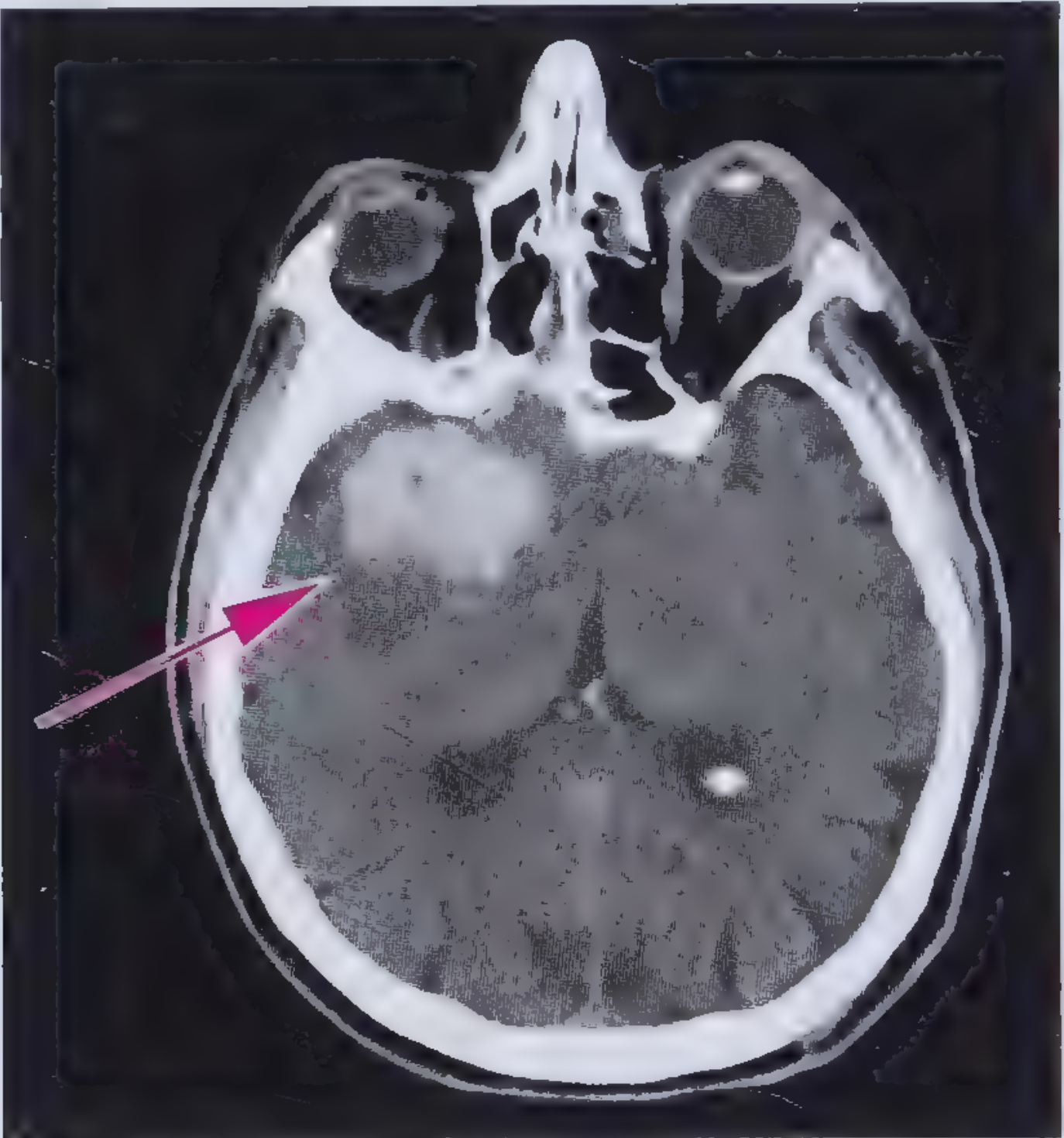
CT appearance: Round or oval hyperdense lesion, 40-80 HU acutely, becoming more homogeneous over hours to days. - Frameworks for Internal Medicine
30-day mortality: ~50%
Risk factors: Hypertension (most common), older age, Black/Asian race, high alcohol use, cerebral amyloid angiopathy (CAA), coagulopathy.
Management of Acute ICH:
1. Blood Pressure Control:
- For SBP 150-220 mmHg: acutely lower to SBP 140 mmHg - this is safe and improves functional outcome at 90 days (INTERACT2 trial).
- For SBP >220 mmHg: aggressive reduction with continuous IV infusion + monitoring.
- Target SBP ~130-139 mmHg may be maximally beneficial (pooled INTERACT2/ATACH2 analysis).
- Rapid drop >60 mmHg within 1 hour is associated with harm.
- Preferred agents: IV nicardipine (calcium channel blocker) or IV labetalol. Avoid nitrates (cerebral vasodilation, elevated ICP). - ROSEN's
2. Reversal of Anticoagulation (immediate):
- Warfarin (INR ≥1.4): IV Vitamin K 10 mg slowly + 4-factor PCC (preferred over FFP - faster correction, lower volume, lower infection risk).
- DOACs (Factor Xa inhibitors: rivaroxaban, apixaban): Andexanet alfa or 4-factor PCC.
- Dabigatran: Idarucizumab (specific reversal agent).
- All antiplatelet agents should also be discontinued acutely.
3. Other measures:
- Identify and correct bleeding diatheses
- Manage blood sugar (avoid hyper- and hypoglycemia)
- Treat seizures with antiepileptic medications
- Elevated ICP: intubation/sedation, head of bed elevation 15-30°, hypertonic saline or mannitol, consider hemicraniectomy
B. Subarachnoid Hemorrhage (SAH)
- Head CT is diagnostic in 90% of SAH within 24 hours; if CT negative and SAH suspected, perform LP and look for xanthochromia.
- Treatment depends on aneurysm anatomy: surgical clipping vs. endovascular coiling.
- Nimodipine (oral calcium channel blocker) is indicated to improve neurological outcomes and reduce the incidence of cerebral infarction from vasospasm, though its exact mechanism remains unclear. - Washington Manual
4. General Medical Management (All Stroke Types)
| Domain | Intervention |
|---|---|
| Aspiration prevention | NPO until formal swallowing assessment by speech pathologist; HOB >30°; frequent suctioning |
| Nutrition | Enteral tube feeding for patients with oropharyngeal dysfunction |
| DVT prophylaxis | LMWH (enoxaparin 40 mg OD) or UFH 5000 units SC BID; IPC if anticoagulation contraindicated |
| Glucose control | Maintain near euglycemia; avoid hyperglycemia and tight control (no clear acute benefit) |
| Urinary catheter | Avoid unless urinary retention; remove ASAP to prevent urosepsis |
| Skin care | Frequent turning, protective dressings, early mobility to prevent pressure sores |
| Cardiac monitoring | Continuous telemetry for 48-72 hours; ECG + troponin on admission |
| Falls prevention | Regular assessment; minimize postprandial hypotension |
| Depression screening | >25% of stroke patients develop depression (more with left frontal infarcts); treat with antidepressants |
5. Secondary Prevention
| Risk Factor | Intervention |
|---|---|
| Hypertension | Long-term antihypertensive therapy (defer until neurologic deficit is stable, usually 1 week) |
| Atrial fibrillation | DOAC (preferred) or warfarin; start 4-14 days post-stroke |
| Carotid stenosis | CEA for symptomatic ≥70% stenosis (reduces 5-year ipsilateral stroke risk significantly) |
| Asymptomatic stenosis ≥60% | CEA reduces 5-year risk in men; individualize for women |
| Carotid stenting (CAS) | Equal efficacy to CEA but higher periprocedural stroke risk (CREST trial) |
| Hyperlipidemia | High-intensity statin therapy |
| Diabetes | Glycemic control |
| Antiplatelet (non-cardioembolic) | Aspirin, clopidogrel, or aspirin/dipyridamole |
6. Rehabilitation
Rehabilitation begins as soon as the diagnosis is established and life-threatening complications are stabilized. Evidence consistently shows benefit from:
- Physical therapy (mobility, gait)
- Occupational therapy (activities of daily living)
- Speech therapy (dysphagia, aphasia, dysarthria)
Shoulder subluxation in hemiparetic patients is minimized with early therapy initiation. Stroke rehabilitation has a clear beneficial impact on post-stroke outcomes. - Washington Manual
ICH on CT
Recent Evidence Updates (2024-2025)
- Endovascular thrombectomy for large core stroke (PMID 40245349, Neurology 2025): Meta-analysis of RCTs confirmed benefit in patients with large ischemic core - expands the eligible population beyond previous strict ASPECTS criteria.
- Anticoagulation in breakthrough stroke on DOACs (PMID 40758940, Neurology 2025): Meta-analysis guides anticoagulation strategies when ischemic stroke occurs despite DOAC use.
- Cervical artery dissection (PMID 38847098, Stroke 2024): No significant difference between anticoagulation and antiplatelet therapy.
Sources: Bradley and Daroff's Neurology in Clinical Practice | ROSEN's Emergency Medicine | Adams and Victor's Principles of Neurology, 12th Ed | Washington Manual of Medical Therapeutics | Frameworks for Internal Medicine | Harrison's Principles of Internal Medicine 22E | Fuster and Hurst's The Heart, 15th Ed
Stroke management
Reading File
Finding Sources
Finding Sources
Searching PubMed
"ischemic stroke" AND "management"
Reading File
Reading File
Finding Sources
Reading File
Reading File
Finding Sources
Reading File
Reading File
Finding Sources
Reading File
Reading File
Searching PubMed
"endovascular thrombectomy" AND "ischemic stroke"
I now have thorough coverage from multiple authoritative sources. Here is the full, structured response.
Stroke Management
Stroke is a neurological emergency defined as a sudden focal (or global) neurological deficit lasting >24 hours due to a vascular cause, or any duration if imaging confirms infarction or hemorrhage. It is the leading cause of adult disability worldwide. Management pivots on the stroke subtype - ischemic (~85%) vs. hemorrhagic (~15%) - and the time elapsed since onset.
1. Immediate Recognition and Triage
FAST / BE-FAST Screening
- Balance loss, Eyes (vision change), Face drooping, Arm weakness, Speech difficulty, Time to call emergency services.
Admission
All patients with TIA or acute stroke, regardless of severity, presenting within 72 hours of onset should be admitted for emergency evaluation - preferably to a dedicated stroke unit or ICU. Stroke unit care correlates with lower mortality, shorter hospital stays, lower nursing-home discharge rates, and reduced cost compared with general ward care. - Bradley and Daroff's Neurology in Clinical Practice
A stroke team with established protocols dramatically expedites care. Every minute counts: "time is brain."
2. Initial Stabilization (All Stroke Types)
Airway, Breathing, Circulation
| Priority | Action |
|---|---|
| Airway | Protect from obstruction, hypoventilation, and aspiration; consider intubation if GCS falls |
| Oxygenation | Pulse oximetry; supplemental O2 only if SpO2 <94%; avoid routine O2 (no benefit shown in normoxic patients) |
| Temperature | Treat fever aggressively - hyperthermia worsens ischemic injury; mild hypothermia is neuroprotective but technically difficult |
| Cardiac monitoring | Continuous telemetry for at least 48-72 hours; new AF detected in 10-15% |
| ECG + Troponin | Obtain immediately; concurrent cerebral and myocardial ischemia occurs in 3-20% of cases |
| IV access + bloods | CBC, coagulation screen, BMP, glucose, group & screen |
Aspiration Prevention
Pneumonia mortality in stroke patients reaches 15-25%. Aspiration was documented by videofluoroscopic modified barium swallow in >1/3 of brainstem strokes, 1/4 of bilateral hemispheric strokes, and 1/10 of unilateral hemispheric strokes. Patients with any oropharyngeal dysfunction should be kept nil by mouth until formally assessed by a speech pathologist. Maintain head of bed >30°; frequent suctioning. - Bradley and Daroff's
3. Emergency Imaging
| Modality | Role |
|---|---|
| Non-contrast CT head | First-line; rapidly distinguishes ischemic from hemorrhagic stroke; diagnostic for SAH in ~90% within 24 h; rate-limiting step for thrombolysis decisions |
| CT Angiography (CTA) ± CT Perfusion | Now standard for all patients with suspected large vessel occlusion (LVO); screens for thrombectomy eligibility up to 24 h; also used in ICH to identify underlying AVM/aneurysm |
| MRI (DWI) | Most sensitive for early ischemic stroke; detects infarct earliest; not always needed if clinical diagnosis is clear |
| MRA | Noninvasive evaluation of cervical and intracranial vasculature |
| Carotid Doppler | Anterior circulation strokes if angiography not already done |
| Echocardiography (TTE/TEE) | Identifies cardioembolic sources: thrombus, vegetations, valvular disease, PFO |
If CT is negative but SAH is highly suspected: perform lumbar puncture, centrifuge CSF, look for xanthochromia (develops after several hours, confirming SAH over traumatic tap). - Washington Manual of Medical Therapeutics
4. Ischemic Stroke
A. IV Thrombolysis - Alteplase (tPA)
Dose: 0.9 mg/kg IV (maximum 90 mg) over 60 minutes; 10% given as a bolus over the first minute. - ROSEN's Emergency Medicine (AHA/ASA Guidelines)
Eligibility by Time Window
| Window | Criteria |
|---|---|
| 0-3 hours | All eligible patients ≥18 years, including age >80, severe stroke, mild disabling stroke |
| 3-4.5 hours | Age ≤80, no history of BOTH diabetes + prior stroke, NIHSS ≤25, not on oral anticoagulants, no ischemic injury >1/3 MCA territory |
BP requirement: Must be <185/110 mmHg before starting (or safely lowered to this level).
Glucose: Eligible if glucose >50 mg/dL.
CT finding: Acceptable with early ischemic changes of mild-moderate extent (not frank hypodensity).
Prior antiplatelets: Benefit of tPA outweighs small increased sICH risk - proceed.
Key Exclusions (0-3 h window)
- Mild non-disabling stroke (NIHSS 0-5)
- Intracranial hemorrhage or mass on CT
- Active bleeding or coagulopathy (INR >3, thrombocytopenia <30k, therapeutic anticoagulation with elevated aPTT)
- SBP >185 or DBP >110 despite treatment
- Prior ICH
After tPA: Hold aspirin and anticoagulants for 24 hours; monitor BP closely (target <180/105 mmHg post-infusion); watch for symptomatic ICH (sICH).
Note on cerebral microbleeds (CMBs): In patients with >10 CMBs on prior MRI, the risk of ICH after thrombolytics is substantially higher (30-47% vs. 1-4%). Benefits are uncertain in this group. - ROSEN's
B. Mechanical Thrombectomy (Endovascular)
This represents the most significant advance in acute stroke therapy since IV tPA was introduced in 1996. Multiple landmark RCTs (MR CLEAN, ESCAPE, SWIFT PRIME, EXTEND-IA, REVASCAT, THRACE) established clear benefit. - Washington Manual
Indication: Acute ischemic stroke with large vessel occlusion (LVO) in the anterior circulation, meeting criteria, within 24 hours of last known well - regardless of whether IV tPA was given.
AHA/ASA Criteria (0-6 hours)
- Pre-stroke mRS ≤1 (no significant disability)
- Occlusion of ICA or M1 segment of MCA
- NIHSS ≥6
- ASPECTS ≥6
Extended Windows
- 6-16 hours: DAWN and DEFUSE-3 trial criteria - CT perfusion-guided penumbra/core mismatch selection
- 16-24 hours: DAWN trial criteria
Timing Matters
Each 1-hour delay to reperfusion is associated with worse functional independence. Benefit becomes nonsignificant after ~7 hours in pooled analyses of patients not selected by perfusion imaging. Pretreatment with IV tPA (if within 4.5 h) is not required before thrombectomy but should not be withheld if otherwise eligible. - ROSEN's
2025 Evidence Update: Two major meta-analyses (PMID 40245349, Neurology; PMID 40245084, PLoS Med) confirm thrombectomy benefit even in patients with large ischemic core (low ASPECTS), expanding eligibility beyond prior strict thresholds.
Cochrane 2025 (PMID 40271574): Endovascular thrombectomy with vs. without IV thrombolysis - bridging therapy remains an active area with ongoing evidence synthesis.
C. Blood Pressure Management in Ischemic Stroke
- BP is commonly elevated post-stroke and typically falls spontaneously over days - do not aggressively lower it.
- Optimal post-stroke SBP appears to be 160-200 mmHg, with lower or higher values associated with larger infarct volumes. - Bradley and Daroff's
- Do NOT lower acutely unless: active MI/CHF, hypertensive emergency with end-organ damage, SBP >220 mmHg, or DBP >120 mmHg.
- If lowering is needed: target 15% reduction in first 24 hours; use IV labetalol (10 mg over 1-2 min, repeat as needed).
- For patients receiving tPA: maintain BP <180/105 mmHg post-infusion.
- Defer treatment of newly identified chronic hypertension until neurological deficit has stabilized (first few days). - Adams and Victor's Principles of Neurology, 12th Ed
D. Antiplatelet Therapy
- Aspirin 325 mg orally within 24-48 hours of ischemic stroke onset (held for 24 h after tPA); reduce to 81 mg in post-acute period.
- Clopidogrel or aspirin/dipyridamole (extended-release) - both superior to aspirin alone for secondary stroke prevention (non-cardioembolic).
- Dual antiplatelet therapy (DAPT) (e.g., aspirin + clopidogrel) - beneficial in select high-risk patients (e.g., minor stroke, high-risk TIA), but increases hemorrhage risk with prolonged use. - Washington Manual
E. Anticoagulation
- Routine heparin/LMWH/warfarin is NOT recommended for acute ischemic stroke (no net benefit, increased hemorrhagic risk).
- Cardioembolic stroke (atrial fibrillation): DOAC (preferred) or warfarin - typically started 4-14 days post-stroke to reduce hemorrhagic transformation risk.
- Cervical artery dissection: 2024 systematic review (PMID 38847098, Stroke) found no significant difference between anticoagulation and antiplatelet therapy - either is appropriate.
F. DVT Prophylaxis
- DVT in hemiparetic limbs is common; VTE risk persists into the post-stroke period.
- LMWH (enoxaparin 40 mg OD) preferred over UFH - superior VTE prevention (PREVAIL study), small increase in extracranial hemorrhage.
- If anticoagulation is contraindicated: intermittent pneumatic compression (IPC) - proven effective in reducing DVT (CLOTS 3 trial). Graduated compression stockings are NOT recommended. - Bradley and Daroff's
5. Hemorrhagic Stroke
A. Intracerebral Hemorrhage (ICH)
30-day mortality: ~50%
CT appearance: Round or oval hyperdense lesion, 40-60 HU initially (heterogeneous); 60-100 HU within hours to days as clot organizes. - Frameworks for Internal Medicine
Risk factors: Hypertension (most common), older age, Black/Asian race, excess alcohol, cerebral amyloid angiopathy (CAA), coagulopathy, AVM/aneurysm.
Blood Pressure Control in ICH
| SBP Range | Target / Action |
|---|---|
| 150-220 mmHg | Acutely lower to SBP 140 mmHg - safe, improves functional outcomes at 90 days (INTERACT2 trial) |
| >220 mmHg | Aggressive IV reduction with continuous infusion; target SBP less clear |
| Optimal target | SBP 130-139 mmHg likely maximally beneficial (pooled INTERACT2/ATACH2 analysis) |
| Avoid | Rapid drop ≥60 mmHg within 1 hour - associated with harm |
- Preferred agents: IV nicardipine (CCB) or IV labetalol - short half-life, easily titrated.
- Avoid nitrates - cerebral vasodilation raises ICP. - ROSEN's
Reversal of Anticoagulation (Immediate)
| Anticoagulant | Reversal Agent |
|---|---|
| Warfarin (INR ≥1.4) | IV Vitamin K 10 mg slowly + 4-factor PCC (preferred over FFP - faster, lower volume, lower infection risk) |
| Dabigatran | Idarucizumab (specific reversal) |
| Factor Xa inhibitors (rivaroxaban, apixaban, edoxaban) | Andexanet alfa or 4-factor PCC |
| Antiplatelet agents | Discontinue immediately (platelet transfusion may be considered in surgical candidates) |
Other ICH Management
- Identify and correct all bleeding diatheses
- Blood sugar: avoid hyper- and hypoglycemia; maintain near-euglycemia
- Seizures: treat with antiepileptics (prophylactic use is controversial)
- Elevated ICP: intubation + sedation; head of bed elevation 15-30°; hypertonic saline or mannitol; consider hemicraniectomy
- Avoid steroids (no benefit, increase infection risk)
B. Subarachnoid Hemorrhage (SAH)
- Typically presents with "thunderclap headache" - worst headache of life.
- Non-contrast CT is diagnostic in ~90% within 24 hours; if negative with high suspicion, LP with xanthochromia testing is required.
- Aneurysm confirmed by CTA or DSA.
- Treatment: surgical clipping vs. endovascular coiling (decision based on aneurysm anatomy, location, patient age, and available expertise).
- Nimodipine (oral, 60 mg every 4 hours for 21 days) - reduces cerebral infarction and improves neurological outcomes; mechanism not fully understood but not via vasospasm prevention.
- Monitor for delayed cerebral ischemia (vasospasm) - typically days 4-14. - Washington Manual
6. General Medical Management (All Stroke Types)
| Domain | Intervention |
|---|---|
| Nutrition | Enteral tube feeding if oropharyngeal dysfunction; assess swallowing before any oral intake |
| Glucose | Maintain near euglycemia; avoid hyperglycemia; avoid excessively tight control (no acute benefit) |
| Urinary catheter | Avoid unless retention; remove at earliest opportunity to prevent urosepsis; treat symptomatic UTI only |
| Skin | Frequent turning, protective dressings, early mobility; ~15% develop pressure sores post-stroke |
| Falls | Regular assessment; postprandial hypotension increases fall risk |
| Shoulder subluxation | Initiate therapy early in hemiplegic patients |
| Depression | Affects >25% of stroke patients; more common with left frontal infarcts; screen and treat with antidepressants |
7. Secondary Prevention
| Risk Factor | Intervention |
|---|---|
| Hypertension | Long-term antihypertensives (defer initiation until deficits stabilize, ~first week) |
| Atrial fibrillation | DOAC (preferred) or warfarin; start 4-14 days post-stroke |
| Symptomatic carotid stenosis ≥70% | Carotid endarterectomy (CEA) - significant reduction in 5-year ipsilateral stroke risk |
| Asymptomatic stenosis ≥60% | CEA reduces 5-year risk in men; individualize for women |
| CAS vs. CEA | CREST trial: equal efficacy, but carotid artery stenting (CAS) carries higher periprocedural stroke risk |
| Hyperlipidemia | High-intensity statin therapy |
| Diabetes | Optimize glycemic control |
| Non-cardioembolic stroke | Antiplatelet therapy (aspirin, clopidogrel, or aspirin/dipyridamole) |
| PFO-related stroke | 2024 ESO guidelines (PMID 38752755) recommend PFO closure in selected patients aged <60 with cryptogenic stroke |
8. Rehabilitation
Stroke rehabilitation begins as soon as diagnosis is established and life-threatening complications are stabilized:
- Physical therapy - mobility, gait, balance
- Occupational therapy - ADLs, fine motor function
- Speech therapy - dysphagia, aphasia, dysarthria
Evidence consistently shows a clear beneficial impact of rehabilitation on post-stroke functional outcomes. Patients with obvious dysphagia, dysarthria, or facial droop should be kept NPO until formal swallowing assessment. - Washington Manual
Quick Reference Summary
ACUTE STROKE PATHWAY
├── CT Head (non-contrast) → differentiate ischemic vs. hemorrhagic
│
├── ISCHEMIC STROKE
│   ├── <4.5 h + eligible → IV Alteplase 0.9 mg/kg (max 90 mg)
│   ├── LVO on CTA + criteria → Mechanical thrombectomy (up to 24 h)
│   ├── BP: do not lower unless SBP >220 or specific indications
│   ├── 24-48 h: Aspirin 325 mg (hold 24 h if tPA given)
│   └── AF: DOAC/warfarin after 4-14 days
│
└── HEMORRHAGIC STROKE (ICH)
    ├── Reverse anticoagulation immediately (PCC/idarucizumab/andexanet)
    ├── SBP 150-220: lower to 140 mmHg (nicardipine/labetalol)
    ├── Treat ICP: HOB↑, hypertonic saline, mannitol, EVD
    ├── Seizure management
    └── SAH: nimodipine + surgical/endovascular aneurysm repair
Sources: Bradley and Daroff's Neurology in Clinical Practice | ROSEN's Emergency Medicine | Adams and Victor's Principles of Neurology, 12th Ed | Washington Manual of Medical Therapeutics | Frameworks for Internal Medicine | Adams and Victor's Principles of Neurology 12th Ed | Fuster and Hurst's The Heart, 15th Ed | Harrison's Principles of Internal Medicine 22E (2025)
This is a shared conversation. Sign in to Orris to start your own chat.