Explain me common 10 heart diseases with ECG changes and ECG images of those diseases
Reading File
Finding Sources
Searching the Web
common heart diseases ECG changes findings with ECG images examples
Finding Sources
Finding Sources
Searching the Web
ECG images STEMI atrial fibrillation heart block ventricular tachycardia LVH example
Finding Sources
Searching the Web
site:litfl.com ECG library STEMI atrial fibrillation heart block WPW hyperkalemia pericarditis
Finding Sources
Loading Image
Loading Image
Loading Image
Loading Image
Searching the Web
litfl.com ECG ventricular tachycardia complete heart block LVH hyperkalemia pulmonary embolism images
Loading Image
Loading Image
Searching the Web
litfl.com ECG ventricular tachycardia LVH left ventricular hypertrophy pulmonary embolism S1Q3T3 images
Loading Image
Loading Image
Loading Image
Loading Image
I now have all the ECG images and content needed to deliver a comprehensive answer. Let me compile the full response.
10 Common Heart Diseases - ECG Changes with Images
Here is a complete guide to the 10 most clinically important heart diseases, their ECG changes, and real ECG examples.
1. ST-Elevation Myocardial Infarction (STEMI)
Pathophysiology: Complete occlusion of a coronary artery causes transmural ischemia, producing a sequence of ECG changes.
ECG Changes (by stage):
- Hyperacute phase (minutes): Tall, broad, peaked "hyperacute" T waves - earliest sign
- Acute phase (hours): ST elevation ≥ 1 mm in ≥ 2 contiguous leads ("tombstone" appearance in severe cases)
- Evolving (hours-days): T wave inversion, development of pathological Q waves (≥ 40 ms wide, ≥ 25% of QRS height)
- Chronic/old MI: Persistent Q waves, T wave normalization
Localization by leads:
| Territory | Leads | Artery |
|---|---|---|
| Anterior | V1-V4 | LAD |
| Inferior | II, III, aVF | RCA |
| Lateral | I, aVL, V5-V6 | LCx |
| Posterior | Tall R in V1-V2, ST depression V1-V3 | RCA/LCx |
Reciprocal changes: ST depression in leads opposite to the infarct zone
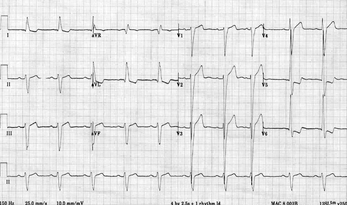
ECG - Inferior STEMI (ST elevation in II, III, aVF with reciprocal changes in I, aVL):
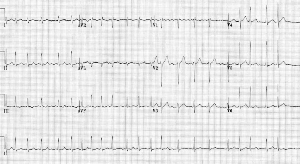
2. Atrial Fibrillation (AF)
Pathophysiology: Chaotic, disorganized atrial electrical activity from multiple micro-reentrant circuits. No organized P waves, irregular ventricular response.
ECG Changes:
- Absent P waves - replaced by irregular fibrillatory baseline (f waves) at 350-600/min
- Irregularly irregular RR intervals - the hallmark finding
- Narrow QRS (unless aberrant conduction or BBB present)
- Ventricular rate typically 100-180/min if uncontrolled
- Fibrillatory baseline is best seen in V1
ECG - Atrial Fibrillation (note irregular RR intervals and absent P waves):
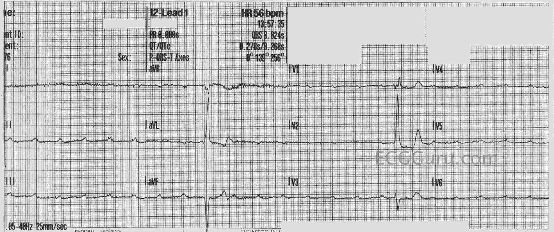
3. Complete Heart Block (3rd Degree AV Block)
Pathophysiology: Complete failure of impulse conduction from atria to ventricles. The atria and ventricles beat independently - AV dissociation.
ECG Changes:
- Regular P waves at normal atrial rate (60-100/min)
- Regular QRS complexes at a slower escape rate
- No relationship between P waves and QRS - they "march through" each other
- Escape rhythm: Junctional escape = narrow QRS at 40-60/min; Ventricular escape = wide QRS at 20-40/min
- PR intervals appear to vary randomly
ECG - Complete Heart Block (P waves dissociated from QRS; ventricular rate ~40 bpm independent of atrial rate):
4. Ventricular Tachycardia (VT)
Pathophysiology: Fast, life-threatening rhythm originating in the ventricles, often from areas of scar (post-MI) or cardiomyopathy.
ECG Changes:
- Wide QRS ≥ 120 ms (usually ≥ 140 ms), bizarre morphology
- Regular rhythm at rate 100-250/min
- AV dissociation - P waves independent of QRS (when visible)
- Fusion beats and capture beats - pathognomonic for VT
- Concordance in precordial leads: all positive (R) or all negative (QS) = strongly suggests VT
- Left axis deviation common
- Brugada criteria: If any one of LBBB morphology + right axis, or RBBB morphology with negative concordance = VT
ECG - Ventricular Tachycardia (wide QRS tachycardia with AV dissociation visible):
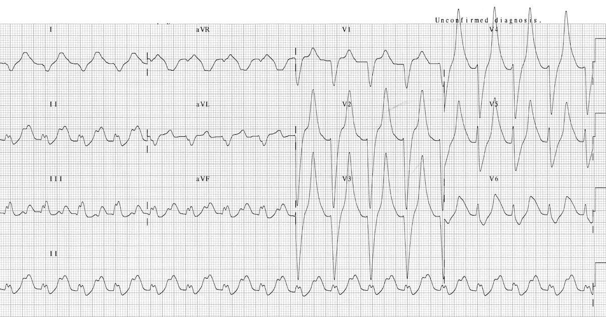
(Above shows wide complex tachycardia pattern with bizarre QRS morphology)
5. Left Ventricular Hypertrophy (LVH)
Pathophysiology: Thickened LV myocardium (from hypertension, aortic stenosis, hypertrophic cardiomyopathy) generates increased electrical forces seen as tall voltages.
ECG Changes (Sokolow-Lyon criteria):
- S in V1 + R in V5 or V6 > 35 mm (voltage criteria)
- R in aVL > 11 mm (alternative criteria)
- LV strain pattern: ST depression + asymmetric T wave inversion in lateral leads (I, aVL, V5-V6) - the "hockey stick" pattern
- ST elevation in right precordial leads V1-3 (discordant to the deep S waves)
- Left axis deviation common
- Prolonged QRS (0.09-0.11 s) due to delayed depolarization
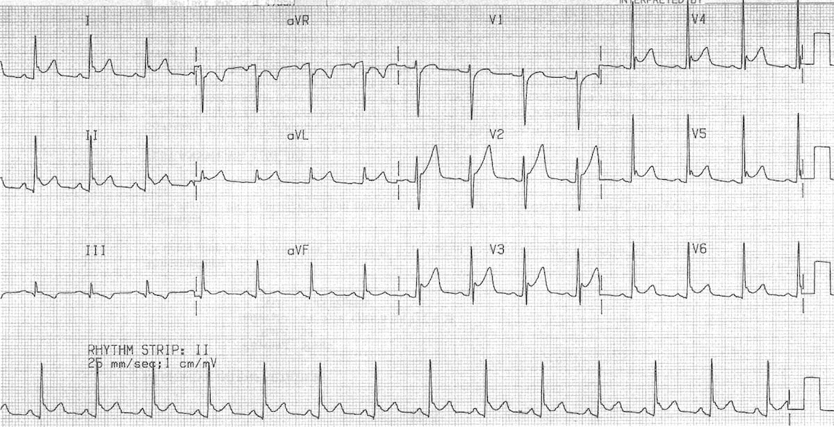
ECG - Left Ventricular Hypertrophy (deep S in V2, tall R in V5 - exceeds 35 mm combined; ST-T changes laterally):
6. Acute Pericarditis
Pathophysiology: Inflammation of the pericardium causes diffuse epicardial irritation and concurrent subepicardial myocarditis - producing widespread ST changes and PR depression.
ECG Changes (4 stages):
- Stage 1 (acute): Diffuse saddle-shaped (concave up) ST elevation in most leads EXCEPT aVR and V1 + PR depression in same leads + PR elevation in aVR - the key differentiator from STEMI
- Stage 2 (days): ST returns to baseline, T waves flatten
- Stage 3: Diffuse T wave inversions
- Stage 4 (weeks): ECG normalizes (or T inversions persist = constrictive pericarditis)
Key differentiators from STEMI:
- ST elevation is concave-up (saddle-shaped) vs convex in STEMI
- Diffuse (not territory-specific)
- PR depression present
- No reciprocal ST depression (except in aVR)
ECG - Acute Pericarditis (diffuse saddle-shaped ST elevation + PR depression):
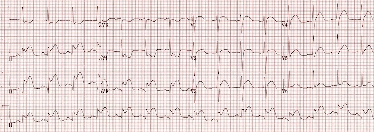
7. Pulmonary Embolism (PE)
Pathophysiology: Acute right heart strain from elevated pulmonary artery pressure causes RV dilatation and right ventricular ischemia.
ECG Changes:
- S1Q3T3 pattern: Deep S wave in I + Q wave in III + T wave inversion in III - classic but only present in ~20% of cases
- Right ventricular strain: T wave inversions in V1-V4 (and sometimes inferior leads II, III, aVF)
- Right axis deviation
- RBBB (new, incomplete or complete) due to RV pressure overload
- Sinus tachycardia - most common finding
- Clockwise rotation - persistent S wave in V6
- T inversion in both lead III AND V1 = most specific finding (specificity up to 99%)
ECG - Pulmonary Embolism (S1Q3T3 + T wave inversions V1-V4 from RV strain):

8. Hyperkalemia
Pathophysiology: Elevated extracellular potassium depolarizes resting membrane potential, slowing conduction. Progressive ECG changes correlate with rising K⁺ levels.
ECG Changes (progressive with rising K⁺):
| K⁺ Level | ECG Change |
|---|---|
| 5.5-6.5 mEq/L | Tall, narrow, peaked (tented) T waves - earliest sign |
| 6.5-7.5 mEq/L | Prolonged PR interval, widened P wave, then P wave flattening/disappearance |
| 7.0-8.0 mEq/L | Wide QRS (> 120 ms), bizarre morphology, right axis shift |
| > 8.5-9.0 mEq/L | "Sine wave" pattern - QRS merges with T wave |
| > 9-10 mEq/L | Ventricular fibrillation or asystole |
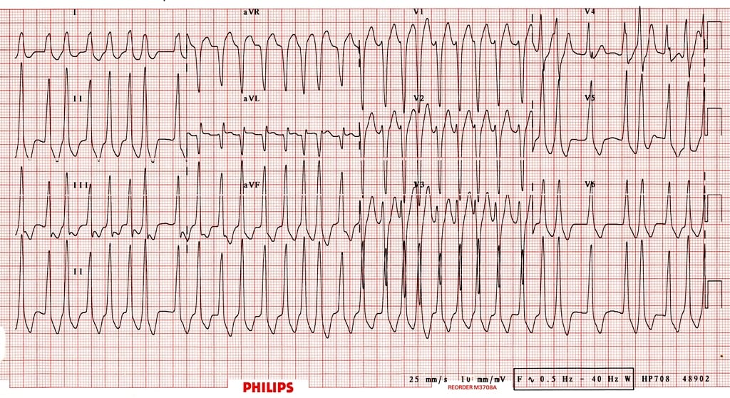
ECG - Hyperkalemia (tall, peaked T waves + widened QRS + bizarre morphology at K⁺ ~9.3):
9. Wolff-Parkinson-White (WPW) Syndrome
Pathophysiology: An accessory pathway (Bundle of Kent) bypasses the AV node and pre-excites the ventricle, creating the delta wave. Risk of life-threatening arrhythmias if AF conducts rapidly down the pathway.
ECG Changes (classic WPW triad):
- Short PR interval (< 120 ms) - fast conduction through accessory pathway
- Delta wave - slurred upstroke at the beginning of QRS (ventricular pre-excitation)
- Wide QRS (> 120 ms) - fusion of normal and pre-excited depolarization
- ST-T changes discordant (opposite) to the QRS polarity
- Pseudo-infarct pattern - negative delta waves can mimic Q waves in inferior leads
WPW + AF = DANGER: Rapid pre-excited AF with wide complex, irregular, extremely fast (> 250/min) rhythm - can degenerate to VF
ECG - WPW with Atrial Fibrillation (rapid pre-excited AF - broad, irregular, bizarre QRS):
10. Heart Failure / Dilated Cardiomyopathy (DCM)
Pathophysiology: Dilated, poorly contracting ventricles cause multiple non-specific ECG abnormalities from chamber enlargement, conduction delays, and fibrosis.
ECG Changes:
- Left bundle branch block (LBBB): QRS > 120 ms, broad notched R in I/V5-V6, broad QS in V1 - present in ~25% of HF patients; indicates dyssynchrony
- Low voltage QRS (if pericardial effusion, amyloid, or severe cardiomyopathy)
- Left axis deviation
- Atrial fibrillation - very common in heart failure
- Ventricular ectopy / PVCs
- Non-specific ST-T changes throughout
- QTc prolongation due to electrolyte disturbances or medications
- Poor R wave progression in precordial leads
Note: LBBB in a heart failure patient is an indication for cardiac resynchronization therapy (CRT/biventricular pacing) when EF ≤ 35%.
Quick Reference Summary Table
| Disease | Key ECG Feature | Rate | Rhythm |
|---|---|---|---|
| STEMI | ST elevation + Q waves | Variable | Regular |
| Atrial Fibrillation | No P waves, irregular RR | 100-180 | Irregularly irregular |
| Complete Heart Block | P waves dissociated from QRS | Atria 60-100; Ventricles 20-40 | Regular (2 independent rhythms) |
| Ventricular Tachycardia | Wide QRS ≥ 120 ms, AV dissociation | 100-250 | Regular |
| LVH | Voltage criteria + strain pattern | Normal | Regular |
| Acute Pericarditis | Diffuse saddle ST elevation + PR depression | Normal | Regular |
| Pulmonary Embolism | S1Q3T3, RV strain (TWI V1-V4) | Tachycardia | Regular |
| Hyperkalemia | Peaked T waves → wide QRS → sine wave | Bradycardia | Progressive |
| WPW | Delta wave + short PR + wide QRS | Variable | Variable |
| DCM/Heart Failure | LBBB, low voltage, AF | Variable | Often irregular |
For an excellent visual overview of killer ECG patterns across multiple conditions, the LITFL ECG Library is a top-tier free resource. The Rhesus Medicine ECG patterns video also covers all these rhythms with clear explanations:
This is a shared conversation. Sign in to Orris to start your own chat.