
Conus vs cauda equina syndrome
conus medullaris cauda equina syndrome comparison anatomy
Conus Medullaris vs. Cauda Equina Syndrome
Anatomy First
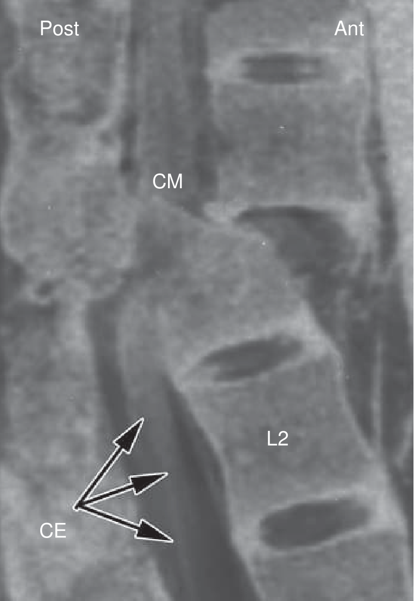
Side-by-Side Comparison
| Feature | Conus Medullaris Syndrome | Cauda Equina Syndrome |
|---|---|---|
| Structure injured | Spinal cord (CNS) | Lumbar/sacral nerve roots (PNS) |
| Vertebral level | T12 - L2 | Below L2 |
| Neuron type | Mixed UMN + LMN | Pure LMN only |
| Motor weakness | Symmetrical | Asymmetrical |
| Muscle tone | Variable (may have spasticity early) | Flaccid, reduced tone |
| Deep tendon reflexes | Absent (areflexic) | Absent (areflexic) |
| Bulbocavernosus reflex | Absent - permanent | Absent - permanent |
| Bladder | Areflexic (spastic/autonomic initially, then low-pressure high-capacity) | Areflexic, flaccid |
| Bowel | Flaccid sphincter, incontinence | Flaccid sphincter, incontinence |
| Sexual dysfunction | Erectile dysfunction, absent reflexogenic erection | Same |
| Saddle anesthesia | Present (bilateral sacral/coccygeal) | Present |
| Pain | Less prominent, may appear late | Prominent radicular pain - often severe, early |
| Symmetry | Bilateral (conus is a compact structure, lesions affect both sides) | Often unilateral or asymmetric |
| Prognosis | Poorer (cord injury, limited regeneration) | Better (nerve roots are resilient and can regenerate; sacral roots are an exception) |
Key Distinguishing Points
Motor Neuron Signature
Symmetry
Pain
Saddle Anesthesia
Reflexes - The Bulbocavernosus Clue
Shared Features
- Saddle anesthesia
- Areflexic bowel and bladder
- Sexual dysfunction
- Absent deep tendon reflexes
- Absent bulbocavernosus reflex
- EMG showing denervation (especially with complete anatomical lesions)
Common Causes
| Conus | Cauda Equina | |
|---|---|---|
| Trauma | Burst fracture / fracture-dislocation at T12-L2 | Burst fracture / fracture-dislocation below L2 |
| Disc | Central disc herniation | Midline disc herniation (most commonly L4-L5) |
| Tumor | Neoplasm (intradural/extradural) | Compressive mass |
| Vascular | Vascular insufficiency | Less common |
| Stenosis | Less common | Central lumbar stenosis |
Imaging
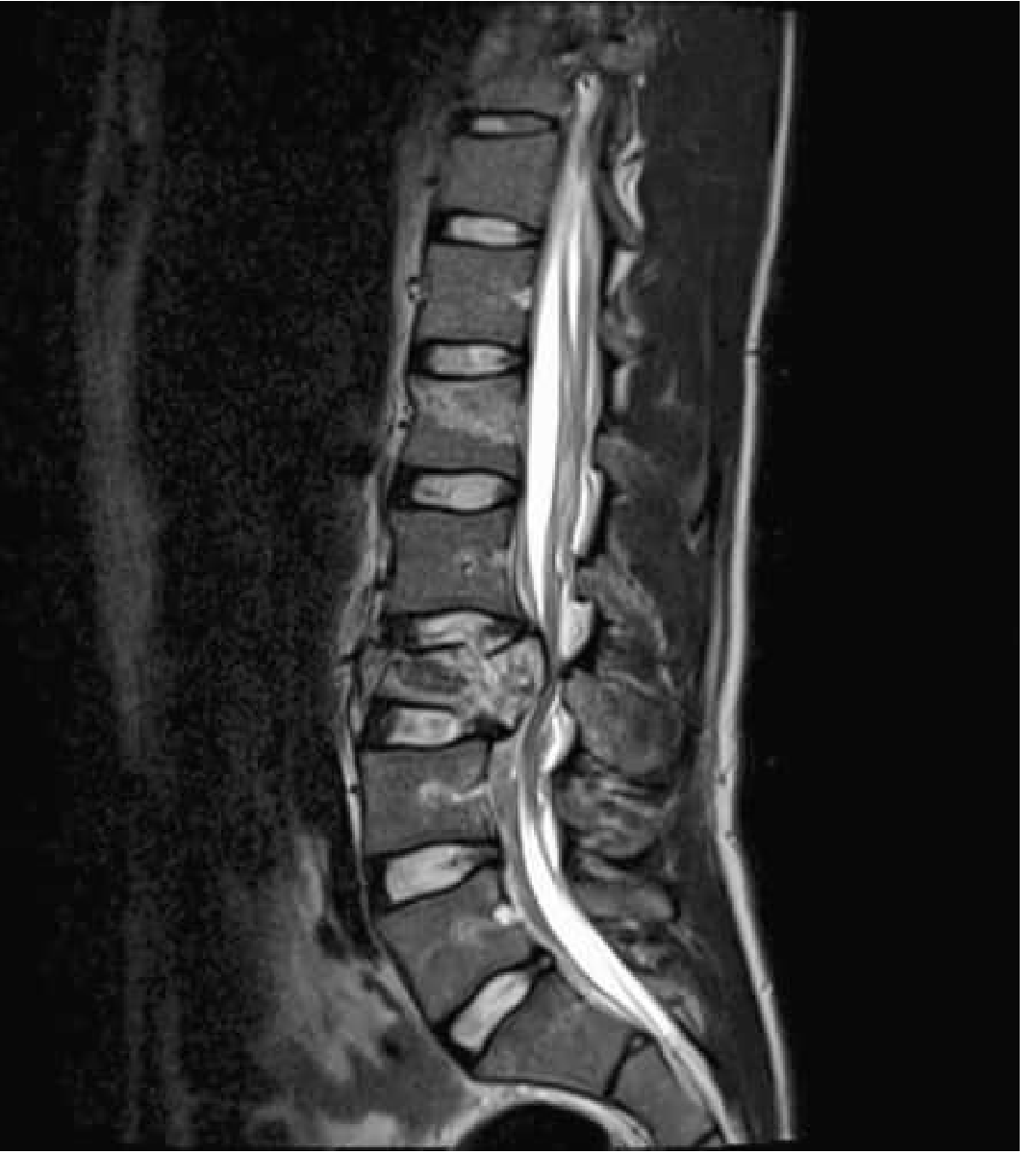
Management & Prognosis
- Cauda equina from disc herniation: early surgical decompression (within 24 hours) is recommended when possible to prevent residual deficits. Recovery is better because peripheral nerve roots can regenerate (note: sacral roots are fragile and injuries may be permanent).
- Conus medullaris: no strong evidence favors surgical vs. non-surgical management, nor early vs. late decompression. Prognosis is generally worse than cauda equina because spinal cord tissue has minimal regenerative capacity.
- In traumatic settings, functional outcomes for both syndromes are broadly similar and there is no compelling evidence that timing of decompression alters outcome in either.
Create a comparison poster of conus vs cauda equina for clinical reference
A clean, professional clinical reference poster comparing Conus Medullaris Syndrome vs Cauda Equina Syndrome. Two-column layout with a clear header. Left column header "CONUS MEDULLARIS SYNDROME" in blue, right column header "CAUDA EQUINA SYNDROME" in teal. Include the following sections with icons or symbols: ANATOMY: Left - "Spinal cord (CNS) T12-L2", Right - "Nerve roots (PNS) Below L2" NEURON TYPE: Left - "Mixed UMN + LMN", Right - "Pure LMN only" MOTOR: Left - "Symmetrical weakness", Right - "Asymmetrical weakness" TONE/REFLEXES: Both sides - "Flaccid, areflexic DTRs absent" BLADDER/BOWEL: Both - "Areflexic bladder, flaccid sphincter, incontinence" PAIN: Left - "Mild, late onset", Right - "Severe radicular pain, early" SENSORY: Both - "Saddle anesthesia (S2-S5)" SEXUAL: Both - "Erectile dysfunction, absent reflexogenic erection" BULBOCAVERNOSUS REFLEX: Both - "Absent, permanent" SYMMETRY: Left - "Bilateral (compact structure)", Right - "Often unilateral/asymmetric" CAUSES: Left - "Burst fracture T12-L2, central disc, tumor, vascular", Right - "Disc herniation L4-L5, fracture below L2, stenosis" PROGNOSIS: Left - "Poorer - cord cannot regenerate", Right - "Better - nerve roots can regenerate (except sacral)" TREATMENT: Left - "No strong evidence for surgical timing", Right - "Early surgery <24h for disc herniation" At bottom: a shared features box labeled "SHARED FEATURES" listing: saddle anesthesia, areflexic bowel/bladder, absent DTRs, absent bulbocavernosus reflex, sexual dysfunction. Footer text: "Clinical Reference - Spinal Cord Injury Syndromes". Use a white background with colored section headers and subtle dividing lines. Medical/clinical aesthetic.
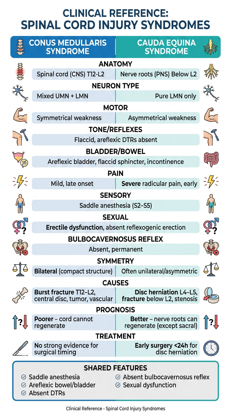
- Anatomy & level - CNS cord (T12-L2) vs. PNS nerve roots (below L2)
- Motor neuron type - Mixed UMN+LMN vs. pure LMN
- Symmetry - bilateral vs. asymmetric weakness
- Pain - mild/late vs. severe radicular/early
- Shared features box at the bottom (saddle anesthesia, areflexic bladder/bowel, absent DTRs, sexual dysfunction)
- Prognosis & treatment differences