
Plz make detailed summmary of topic coronary circulation from guyton
Reading File
Now let me search the Guyton textbook for the coronary circulation topic.
Finding Sources
Finding Sources
Found Guyton's coronary circulation section. Now let me get the full TOC and read the content.
Finding Sources
Reading File
Good start. Let me read more of this section and also get the coronary circulation headings.
Reading File
Finding Sources
Reading File
Reading File
Reading File
Reading File
Now let me get the middle section I'm missing (local control, oxygen metabolism, and the transition into ischemic heart disease).
Reading File
I now have all the key content from Guyton. Let me also fetch the coronary anatomy diagram image and compose the full summary.
Loading Image
Loading Image
I now have all the content needed for a detailed summary. Let me compile the full summary from Guyton.
Coronary Circulation - Detailed Summary (Guyton and Hall Textbook of Medical Physiology)
Introduction
About one-third of all deaths in industrialized Western countries result from coronary artery disease, and most older adults have at least some impairment of coronary artery circulation. This makes coronary circulation one of the most important topics in medicine.
1. Physiological Anatomy of the Coronary Blood Supply
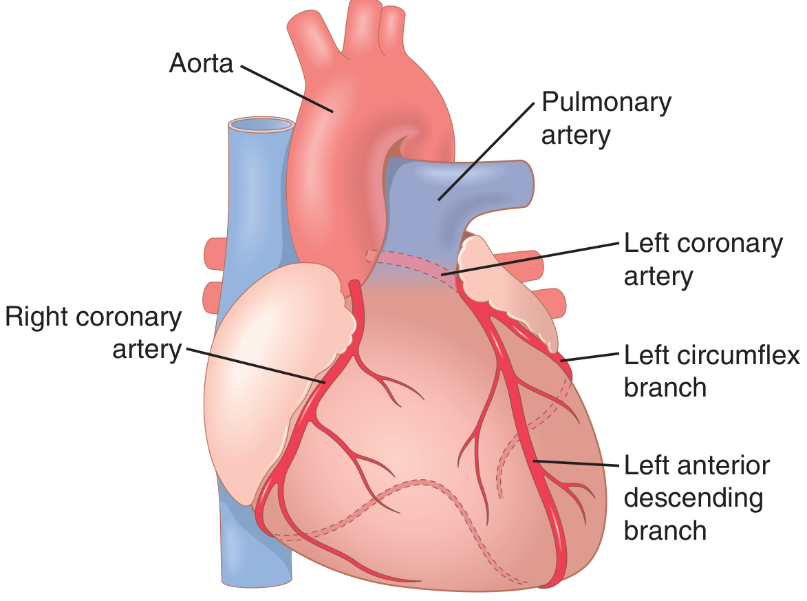
Fig. 21.3 - Coronary arteries showing the aorta, pulmonary artery, right coronary artery, left coronary artery, left circumflex branch, and left anterior descending branch.
Key points:
- The main coronary arteries lie on the surface of the heart (epicardial). Smaller arteries then penetrate from the surface into the cardiac muscle mass.
- Nutritive blood supply is almost entirely through these arteries. Only the inner 0.1 mm of the endocardial surface can obtain significant nutrition directly from blood inside the cardiac chambers - so this contribution is miniscule.
Distribution:
| Artery | Territory Supplied |
|---|---|
| Left coronary artery | Anterior and left lateral portions of the left ventricle |
| Right coronary artery | Most of the right ventricle + posterior part of left ventricle (80-90% of people) |
Venous drainage:
- About 75% of coronary venous blood from the left ventricle returns to the right atrium via the coronary sinus
- Coronary venous blood from the right ventricle returns through small anterior cardiac veins directly into the right atrium (not via coronary sinus)
- A very small amount flows back via thebesian veins, which empty directly into all chambers of the heart
2. Normal Coronary Blood Flow Averages 5% of Cardiac Output
- At rest: approximately 70 mL/min per 100 g of heart weight, or about 225 mL/min total - roughly 4% to 5% of total cardiac output
- Coronary blood flow per gram of heart weight is typically higher in women than in men, although women's hearts are smaller overall
- During strenuous exercise, cardiac work output may increase 6 to 9 times normal, and coronary blood flow must increase proportionately to provide the necessary nutrients
3. Phasic Changes in Coronary Blood Flow - Systole vs. Diastole
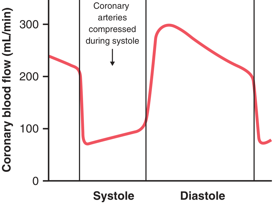
Fig. 21.4 - Phasic flow of blood through the coronary capillaries of the human left ventricle during cardiac systole and diastole.
Left ventricle:
- Coronary capillary blood flow falls to a low value during systole - opposite to most other vascular beds in the body
- This is because intramuscular blood vessels are compressed by the contracting left ventricular muscle during systole
- During diastole, the muscle relaxes, vessels open, and blood flows rapidly throughout diastole
Right ventricle:
- Also shows phasic changes, but the inverse changes are only partial because the force of contraction of the right ventricular muscle is far less than the left
Clinical significance of subendocardial blood flow:
- The epicardial coronary arteries supply most of the muscle from the outside, while a subendocardial arterial plexus lies just beneath the endocardium
- During systole, intramuscular vessel compression is greatest in the subendocardium
- The extra vessels of the subendocardial plexus normally compensate for this reduction
- In coronary ischemia, the subendocardium is most vulnerable - this explains why ischemia tends to affect the inner (subendocardial) layers first
4. Control of Coronary Blood Flow
4a. Local Muscle Metabolism - Primary Controller
Blood flow through the coronary system is regulated mostly by local arteriolar vasodilation in response to the metabolic needs of cardiac muscle:
- Increased vigor of cardiac contraction → increased coronary blood flow
- Decreased heart activity → decreased coronary flow
- This is similar to local flow regulation in skeletal muscles and other tissues
4b. Oxygen Demand as the Key Regulatory Signal
- Normally, about 70% of oxygen in coronary arterial blood is removed as it flows through the heart muscle
- Very little additional oxygen can be supplied by increasing oxygen extraction alone - so the heart must increase flow when it needs more oxygen
- Coronary blood flow increases almost in direct proportion to any additional metabolic oxygen consumption by the heart
Vasodilator mediators released during hypoxia/increased activity:
| Substance | Mechanism |
|---|---|
| Adenosine | Major vasodilator; released when ATP degrades to AMP in low-oxygen conditions; diffuses out of cardiac cells and causes arteriolar dilation |
| Adenosine phosphate compounds | Additional vasodilator effect |
| Potassium ions | Released during rapid cardiac contraction |
| Hydrogen ions | Produced during anaerobic metabolism |
| Carbon dioxide | Metabolic by-product causing vasodilation |
| Prostaglandins | Local hormones with vasodilator effect |
| Nitric oxide | Endothelium-derived relaxing factor |
Important note: Agents that block adenosine do not completely prevent coronary vasodilation caused by increased heart activity, confirming that multiple vasodilator mechanisms act together.
4c. Nervous Control of Coronary Blood Flow
Autonomic nerves affect coronary flow both directly (on vessel walls) and indirectly (via changes in cardiac metabolism).
Indirect effects (dominant):
- Sympathetic stimulation → releases norepinephrine (from nerves) and epinephrine/norepinephrine (from adrenal medulla) → increases heart rate and contractility → increases cardiac metabolism → local metabolic vasodilation dominates, and coronary flow increases in proportion to metabolic needs
- Vagal stimulation → releases acetylcholine → slows heart, mildly depresses contractility → decreases oxygen consumption → indirect coronary vasoconstriction
Direct effects (secondary):
- Parasympathetic (vagal) fibers to the ventricular coronary system are sparse. Acetylcholine has a direct dilatory effect on coronary vessels.
- Sympathetic fibers release norepinephrine, which acts on alpha-adrenergic receptors to cause direct vasoconstriction of coronary arteries
- However, sympathetic stimulation also activates beta-adrenergic receptors in the heart muscle, which increase cardiac metabolism, and the resulting increase in local vasodilators overwhelms the direct alpha-constrictor effect
- Net result: sympathetic stimulation increases coronary blood flow overall, via the metabolic (indirect) pathway
5. Oxygen Metabolism of the Heart and Coronary Ischemia
- More than 95% of metabolic energy in the heart is liberated from foods using ATP in the mitochondria
- ATP → adenosine diphosphate (ADP) → adenosine monophosphate (AMP) → adenosine during severe ischemia
- The cardiac cell membrane is slightly permeable to adenosine, so it diffuses out into blood - causing coronary vasodilation (beneficial), but also causing loss of adenine base from cardiac cells (harmful)
Critical timeframe:
- Within as little as 30 minutes of severe coronary ischemia, about half of the adenine base can be lost from the affected cardiac muscle cells
- New adenine is synthesized at only 2% per hour - extremely slow replacement
- Therefore, once ischemia has persisted for 30 minutes or longer, relieving the ischemia may be too late to prevent injury and death of cardiac cells
- This is a major reason cardiac cells die during myocardial infarction
6. Ischemic Heart Disease
Epidemiology:
- Most common cause of death in men and women in Western countries
- About 35% of people in the United States aged 65 and older die of ischemic heart disease
- Encompasses both obstructive and nonobstructive coronary artery disease
- Deaths may occur suddenly (acute occlusion, fibrillation) or slowly over weeks to years (progressive cardiac weakening)
6a. Atherosclerosis as a Major Cause
- Excess cholesterol deposits beneath the endothelium in people with genetic predisposition, obesity, sedentary lifestyle, hypertension, or endothelial damage
- Deposits are invaded by fibrous tissue and may become calcified → atherosclerotic plaques that protrude into and obstruct vessel lumens
- Common site: first few centimeters of the major coronary arteries
- Women have lower prevalence of obstructive coronary atherosclerosis than men, but this difference is attenuated in older adults
- Coronary microvascular dysfunction and coronary vasospasm are common causes when no obstruction can be detected, especially in women
6b. Acute Coronary Artery Occlusion
Usually occurs in a person who already has underlying atherosclerotic disease. Two main mechanisms:
- Atherosclerotic plaque rupture → local blood clot (thrombus) → acute occlusion
- Prolonged coronary spasm of an already narrowed artery
Consequences of acute occlusion:
- Immediate: Severe cardiac pain due to release of acidic substances (lactic acid, histamine, kinins, proteolytic enzymes) that stimulate pain nerve endings
- Anaerobic glycolysis begins but cannot sustain cardiac function for long
- Blood flow to ischemic areas may partially be restored by collateral circulation (pre-existing small anastomotic vessels that enlarge over weeks to months)
Myocardial infarction:
- When blood flow is critically reduced or blocked long enough, infarction (irreversible necrosis) of cardiac muscle occurs
- The infarcted muscle may be surrounded by an ischemic zone that potentially can recover if blood flow is restored
7. Effects of Myocardial Infarction
The four most common causes of death after acute MI:
- Decreased cardiac output (systolic dysfunction)
- Pulmonary edema (from blood damming in the pulmonary vessels)
- Ventricular fibrillation
- Cardiac rupture (rare)
7a. Systolic Stretch and Cardiogenic Shock
- Systolic stretch: When normal portions of the ventricular muscle contract, the ischemic (non-functional) portion bulges outward passively instead of contracting. This wastes pumping force.
- Cardiogenic shock (coronary shock) occurs when the heart cannot pump sufficient blood into the peripheral arterial tree
- Occurs when >40% of the left ventricle is infarcted
- Short-term mortality is 40% to 50% once cardiac shock develops
7b. Ventricular Fibrillation
- Most likely during two dangerous periods:
- First 10 minutes after the infarction
- A second period beginning 1 hour later and lasting for a few more hours
- Can also occur many days after the infarct, but less commonly
- Some patients with chronic coronary insufficiency die suddenly of fibrillation without any acute infarction
7c. Renal Retention of Fluid
- Cardiac output reduced → renal blood flow reduced → oliguria or anuria when cardiac output falls to 50-60% of normal
- Progressive fluid retention → increased blood volume → initially compensatory but eventually worsens pulmonary edema and cardiac workload
8. Heart Function After Recovery from MI
- The heart ordinarily recovers rapidly during the first few days and weeks
- Most of the final recovery state is achieved within 5 to 7 weeks, though mild additional recovery can continue for months
- Recovery mechanisms include:
- New collateral blood supply penetrating peripheral portions of the infarcted area
- Hypertrophy of undamaged muscle to partially offset cardiac damage
- Some patients may regain almost full function; more frequently, pumping capability remains permanently reduced
- Because the normal heart has a cardiac reserve of 300-400%, even significant damage may leave the patient able to perform normal daily activities at rest
9. Cardiac Pain in Coronary Heart Disease
Mechanism:
- Ischemia causes the muscle to release acidic substances (lactic acid) and pain-promoting products (histamine, kinins, cellular proteolytic enzymes)
- These are not removed quickly enough by slow coronary blood flow
- High concentrations stimulate pain nerve endings in cardiac muscle
- Pain impulses travel via sensory afferent nerve fibers to the CNS
Angina Pectoris
- Progressive coronary narrowing → cardiac pain when metabolic demands exceed available coronary flow
- Location: typically beneath the upper sternum
- Referred pain: classically to the left arm and left shoulder, also the neck and face
- Reason: during embryonic life, the heart originates in the neck region (same as the arms), so both areas share the same spinal cord pain fiber segments
- Triggers: exercise, emotional stress, cold temperatures, full stomach (all increase cardiac workload or cause sympathetic vasoconstriction)
- Pain character: hot, pressing, and constricting - severe enough to stop all unnecessary activity
Drug Treatment of Angina
- Vasodilator drugs provide immediate relief during acute attacks (e.g., nitrates, calcium channel blockers)
- Beta-blockers: reduce heart rate and contractility, thereby reducing cardiac metabolic demand
10. Cardiac Rehabilitation After Myocardial Infarction
- Rest immediately after MI is essential to reduce cardiac workload
- Prescribed aerobic exercise is a core component of cardiac rehabilitation once the patient is clinically stable
- Exercise training provides favorable effects including:
- Improved endothelial function in coronary arteries
- Reduced inflammation
- Improved contractile function of surviving cardiomyocytes
- Improved physical fitness can allow at least partial recovery of cardiac reserve and exercise tolerance
Summary Table
| Topic | Key Point |
|---|---|
| Normal coronary flow | ~225 mL/min (~5% of cardiac output) at rest |
| Left ventricular flow | Primarily during diastole (vessels compressed during systole) |
| Right ventricular flow | Phasic changes present but less marked |
| Most vulnerable region | Subendocardial (highest compression during systole) |
| Primary flow regulator | Local metabolic vasodilation (oxygen demand) |
| Key vasodilator | Adenosine (+ K⁺, H⁺, CO₂, nitric oxide, prostaglandins) |
| Oxygen extraction | ~70% extracted at rest (very high - near maximum) |
| Critical ischemia time | 30 minutes before irreversible adenine loss begins |
| Atherosclerosis site | First few cm of major coronary arteries |
| Cardiogenic shock threshold | >40% of left ventricle infarcted |
| Angina pain referral | Left arm, left shoulder, neck (embryological proximity) |
Source: Guyton and Hall Textbook of Medical Physiology, Chapter 21 - Coronary Circulation, pp. 267-274
This is a shared conversation. Sign in to Orris to start your own chat.