

IMG_7603.jpeg
There is this white bump in eye
analyse_media
Reading File
Finding Sources
Searching the Web
pinguecula eye white bump symptoms treatment
Reading File
Reading File
Reading File
Loading Image
Based on the image and medical textbook sources, here is a thorough breakdown:
What Is That White Bump?
The image shows a yellowish-white, slightly raised nodule on the bulbar conjunctiva (the white of the eye), located in the interpalpebral zone (the area exposed when the eyelids are open) adjacent to the cornea. There is also visible redness and fine blood vessels around it, suggesting some local irritation.
This is most consistent with a pinguecula - and since the area looks inflamed, the condition may be called pingueculitis.
Here is a textbook photo of a pterygium (a related but more advanced lesion) for comparison:
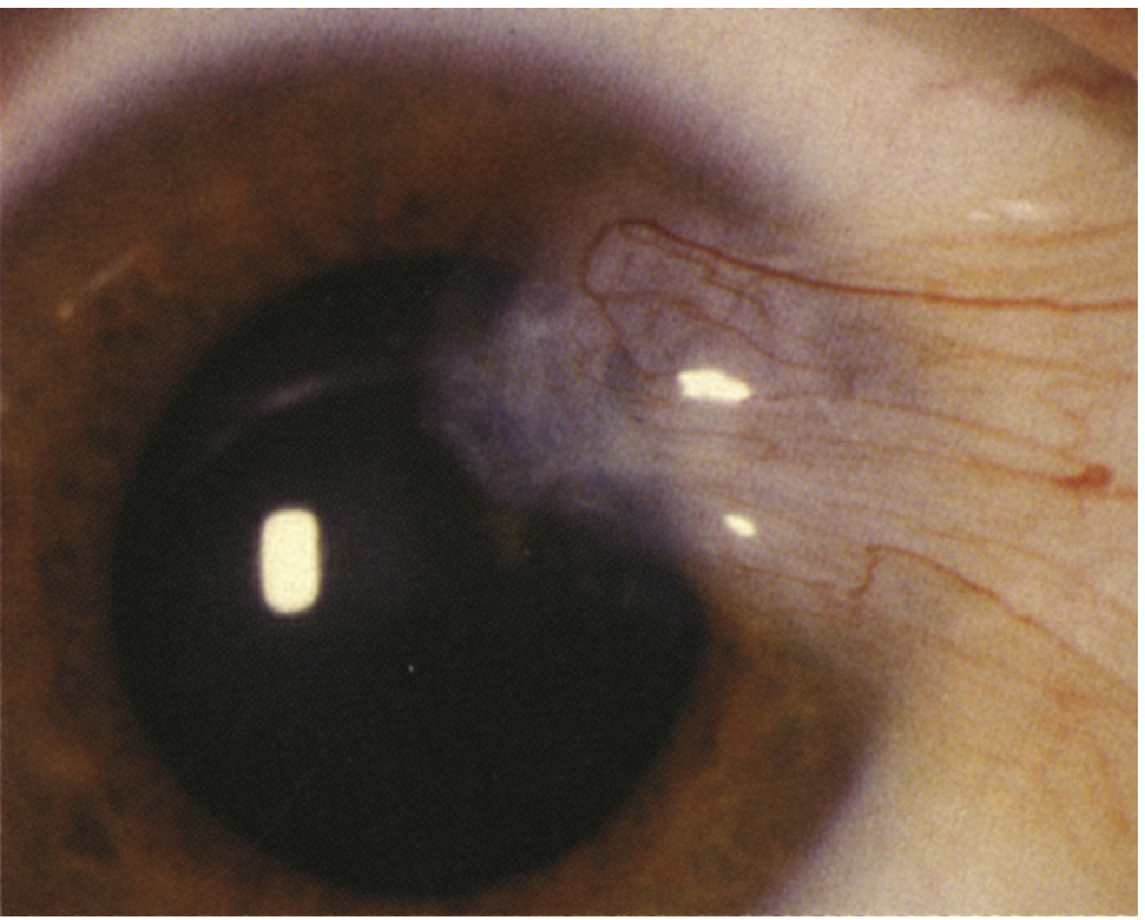
Pterygium (Wills Eye Manual, Figure 4.9.1) - note the tissue extending onto the cornea, which is NOT seen in your image
Pinguecula vs. Pterygium - Key Difference
| Feature | Pinguecula | Pterygium |
|---|---|---|
| Location | Stays on white of eye (conjunctiva) | Grows onto the cornea |
| Shape | Flat or slightly raised yellow-white nodule | Wing/wedge-shaped fleshy tissue |
| Vision risk | Generally none | Can cause astigmatism if large |
| Common name | - | "Surfer's Eye" |
Your image shows the lesion not encroaching on the cornea, which favors a pinguecula.
What Causes It?
- Chronic UV light (sun) exposure is the primary cause
- Wind, dust, and dry environments also contribute
- More common in people who spend time outdoors or live in equatorial regions
- It represents elastotic degeneration of the deep conjunctival layers, causing fibrovascular tissue to accumulate
(Goldman-Cecil Medicine, p. 4126)
Symptoms You Might Notice
- A gritty or "foreign body" sensation (feeling like something is in the eye)
- Redness and irritation - especially when inflamed (pingueculitis)
- Dryness or mild discomfort
- Often completely asymptomatic
Treatment
According to the Wills Eye Manual (p. 192-194):
- Protect the eye - wear UV-blocking sunglasses, especially outdoors
- Artificial tears (preservative-free preferred) - 4 to 8 times per day to reduce irritation
- If inflamed (like yours appears to be):
- Mild inflammation: artificial tears 4x/day
- Moderate-to-severe: a mild topical steroid (e.g., fluorometholone 0.1% or loteprednol) or anti-inflammatory drops (e.g., ketorolac 0.4%) can reduce symptoms - these require a prescription
- Surgery is only indicated if:
- The lesion grows onto the cornea and threatens vision
- Persistent irritation not relieved by drops
- Significant cosmetic concern
Should You See a Doctor?
Yes, you should get this checked by an ophthalmologist or optometrist. The redness and blood vessel congestion visible in your photo suggest active inflammation. A slit-lamp exam is needed to:
- Confirm whether it is a pinguecula or an early pterygium
- Rule out less common causes like conjunctival intraepithelial neoplasia (CIN), which can occasionally look similar
- Prescribe appropriate anti-inflammatory drops if needed
Important note: This is informational only and not a diagnosis. An in-person eye exam with a slit lamp is needed for an accurate diagnosis.
This is a shared conversation. Sign in to Orris to start your own chat.