
Difference between laceration and incised wound
laceration vs incised wound comparison forensic
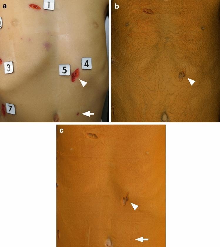
This forensic clinical comparison figure displays multiple stab wounds on a human torso using three imaging modalities: (a) a clinical photograph, (b) 3D computed tomography (CT) with volume rendering (VR), and (c) 3D CT with global illumination rendering (GIR). Panel (a) shows numerous sharp-force injuries with visible hemorrhage and numbered forensic markers. The arrowhead points to a prominent laceration. Panel (b) illustrates conventional VR, characterized by prominent contour-like striping artifacts on the skin surface and lower definition of wound margins. Panel (c) demonstrates GIR, which utilizes advanced lighting models to provide a smoother, more photorealistic skin surface. Comparison highlights the diagnostic superiority of GIR in forensic pathology; it successfully visualizes a small, shallow stab wound (arrow) and resolves adjacent injuries that appear coalesced or are undetectable in the VR image. The figure serves as an educational tool for comparing post-mortem radiological reconstruction techniques in trauma and forensic medicine.
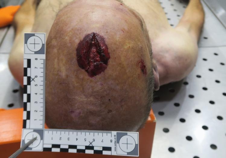
This forensic clinical photograph depicts a close-up, superior view of a human scalp with a significant blunt-force trauma injury, located anterior to the cranial apex in the midline fronto-parietal region. The primary injury is a deep, vertical, roughly triangular laceration measuring approximately 4.5 cm in length. The laceration features irregular, gaping edges with visible blood and subcutaneous tissue. Surrounding the central laceration is a prominent, oval-shaped abrasion and contusion measuring approximately 5 x 3.5 cm, characterized by reddish-purple discoloration and epidermal denudation. A second, smaller, irregular abrasion is visible lateral to the primary wound, anterior to the right ear. A forensic L-shaped scale is placed adjacent to the injuries for dimensional reference. This visual evidence is characteristic of a high-energy impact to the top of the head, often associated in forensic medicine with axial loading injuries such as a Jefferson fracture of the atlas (C1) or other cervical spine traumas.
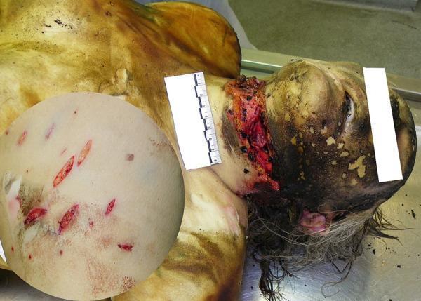
This forensic clinical photograph depicts the upper torso and head of a deceased individual with significant sharp force trauma and thermal injuries. A prominent, deep incised (slash) wound is visible across the anterior-lateral neck, demonstrating irregular edges and exposure of deep cervical structures including muscle and vascular tissue. A metric scale is placed adjacent to the neck wound for size reference. The head and face exhibit signs of thermal damage, including skin charring, soot deposition, and singed hair. An inset circular magnification on the left side of the image highlights the chest region, revealing multiple discrete, spindle-shaped (fusiform) stabbing-cutting injuries. These wounds are characterized by sharp margins and a linear distribution, consistent with a single-edged bladed instrument. The primary educational focus is the visual documentation of perimortem or postmortem trauma and thermal artifacts in a forensic pathology context, illustrating the morphological differences between large incised wounds and smaller stab wounds.
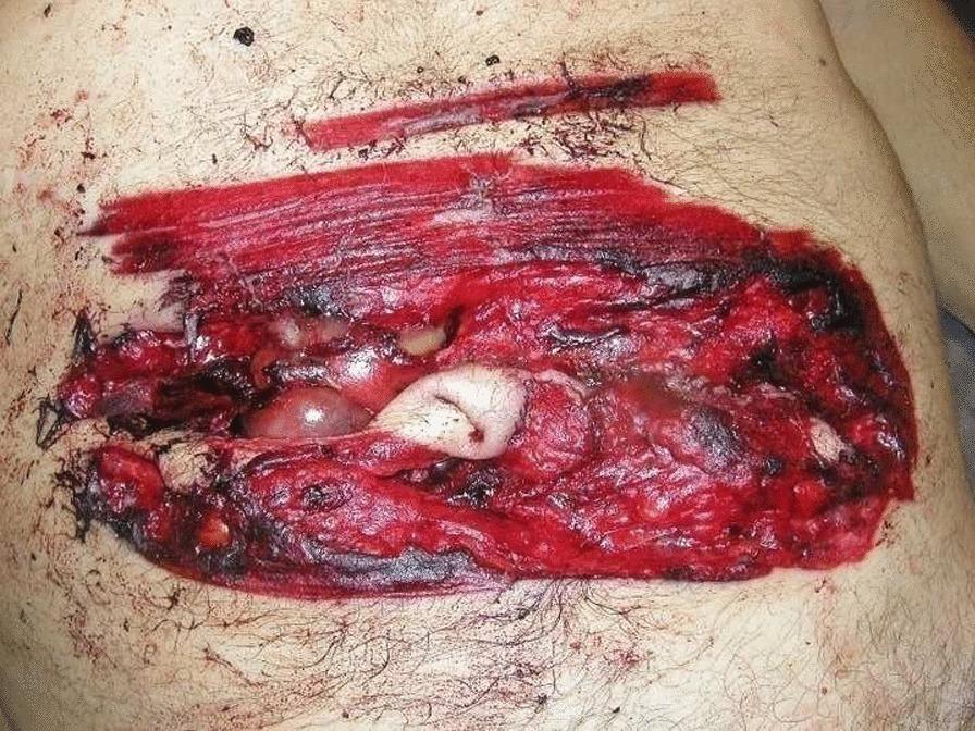
This clinical photograph from a forensic pathology context displays a large, complex abdominal wound. The primary lesion is a horizontal, deep incised wound located in the mesogastrium. The wound morphology is characterized by irregular, torn edges with focal areas of grazing, differing from a typical clean surgical incision. Centrally, the wound penetrates through the skin and subcutaneous fat layers, exposing internal structures including glistening, pinkish-white intestinal loops. Surrounding the deep defect is an extensive, well-demarcated area of reddish-brown abrasion and cutaneous bruising, measuring approximately 23 cm by 12 cm. There is evidence of peripheral blackish discoloration and hemorrhagic infiltration within the exposed tissue. Superior to the main lesion, a smaller, superficial linear 'trial' or hesitation wound is visible. The image illustrates severe abdominal trauma and is used in medical education to differentiate between standard incised wounds and those caused by high-velocity mechanical instruments like chainsaws.
Laceration vs. Incised Wound
Definitions
Laceration
- KS Narayan Reddy's Essentials of Forensic Medicine and Toxicology, 36th ed., p. 194
Incised Wound
- KS Narayan Reddy's Essentials of Forensic Medicine and Toxicology, 36th ed., p. 196
Detailed Comparison Table
| Trait | Incised Wound | Lacerated Wound |
|---|---|---|
| Manner of production | By sharp objects (knife, razor, scalpel, scissors, sword) | By blunt objects; crushing or tearing force |
| Site | Anywhere on the body | Usually over bony prominences |
| Margins | Smooth, even, clean-cut and everted | Irregular, ragged, often undermined |
| Abrasion on edges | Absent | Usually present |
| Bruising | No adjacent bruising of soft tissues | Bruising of surrounding and underlying tissues |
| Shape | Linear or spindle-shaped (skin retracts due to elasticity) | Varies; usually irregular, stellate, or jagged |
| Dimensions | Longer than deep; often gaping | Usually longer than deep |
| Tissue bridges at base | Absent - structures are cleanly cut to the depth | Present - small strands of tissue bridge across the margins |
| Hemorrhage | Usually profuse and external; spurting may be seen | Slight except in scalp wounds; mostly external |
| Hair bulbs | Cleanly cut | Crushed or torn |
| Bones | May be cut | May be fractured |
| Foreign bodies in wound | Absent | Usually present (dirt, grit) |
| Clothes | May be cut | May be torn |
Key Distinguishing Features (Memory Aids)
- Tissue bridging - the single most important differentiating feature. Lacerations always show tissue bridges (nerves, vessels, connective tissue strands) crossing the base of the wound. Incised wounds have a completely clean base.
- Edge condition - incised wounds have sharp, clean margins; lacerations have ragged, bruised, abraded margins.
- Bleeding - incised wounds bleed profusely (clean vessel cut); lacerations bleed less (vessels are crushed and tend to thrombose).
- Hair bulbs - crushed/torn in lacerations, cleanly cut in incised wounds (useful for microscopic examination).
Incised-Looking Lacerations (Forensically Important)
- Tissue bridges at the base
- Abraded/bruised margins
- Crushed hair follicles
Medicolegal Significance
- Incised wounds indicate use of a sharp-edged weapon; may suggest suicide (hesitation cuts on the wrist/throat) or homicide.
- Lacerations indicate blunt force trauma; common in accidents and assaults.
- Wound age estimation, direction of force, and nature of weapon can all be inferred from these features.
- KS Narayan Reddy's Essentials of Forensic Medicine and Toxicology, 36th ed., pp. 194-201 (Table 8.9)
- PC Dikshit's Textbook of Forensic Medicine and Toxicology, Table 13.1, pp. 179-180
- Parikh's Textbook of Medical Jurisprudence, Forensic Medicine and Toxicology