
Femtosecond laser in ophthalmology
I'll start by reading the medical library skill, then search for femtosecond laser content in parallel.
Searching sources...
Reading File
Now let me search the library and web in parallel for comprehensive content.
Finding Sources
Searching PubMed
femtosecond laser ophthalmology
The heading search returned nothing. Let me do a paragraph FTS search and web search in parallel.
Finding Sources
Searching the Web
femtosecond laser ophthalmology applications LASIK FLACS cornea 2024
Searching Images
femtosecond laser cataract surgery lens fragmentation diagram
Good. Now let me read the key textbook sections in parallel.
Reading File
Reading File
Reading File
Excellent - I have rich content. Now let me also read the Wills Eye section and look for relevant images from the textbook.
Reading File
Reading File
Finding Sources
I now have comprehensive material. Let me fetch the LASIK surgical image and compile the full answer.
Loading Image
Loading Image
I now have all the content needed for a thorough, well-illustrated answer.
Femtosecond Laser in Ophthalmology
Physics and Mechanism
A femtosecond (FS) laser emits near-infrared pulses of extremely short duration (10⁻¹⁵ seconds). When focused within ocular tissue, each pulse delivers enough energy to cause photodisruption - plasma formation and cavitation bubble expansion that cleave tissue at the focal plane with minimal thermal spread to surrounding structures. This "cold cutting" is what makes FS lasers fundamentally different from the thermal ablation of the excimer laser. Pulse duration is far shorter than nanosecond or picosecond lasers, which dramatically reduces collateral tissue damage. The first ophthalmic FS laser application was FDA-approved in 2001 for LASIK flap creation.
Applications
1. LASIK Flap Creation
This was the original and most widespread use. Traditionally, a mechanical microkeratome blade created the hinged corneal flap. FS laser replaced the blade and now creates flaps with uniform thickness of 100-120 μm, with significantly less deviation from the target thickness compared to microkeratomes.
Advantages over microkeratome:
- Much lower risk of flap complications (buttonholing, amputation, irregular flap, anterior chamber penetration)
- Thinner, more predictable flaps - enabling treatment in thinner corneas and higher ametropia
- Reduced percent of tissue altered (PTA), lowering the risk of post-LASIK ectasia
- Better biomechanical preservation of the corneal stroma
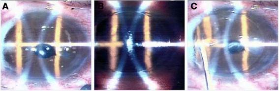
After flap creation, an excimer laser reshapes the stromal bed, and the flap is repositioned without sutures.
Surgical sequence: (A) flap elevated, (B) stromal ablation, (C) flap repositioned with irrigation:
Variants of LASIK ablation:
- Wavefront-guided LASIK - excimer ablation pattern based on wavefront aberrometry measurements
- Topography-guided LASIK - ablation guided by corneal topography map
- Optimised LASIK - larger optical zones, better asphericity to reduce glare/haloes
- LASIK Plus (LASIK + CXL) - riboflavin applied to stromal bed before flap closure, then UV cross-linking; considered in eyes at risk of ectasia (e.g., >-7D)
2. SMILE (Small Incision Lenticule Extraction)
SMILE uses a FS laser (Carl Zeiss VisuMax platform) to create a lenticule - a lens-shaped disc of stromal tissue - entirely within the intact cornea. The lenticule is then mechanically dissected and extracted through a small 4 mm peripheral incision, without creating a full corneal flap.
When the lenticule is removed via a LASIK-style flap instead, the procedure is called ReLex (Refractive Lenticular Extraction).
Advantages of SMILE:
- No flap - eliminates all flap-related complications
- Better corneal biomechanical stability (more anterior stromal lamellae preserved)
- Faster recovery of dry eye symptoms
- Quicker corneal reinnervation
- Minimal surface disturbance
Refractive results are comparable to LASIK. Complications include interface inflammation, epithelial ingrowth into the interface, and irregular astigmatism.
3. Femtosecond Laser-Assisted Cataract Surgery (FLACS)
FS lasers have been incorporated into phacoemulsification to automate several manual steps:
| Step | FS Laser Role |
|---|---|
| Corneal incisions | Main wound + side port - more precise geometry and self-sealing architecture |
| Capsulorhexis (capsulotomy) | Automated circular anterior capsulotomy - more precise size, shape, and centration than manual technique |
| Lens fragmentation | Pre-softening/fragmenting the nucleus - reduces phacoemulsification energy (ultrasound time) delivered to the eye |
| Astigmatic incisions (AK) | Arcuate corneal incisions to correct pre-existing astigmatism |
Potential advantages:
- Greater incision precision and integrity
- Reduced cumulative dissipated energy (CDE) / phaco energy
- More precise capsulorhexis placement - may improve refractive outcomes with premium IOLs
- Better overlap of capsulotomy over optic edge (reduces PCO risk)
Disadvantages:
- Substantially higher cost (equipment + disposables)
- Longer total operating time
- Challenging in small pupils, dense white cataracts, or significant zonular weakness
- Significant learning curve
- Supplemental oxygen is contraindicated during FS laser use due to fire risk (Miller's Anesthesia)
Recent evidence note: Three 2024-2025 meta-analyses (PMIDs 40731148, 39043258, 38291620) comparing FLACS vs. conventional phacoemulsification show broadly similar visual outcomes, with FLACS reducing phaco energy but not consistently demonstrating superior BCVA. A 2025 systematic review (PMID 40345360) found FS arcuate keratotomy during cataract surgery to be effective for astigmatism correction.
4. Keratoplasty
FS lasers have transformed corneal transplantation by enabling precisely shaped incisions in both donor and recipient tissue:
| Procedure | FS Laser Role |
|---|---|
| Penetrating keratoplasty (PKP) | Custom trephination shapes (zigzag, top-hat, mushroom) - increase wound contact area, improve self-sealing, reduce suture dependency |
| DALK (Deep Anterior Lamellar Keratoplasty) | Donor and recipient trephination and dissection |
| DSAEK / DMEK (Endothelial keratoplasty) | Donor button preparation for Descemet's stripping automated endothelial keratoplasty |
| Bowman Layer Transplantation (BLT) | Donor Bowman graft preparation |
5. Other Corneal Applications
- Intracorneal ring segment (ICRS) tunnels - channel creation for ICRS implantation in keratoconus, replacing manual dissection with a Sinskey hook
- Astigmatic keratotomy (AK) - intrastromal arcuate incisions for astigmatism correction
- Intrastromal pockets - for corneal inlay implantation (presbyopia correction, e.g., KAMRA, Raindrop)
- INTRACOR - intrastromal incision pattern to treat presbyopia
- Stromal keratophakia - intrastromal lenticule implantation
- Pterygium and conjunctival surgery - emerging application
Complications Specific to FS Laser Use
Transient Light Sensitivity Syndrome (TLSS)
- Occurs 2-8 weeks after surgery, more frequent after hyperopic LASIK (9%) vs. myopic LASIK (5%) vs. SMILE (1%)
- Responds to topical steroids
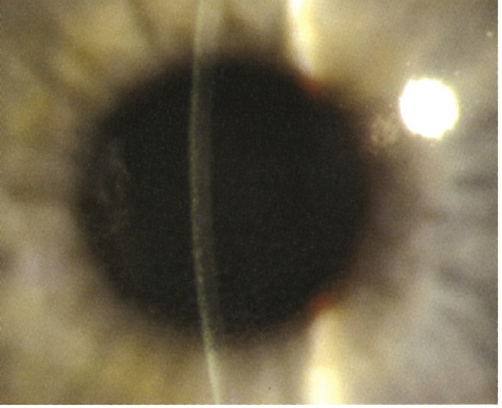
Diffuse Lamellar Keratitis (DLK) - "Sands of the Sahara"
- Granular deposits at the flap interface
- Develops 1-7 days post-LASIK
- Treatment: intensive topical antibiotic and steroid
Other FS-Related Complications
- Opaque bubble layer (OBL) - residual gas bubbles from photodisruption within the stromal lamellae; can interfere with subsequent excimer ablation
- Suction loss during FS delivery - may cause incomplete cut
- Rainbow glare - diffraction phenomenon unique to FS-created flaps, due to regular lamellar microstructure
- Epithelial ingrowth (1-2%) under flap
- Fire risk - if supplemental O₂ is present during FS laser application (anesthesia consideration)
FS Laser vs. Microkeratome: Summary
| Parameter | Microkeratome | Femtosecond Laser |
|---|---|---|
| Flap uniformity | Variable | Highly uniform (±5 μm) |
| Flap complication rate | Higher | Significantly lower |
| Buttonhole / amputation | Possible | Very rare |
| Cost | Lower | Higher |
| OBL | None | Possible |
| Rainbow glare | Absent | Possible |
| Biomechanics | Slightly less favorable | Better preservation |
Platforms Available
- IntraLase (J&J Vision) - first FDA-approved FS laser for LASIK
- VisuMax (Carl Zeiss Meditec) - used for LASIK and SMILE
- WaveLight FS200 (Alcon)
- LenSx, LensAR, Catalys (various) - FLACS platforms
- iFS (Abbott/J&J)
Summary of Key Points
- FS laser achieves tissue cleavage by photodisruption, not thermal ablation
- Primary uses: LASIK flap creation, SMILE lenticule dissection, FLACS (capsulorhexis + nucleus fragmentation + incisions), keratoplasty, ICRS tunnels, AK, intrastromal pockets
- Compared to microkeratome: more uniform flaps, fewer flap complications, but higher cost and OBL risk
- SMILE offers flap-free refractive correction with better biomechanics and dry eye profile
- FLACS reduces phaco energy but current meta-analyses show comparable visual outcomes to conventional phaco
- Supplemental O₂ is contraindicated during FS laser use (fire hazard)
Sources: Kanski's Clinical Ophthalmology 10th Ed., p. 305, 339 | Wills Eye Manual | Miller's Anesthesia 10th Ed. | EyeWiki - Femtosecond Lasers in LASIK | Femtosecond laser applications in corneal surgery - PMC
Preservatives in eyedrops , side effects and treatment
Quick answer: Benzalkonium chloride (BAK) is the most common eyedrop preservative and the main culprit for ocular surface toxicity. It disrupts the tear film lipid layer, damages corneal and conjunctival epithelium, and triggers inflammation. Management centers on switching to preservative-free formulations or using newer, less toxic preservatives.
Searching sources...
Reading File
Finding Sources
Finding Sources
Searching PubMed
benzalkonium chloride ocular surface toxicity preservative-free eyedrops
Searching the Web
preservatives in eyedrops benzalkonium chloride ocular surface disease side effects treatment 2024
Good results. Let me now search the textbook library more broadly, and extract the key EyeWiki and review content in parallel.
Finding Sources
Reading Web Page
https://eyewiki.org/Preservatives_in_Topical_Ophthalmic_Medi…
Reading Article
PMID: 39098762
Excellent - very rich data collected. Let me get the Kanski tear substitute section and a glaucoma/dry eye textbook section, plus search for images.
Reading File
Finding Sources
Searching Images
benzalkonium chloride ocular surface toxicity corneal epithelium damage superficial punctate keratitis
I have comprehensive data from textbooks, expert reviews, and recent literature. Now I'll compile the full answer.
Preservatives in Eyedrops: Types, Side Effects & Management
Why Preservatives Are Used
Preservatives are added to multi-dose ophthalmic bottles to prevent microbial contamination (bacteria, fungi, acanthamoeba) between uses, protect drug potency, and prevent biodegradation. Without them, a multi-dose bottle would become a bacterial culture medium within days. However, with chronic use - particularly in glaucoma, dry eye, and allergy patients - the cumulative toxic load on the ocular surface becomes clinically significant.
Types of Ophthalmic Preservatives
1. Benzalkonium Chloride (BAK) - Most Common
BAK has been used since 1940 and is found in approximately 70% of all topical ophthalmic eyedrops. It is a quaternary ammonium compound with both hydrophilic and hydrophobic properties.
Mechanism of antimicrobial action: Acts as a detergent - disrupts the lipid component of microbial cell walls, increasing permeability and causing cell lysis. Broad-spectrum activity against gram-positive, gram-negative bacteria, fungi, and acanthamoeba.
Concentrations used: 0.003-0.02% in commercial formulations. Toxicity threshold estimated at as low as 0.005% - often lower than the concentrations actually present in eyedrops.
Common eyedrops containing BAK: Beta-blockers (timolol), prostaglandin analogues (latanoprost, bimatoprost), alpha-2 agonists (brimonidine), carbonic anhydrase inhibitors (dorzolamide), antihistamines, antibiotics, NSAIDs.
2. Alternative / "Softer" Preservatives
| Preservative | Examples of Use | Mechanism | Relative Safety |
|---|---|---|---|
| Polyquaternium-1 (Polyquad) | Travatan Z (travoprost) | Larger molecule, less cell penetration | Lower toxicity than BAK |
| Purite (stabilized oxychloro complex) | Alphagan P (brimonidine) | Breaks down to water + NaCl on exposure to light | Better tolerated |
| SofZia (ionic buffered system) | Travatan Z alternate formulation | pH/ion-based antimicrobial | Low toxicity profile |
| Sodium perborate | Some artificial tears | Breaks down to water + O₂ on contact with tear film | Generally well tolerated |
| Thimerosal | Older formulations | Organomercury compound | High sensitization rate - largely discontinued |
| Chlorobutanol | Some antibiotics, steroids | Halogenated alcohol | Corneal toxicity; slower degradation |
| Sorbic acid / potassium sorbate | Some lubricants | Weak organic acid | Mild; may cause stinging |
| EDTA (ethylenediaminetetraacetic acid) | Usually co-preservative | Chelates Ca²⁺, disrupts cell membranes | Synergistic with BAK - additive toxicity |
Key principle: Polyquad and Purite exhibit lower ocular surface toxicity than BAK and should be favored when preservative-free is not available. - Kanski's Clinical Ophthalmology 10th Ed.
Pathophysiology of BAK Toxicity
BAK causes ocular surface damage through three main mechanisms:
1. Detergent / Surfactant Effect
- Disrupts the lipid layer of the tear film, reducing tear break-up time (TBUT)
- Destabilizes the entire three-layer tear film (lipid/aqueous/mucin)
- Increases tear film osmolarity
2. Direct Cellular Cytotoxicity
- Corneal epithelial barrier disruption - increases paracellular permeability
- Concentration-dependent decrease in cellular viability - demonstrated in vitro
- Pro-apoptotic effects on corneal and conjunctival epithelial cells
- Goblet cell loss - goblet cells produce MUC5AC mucin, essential for tear film stability; their loss worsens dry eye
- Delayed corneal wound healing
- Trabecular meshwork toxicity - relevant in glaucoma patients
3. Inflammatory Cascade
- Activates NF-κB and other inflammatory pathways
- Elevated inflammatory cytokines and cell markers in ocular tissues
- Subconjunctival fibrosis with chronic exposure - worsens outcomes of subsequent filtration (glaucoma) surgery
- Stimulates mast cell degranulation contributing to allergic-like responses
Clinical Side Effects
Symptoms (Subjective)
- Burning and stinging on instillation
- Foreign body sensation
- Itching
- Redness / irritation
- Dryness and grittiness
- Increased tearing (reflex)
- Photophobia with severe toxicity
Signs (Objective)
| Sign | Notes |
|---|---|
| Superficial punctate keratopathy (SPK) | Rose bengal / lissamine green / fluorescein staining of corneal epithelium; most common sign |
| Reduced tear break-up time (TBUT) | Lipid layer disruption; TBUT < 10 seconds |
| Conjunctival hyperemia | Chronic injection |
| Lower Schirmer scores | Reduced aqueous secretion |
| Goblet cell density reduction | Measured by impression cytology |
| Subconjunctival fibrosis | Late sign in heavy users; seen as fornix foreshortening |
| Corneal staining | Diffuse / inferiorly distributed in mild cases |
| Blepharitis / lid margin inflammation | Secondary to tear film instability |
| Increased tear osmolarity | >308 mOsm/L |
| Meibomian gland dysfunction (MGD) | Chronic inflammation worsens meibomian gland obstruction |
"Excessive use of preserved lubricating eye drops can result in corneal toxicity." - Kanski's Clinical Ophthalmology 10th Ed.
High-Risk Groups
- Glaucoma patients on multiple preserved drops (most common and most studied group)
- Pre-existing dry eye disease
- Post-cataract or post-refractive surgery (corneal nerve disruption)
- Contact lens wearers (BAK adsorbs onto soft lens material)
- Elderly patients (reduced baseline tear volume)
- Post-LASIK patients (reduced corneal sensation for 3-12 months)
Diagnosis / Assessment of Preservative Toxicity
- History - number of drops used/day, duration, number of different medications
- Slit-lamp examination - assess for SPK, conjunctival injection, lid changes
- Fluorescein staining + TBUT measurement (TBUT <10 s = abnormal)
- Rose bengal / lissamine green staining - conjunctival and corneal staining
- Schirmer test - strips after 5 minutes (< 5 mm = severe aqueous deficiency)
- Tear osmolarity - >308 mOsm/L suggests hyperosmolar state
- Impression cytology - goblet cell density (research/tertiary setting)
- Meibography - assess meibomian gland structure
Management / Treatment
Step 1 - Reduce BAK Exposure (Root Cause Treatment)
The most important intervention. Strategies in order of preference:
| Strategy | Details |
|---|---|
| Switch to preservative-free (PF) formulations | Best option when available. Tafluprost PF (Zioptan), latanoprost PF (Iyuzeh, Xelpros), dorzolamide/timolol PF (Cosopt PF), PF artificial tears |
| Switch to less toxic preservatives | Polyquad (Travatan Z), Purite (Alphagan P), SofZia |
| Use fixed-dose combination drops | Reduces total number of drops/day (e.g., combined beta-blocker + prostaglandin instead of two separate bottles) |
| Reduce instillation frequency | Once-daily agents preferred where therapeutically equivalent |
| Procedural IOP reduction (glaucoma) | Laser trabeculoplasty (SLT) or surgical filtering procedures to reduce or eliminate need for drops altogether |
"For patients receiving multiple topical preserved drugs, best practice is to switch to nonpreserved equivalents wherever feasible, regardless of OSD severity." - Kahook et al., The Ocular Surface, 2024 (PMID 39098762)
Key clinical rule from Wills Eye Manual: "Always use preservative-free artificial tears if dosing is more frequent than QID to prevent preservative toxicity."
Step 2 - Symptomatic Ocular Surface Treatment
Tear Substitutes
- Preservative-free artificial tears (cellulose derivatives, sodium hyaluronate, carbomer gels, PVA) - cornerstone therapy
- Cellulose derivatives (hypromellose, methylcellulose) - mild cases
- Carbomer gels - longer-lasting, preferred in moderate-severe cases
- Sodium hyaluronate - viscoelastic, good retention, preferred by many
- PVA - useful in mucin deficiency
- Liposome-based eyelid sprays - stabilize lipid layer
- Petrolatum ointments - bedtime use to supplement drops; too blurring for daytime
- Artificial tear inserts - once/twice daily, for extended duration
Anti-inflammatory Therapy
- Topical cyclosporine A 0.05% (Restasis) or 0.09% (Cequa) - reduces T-cell mediated inflammation; preferred for chronic OSD
- Lifitegrast 5% (Xiidra) - LFA-1 antagonist, blocks T-cell adhesion
- Short-course topical steroids - for acute exacerbations; use cautiously given IOP risk
- Medroxyprogesterone acetate 1% - for prolonged anti-inflammatory need (avoids steroid-induced IOP rise)
Secretagogues
- Diquafosol - P2Y2 receptor agonist; topical secretagogue that stimulates mucin and aqueous secretion
- Rebamipide - mucin secretagogue (available in some countries)
Punctal Occlusion
- Reduces tear drainage, conserves natural tears, prolongs artificial tear effect
- Performed with collagen plugs (temporary, dissolve in days-weeks) or silicone plugs (semi-permanent)
- Thermal or laser punctoplasty for permanent occlusion in severe cases
- Caution: In eyes on multiple preserved medications, punctal occlusion increases contact time of preservatives on the ocular surface - worsens toxicity. Must reduce/eliminate BAK-preserved drops before occluding puncta.
Haemoderivative Therapy
- Autologous serum eye drops (20-100%) - contain growth factors (EGF, TGF-β), fibronectin, vitamins; promote epithelial healing
- Indicated in: severe OSD, graft-versus-host disease, Sjögren's-related dry eye, post-LASIK neurotrophic keratopathy, persistent epithelial defects
Mucolytics
- Acetylcysteine 5% - dissolves corneal filaments and mucous plaques; may sting
MGD / Lid Hygiene
- Warm compresses + lid massage
- Omega-3 fatty acid supplementation
- Intense pulsed light (IPL) therapy for meibomian gland dysfunction
Preservative-Free (PF) Formulations Available
| Drug Class | PF Options |
|---|---|
| Prostaglandin analogues | Tafluprost (Zioptan), Latanoprost (Iyuzeh, Xelpros) |
| Combined glaucoma | Dorzolamide/Timolol (Cosopt PF) |
| Artificial tears | Systane PF, Refresh PF, Hylo, many others |
| Steroids | Various unit-dose preparations |
| Antibiotics | Single-use unit dose preparations |
| NSAIDs | Some formulations available |
Summary of Key Points
- BAK is in ~70% of all ophthalmic drops and is the leading cause of iatrogenic ocular surface disease (OSD)
- BAK acts by disrupting the tear film lipid layer, inducing epithelial apoptosis, destroying goblet cells, and triggering inflammation - all at concentrations as low as 0.005%
- Clinical picture: SPK, reduced TBUT, conjunctival staining, hyperemia, burning, foreign body sensation, subconjunctival fibrosis in chronic cases
- Glaucoma patients on multiple preserved drops are the highest-risk group
- Root cause treatment = reduce/eliminate BAK - switch to PF formulations or less toxic preservatives (Polyquad, Purite)
- Never use punctal occlusion while patient is still on preserved drops
- PF artificial tears should be used if dosing exceeds QID; symptomatic treatment with anti-inflammatories (cyclosporine, lifitegrast) addresses underlying inflammation
- SLT or glaucoma surgery can eliminate drop dependence entirely in suitable candidates
Sources: Kanski's Clinical Ophthalmology 10th Ed., p. (dry eye/tear substitutes section) | Wills Eye Manual | Kahook et al. The Ocular Surface 2024 (PMID 39098762) | EyeWiki - Preservatives in Topical Ophthalmic Medications | Goldstein et al. Eye 2022 (PMID 34262161) | Vazirani et al. Seminars in Ophthalmology 2026 (PMID 40937610)
This is a shared conversation. Sign in to Orris to start your own chat.