
can you do a plan like you show me 20 cases daily
What subject or field are these cases for? · What is your main goal? · What format would you like for each case?
so your not able to remember conversations
- I can only see what's written in this current conversation
- Previous sessions, preferences, plans, or cases we went through are not accessible to me
- Each time you start a new chat, you'd need to give me context again
- Keep the same chat open - within a single session, I remember everything said so far
- Paste a summary at the start of each new chat (e.g., "We've covered Day 1-5 of X topic, continue from Day 6")
- Save a running document with your progress, then share it with me each session
- Ask me to generate a full multi-day plan in one go, so you can save it and follow it yourself
imm fcps toacs radiology
What would you like me to create for your FCPS TOACS Radiology prep? · How many days do you want the plan to cover?
FCPS TOACS radiology exam format topics cases Pakistan
https://radiopaedia.org/articles/fellow-of-college-of-physic…
mkdir -p /tmp/workspace/fcps-toacs-radiology && cat > /tmp/workspace/fcps-toacs-radiology/60day_plan.md << 'ENDOFFILE' # FCPS TOACS Radiology — 60-Day Study Plan ## 20 Cases Per Day | Start Date: August 10, 2026 --- ## HOW TO USE THIS PLAN - Each day lists **20 case topics** to review. - For each case: identify the **modality**, **key findings**, **diagnosis**, and **differential diagnoses**. - Use **Radiopaedia.org** to look up each case with real images. - For TOACS format: practice describing the image → giving a diagnosis → explaining next steps. - Revise previous days on weekends (Day 7, 14, 21, etc.). - **Tip:** Save your progress in a notebook. Mark each case ✅ when done. --- ## SYSTEM DISTRIBUTION (60 days × 20 cases = 1200 cases total) | System | Days | Cases | |---|---|---| | Chest (CXR + CT) | Days 1–8 | 160 | | Abdomen & GI | Days 9–15 | 140 | | Hepatobiliary & Pancreas | Days 16–20 | 100 | | Musculoskeletal | Days 21–28 | 160 | | Neuroradiology (Brain) | Days 29–35 | 140 | | Neuroradiology (Spine) | Days 36–38 | 60 | | Head & Neck | Days 39–41 | 60 | | Genitourinary | Days 42–46 | 100 | | Obstetrics & Gynecology | Days 47–50 | 80 | | Paediatric Radiology | Days 51–54 | 80 | | Breast Radiology | Days 55–57 | 60 | | Vascular & Interventional | Days 58–59 | 40 | | Mixed Revision + High-Yield | Day 60 | 20 | --- ## WEEK 1 — CHEST RADIOLOGY (CXR Focus) ### DAY 1 — Basic CXR: Consolidation & Collapse 1. Lobar pneumonia — right lower lobe consolidation 2. Left upper lobe collapse (Luftsichel sign) 3. Right upper lobe collapse (Golden S sign) 4. Left lower lobe collapse (sail sign) 5. Right middle lobe collapse (loss of right heart border) 6. Bilateral consolidation — COVID-19 pneumonia 7. Aspiration pneumonia — right lower zone 8. Lobar pneumonia — left lower lobe 9. Bronchopneumonia — patchy bilateral infiltrates 10. Pulmonary oedema — bat-wing appearance 11. Cardiogenic vs non-cardiogenic pulmonary oedema 12. Interstitial pneumonia — reticular pattern 13. Miliary pattern — tuberculosis 14. Atelectasis — linear/discoid (subsegmental) 15. Right middle lobe syndrome 16. Round pneumonia — children 17. Lipoid pneumonia 18. Pneumocystis jirovecii pneumonia (PCP) 19. Organising pneumonia (COP/BOOP) 20. Eosinophilic pneumonia ### DAY 2 — Chest: Pleural Disease & Air Spaces 1. Simple pneumothorax — right side 2. Tension pneumothorax — tracheal deviation 3. Hydropneumothorax 4. Transudative pleural effusion (bilateral — CCF) 5. Exudative pleural effusion — empyema 6. Loculated pleural effusion 7. Pleural effusion — subpulmonic 8. Haemothorax 9. Chylothorax 10. Mesothelioma — pleural thickening 11. Pleural plaques — asbestosis 12. Pneumomediastinum 13. Subcutaneous emphysema 14. Surgical emphysema post-procedure 15. Pneumopericardium 16. Right-sided pleural effusion — liver abscess cause 17. Pleural effusion — malignant (metastatic) 18. Spontaneous pneumothorax — tall thin male 19. Iatrogenic pneumothorax — post-biopsy 20. Bilateral pleural effusions — nephrotic syndrome ### DAY 3 — Chest: Cardiac & Mediastinal 1. Cardiomegaly — CCF 2. Mitral stenosis — double right heart border 3. Left ventricular enlargement — aortic regurgitation 4. Right ventricular enlargement — pulmonary hypertension 5. Pericardial effusion — globular heart 6. Calcified pericardium — constrictive pericarditis 7. Widened mediastinum — aortic dissection 8. Widened mediastinum — lymphoma 9. Anterior mediastinal mass — thymoma (4 T's) 10. Anterior mediastinal mass — teratoma 11. Anterior mediastinal mass — thyroid goitre 12. Middle mediastinal mass — lymph node enlargement 13. Posterior mediastinal mass — neurogenic tumour 14. Posterior mediastinal mass — oesophageal duplication cyst 15. Aortic knuckle — unfolded aorta 16. Aortic dissection — CXR findings 17. Coarctation of aorta — rib notching 18. Pulmonary arterial hypertension 19. Dextrocardia with situs inversus 20. Dextroposition of heart ### DAY 4 — Chest: Pulmonary Masses & Nodules 1. Solitary pulmonary nodule — benign calcification patterns 2. Hamartoma — popcorn calcification 3. Primary bronchogenic carcinoma — peripheral mass 4. Primary bronchogenic carcinoma — central (hilar) mass 5. Pancoast tumour — apical mass 6. Cavitating lung carcinoma 7. Small cell lung cancer — mediastinal widening 8. Pulmonary metastases — cannonball 9. Pulmonary metastases — miliary 10. Lymphangitis carcinomatosa 11. Pulmonary carcinoid tumour 12. Lung abscess — thick-walled cavity with air-fluid level 13. Aspergilloma — air crescent sign 14. Hydatid cyst — water lily sign 15. Pulmonary AVM (arteriovenous malformation) 16. Bronchial carcinoid — hilar mass 17. Pulmonary sequestration 18. Bronchogenic cyst 19. Mucoid impaction — ABPA 20. Multiple pulmonary nodules — differential ### DAY 5 — Chest CT: HRCT Patterns 1. UIP pattern — usual interstitial pneumonia (IPF) 2. NSIP pattern — non-specific interstitial pneumonia 3. Sarcoidosis — bilateral hilar lymphadenopathy + nodules 4. Hypersensitivity pneumonitis — upper lobe ground glass 5. DIP — desquamative interstitial pneumonia 6. LIP — lymphoid interstitial pneumonia 7. RBILD — respiratory bronchiolitis ILD 8. Emphysema — centrilobular 9. Emphysema — panlobular (alpha-1 antitrypsin) 10. Emphysema — paraseptal / bullae 11. Bronchiectasis — tram-track / signet ring signs 12. Cystic fibrosis — bronchiectasis pattern 13. Pulmonary Langerhans cell histiocytosis — cysts + nodules 14. LAM — lymphangioleiomyomatosis (diffuse thin-walled cysts) 15. Crazy-paving pattern — alveolar proteinosis 16. Tree-in-bud pattern — endobronchial spread TB 17. Air trapping — mosaic attenuation 18. Traction bronchiectasis 19. Honeycombing — end-stage fibrosis 20. Pleuroparenchymal fibroelastosis ### DAY 6 — Chest: Tuberculosis & Infections 1. Primary TB — hilar lymphadenopathy 2. Post-primary TB — upper lobe fibrocavitary 3. Miliary TB 4. TB pleurisy — pleural effusion 5. TB empyema with calcification 6. Ghon focus + Ranke complex 7. TB — endobronchial spread (tree-in-bud) 8. Aspergillosis — invasive (CT halo sign) 9. Mucormycosis — reverse halo/atoll sign 10. Histoplasmosis — mediastinal fibrosis 11. Viral pneumonia — bilateral GGO 12. Pneumococcal lobar pneumonia 13. Klebsiella pneumonia — upper lobe + bulging fissure 14. Staphylococcal pneumonia — pneumatocoeles 15. Legionella pneumonia — rapidly progressive consolidation 16. Nocardiosis — cavitating consolidation 17. Actinomycosis — chest wall involvement 18. Paragonimiasis — pleural + lung cysts 19. Echinococcus — pulmonary hydatid 20. COVID-19 pneumonia — peripheral GGO + consolidation ### DAY 7 — WEEKEND REVISION DAY 1–6 + Chest Trauma 1. Rib fractures — counting ribs CXR 2. Pneumothorax post-trauma 3. Pulmonary contusion 4. Pulmonary laceration — traumatic pneumatocoele 5. Haemothorax post-trauma 6. Traumatic aortic injury — CXR signs 7. Diaphragmatic rupture — left side 8. Diaphragmatic rupture — right side 9. Sternal fracture — lateral CXR 10. Tracheobronchial tear 11. **REVISION:** Left lower lobe collapse 12. **REVISION:** Tension pneumothorax 13. **REVISION:** Pulmonary oedema 14. **REVISION:** Miliary TB 15. **REVISION:** Pancoast tumour 16. **REVISION:** UIP pattern on HRCT 17. **REVISION:** Mesothelioma 18. **REVISION:** Aortic dissection CXR 19. **REVISION:** Aspergilloma 20. **REVISION:** COVID-19 HRCT pattern ### DAY 8 — Chest: Vascular & Miscellaneous 1. Pulmonary embolism — CXR findings (Westermark sign) 2. Pulmonary embolism — Hampton hump 3. Pulmonary infarction 4. CT pulmonary angiography — saddle embolus 5. Chronic thromboembolic pulmonary hypertension 6. Pulmonary arteriovenous fistula 7. Superior vena cava syndrome 8. Azygous lobe — normal variant 9. Accessory fissures — azygous fissure 10. Diaphragmatic eventration 11. Hiatus hernia — retrocardiac mass 12. Rolling hiatus hernia 13. Bochdalek hernia — posterior (infant) 14. Morgagni hernia — anterior 15. Diaphragmatic phrenic nerve palsy 16. Scimitar syndrome 17. Pulmonary sequestration — intralobar 18. Pulmonary sequestration — extralobar 19. Poland syndrome — absent pectoralis 20. Cervical rib — CXR finding --- ## WEEK 2 — ABDOMINAL RADIOLOGY ### DAY 9 — Abdomen Plain X-Ray 1. Dilated small bowel — mechanical obstruction (valvulae conniventes) 2. Dilated large bowel — colonic obstruction (haustra) 3. Sigmoid volvulus — coffee bean sign 4. Caecal volvulus 5. Paralytic ileus — generalised 6. Pneumoperitoneum — erect CXR (subdiaphragmatic gas) 7. Pneumoperitoneum — lateral decubitus 8. Toxic megacolon 9. Appendicolith — RIF calcification 10. Gallstones — calcified (10% visible on AXR) 11. Renal calculi — nephrolithiasis 12. Staghorn calculus 13. Porcelain gallbladder 14. Pancreatic calcification — chronic pancreatitis 15. Aortic calcification — AAA 16. Psoas shadow obliteration — retroperitoneal pathology 17. Free air under diaphragm — perforated viscus 18. Rigler's sign (double-wall sign) 19. Football sign — massive pneumoperitoneum (child) 20. Stepladder pattern — SBO ### DAY 10 — Abdominal CT: GI Tract 1. Acute appendicitis — CT findings 2. Perforated appendicitis with abscess 3. Diverticulitis — sigmoid 4. Diverticular abscess 5. Small bowel obstruction — adhesions 6. Large bowel obstruction — carcinoma 7. Crohn's disease — string sign, skip lesions, fistulae 8. Ulcerative colitis — lead pipe colon 9. Ischaemic colitis — thumbprinting sign 10. Mesenteric ischaemia — pneumatosis intestinalis 11. Portal venous gas 12. Colorectal carcinoma — apple-core lesion (barium) 13. Colorectal carcinoma — CT staging 14. GIST — gastrointestinal stromal tumour 15. Carcinoid tumour — small bowel with desmoplastic reaction 16. Intussusception — target sign 17. Volvulus — whirlpool sign CT 18. Internal hernia — post-gastric bypass 19. Meckel's diverticulum on nuclear scan 20. Caecal carcinoma vs appendicitis ### DAY 11 — Abdominal CT: Solid Organs Overview 1. Normal CT anatomy — liver segments (Couinaud) 2. Normal CT anatomy — pancreatic anatomy 3. Normal CT anatomy — splenic anatomy 4. Hepatomegaly — differential on CT 5. Splenomegaly — differential on CT 6. Fatty liver — diffuse steatosis (CT attenuation <40 HU) 7. Focal fatty sparing — pseudolesion 8. Focal fatty infiltration 9. Iron overload — haemochromatosis (liver) 10. Wilson's disease — hepatic CT 11. Haemosiderosis — spleen 12. Budd-Chiari syndrome 13. Portal hypertension — varices, splenomegaly 14. Cirrhosis — nodular liver, ascites 15. Ascites — free fluid CT 16. Peritoneal carcinomatosis — omental caking 17. Mesenteric lymphadenopathy — differential 18. Retroperitoneal fibrosis 19. Mesenteric panniculitis 20. Sclerosing mesenteritis ### DAY 12 — Abdomen: Hepatobiliary Masses 1. Simple hepatic cyst 2. Biliary hamartoma (von Meyenburg complexes) 3. Haemangioma — peripheral nodular enhancement 4. Focal nodular hyperplasia (FNH) — central scar 5. Hepatic adenoma — young female on OCP 6. Hepatocellular carcinoma (HCC) — washout on CT 7. HCC — portal vein thrombosis 8. Fibrolamellar HCC — young patient, central scar 9. Cholangiocarcinoma — intrahepatic (mass-forming) 10. Perihilar cholangiocarcinoma (Klatskin tumour) 11. Hepatic metastases — hypovascular (CRC) 12. Hepatic metastases — hypervascular (neuroendocrine) 13. Hepatic abscess — pyogenic (cluster sign) 14. Amoebic liver abscess 15. Hydatid cyst liver — daughter cysts 16. Hydatid cyst — calcified (dead) 17. Hepatic lymphoma 18. Angiosarcoma — liver 19. Epithelioid haemangioendothelioma 20. Infantile hepatic haemangioma ### DAY 13 — Abdomen: Gallbladder & Biliary 1. Acute cholecystitis — US findings 2. Gangrenous cholecystitis 3. Emphysematous cholecystitis 4. Gallbladder perforation 5. Chronic cholecystitis — thick wall 6. Gallbladder polyp — <1cm benign 7. Gallbladder carcinoma — CT features 8. Choledocholithiasis — CBD stone 9. Primary sclerosing cholangitis (PSC) 10. Primary biliary cholangitis (PBC) 11. Choledochal cyst — Type I (Todani classification) 12. Caroli disease 13. Biliary cystadenoma / cystadenocarcinoma 14. Mirizzi syndrome 15. Biliary stricture — post-op vs malignant 16. Pneumobilia — biliary fistula 17. Acute cholangitis — Charcot's triad 18. Biliary obstruction — dilated CBD on US 19. Biliary hamartoma vs metastases 20. MRCP — normal anatomy and technique ### DAY 14 — Abdomen: Pancreas & Spleen 1. Acute pancreatitis — CT severity index (Balthazar) 2. Necrotising pancreatitis 3. Pancreatic pseudocyst 4. Walled-off necrosis (WON) 5. Chronic pancreatitis — ductal dilatation, calcification 6. Pancreatic ductal adenocarcinoma (PDAC) — double duct sign 7. Pancreatic neuroendocrine tumour (PNET) — insulinoma 8. Mucinous cystic neoplasm (MCN) — females, body/tail 9. Serous cystadenoma — honeycomb / sunburst calcification 10. IPMN — intraductal papillary mucinous neoplasm 11. Solid pseudopapillary tumour — young female 12. Pancreatic metastases 13. Splenic cyst — simple 14. Splenic haemangioma 15. Splenic abscess 16. Splenic infarction 17. Splenic lymphoma 18. Splenic metastases 19. Wandering spleen 20. Accessory spleen — vs pancreatic tail mass ### DAY 15 — Abdomen: Adrenal Glands 1. Adrenal adenoma — CT washout >60% 2. Adrenal myelolipoma — fat density 3. Adrenal cyst 4. Adrenal haemorrhage — neonate vs adult 5. Adrenal phaeochromocytoma — CT/MRI 6. Adrenocortical carcinoma — large, calcified 7. Adrenal metastases — bilateral 8. Primary hyperaldosteronism (Conn's) — adenoma 9. Cushing's syndrome — bilateral hyperplasia 10. Neuroblastoma — child, crosses midline 11. Ganglioneuroma — posterior mediastinum / retroperitoneum 12. Adrenal incidentaloma — workup protocol 13. Extra-adrenal paraganglioma 14. Retroperitoneal lymphadenopathy 15. Retroperitoneal sarcoma 16. IVC tumour thrombus — renal cell ca 17. Retroperitoneal haematoma 18. Psoas abscess 19. Desmoid tumour — FAP 20. Liposarcoma — retroperitoneal --- ## WEEK 3 — MUSCULOSKELETAL RADIOLOGY ### DAY 16 — MSK: Fracture Principles + Shoulder 1. Fracture description — systematic approach (ABCS) 2. Clavicle fracture — middle third 3. Acromioclavicular joint dislocation — grades 4. Shoulder dislocation — anterior (Hill-Sachs, Bankart) 5. Posterior shoulder dislocation — lightbulb sign 6. Luxatio erecta — inferior dislocation 7. Proximal humerus fracture — Neer classification 8. Greater tuberosity fracture 9. Humeral neck fracture — valgus impacted 10. Rotator cuff tear — MRI findings 11. Supraspinatus tear — full vs partial thickness 12. SLAP lesion — MRI arthrogram 13. Bankart lesion — osseous vs fibrocartilaginous 14. Hill-Sachs defect — post dislocation 15. Subacromial impingement 16. Calcific tendinitis — shoulder 17. Adhesive capsulitis (frozen shoulder) — MRI 18. Biceps tendon tear — long head 19. AC joint OA 20. Glenohumeral OA ### DAY 17 — MSK: Elbow, Wrist & Hand 1. Radial head fracture — sail sign (fat pad) 2. Monteggia fracture-dislocation 3. Galeazzi fracture-dislocation 4. Nursemaid's elbow (pulled elbow) 5. Supracondylar fracture — child (anterior humeral line) 6. Lateral condyle fracture — child 7. Olecranon fracture 8. Distal radius fracture — Colles 9. Distal radius fracture — Smith's (reversed Colles) 10. Scaphoid fracture — waist 11. Scaphoid AVN — Preiser disease 12. Perilunate dislocation 13. Lunate dislocation 14. Carpal tunnel syndrome — US/MRI 15. De Quervain's tenosynovitis 16. Boxer's fracture — 5th metacarpal neck 17. Bennett's fracture — 1st metacarpal base 18. Rolando fracture 19. Finger fracture — phalangeal 20. Mallet finger ### DAY 18 — MSK: Hip & Pelvis 1. NOF fracture — Garden classification 2. Subcapital fracture — AVN risk 3. Intertrochanteric fracture 4. Subtrochanteric fracture 5. Acetabular fracture — Judet views 6. Hip dislocation — posterior (dashboard injury) 7. Hip dislocation — anterior 8. Developmental dysplasia of hip (DDH) — US infant 9. Perthes disease — avascular necrosis (child) 10. SUFE — slipped upper femoral epiphysis 11. Femoral AVN (adult) — crescent sign 12. Pelvic ring fracture — Tile classification 13. Pubic rami fractures — elderly 14. Sacral fractures — Denis zones 15. Greater trochanter avulsion fracture 16. Snapping hip — iliotibial band 17. Femoroacetabular impingement (FAI) 18. Hip joint effusion — US 19. Total hip replacement — complications (loosening, dislocation) 20. Iliopsoas bursitis ### DAY 19 — MSK: Knee 1. ACL tear — MRI (Segond fracture association) 2. PCL tear — MRI 3. Medial meniscus tear — types 4. Lateral meniscus tear 5. Discoid meniscus 16. MCL sprain/tear — Pellegrini-Stieda 7. LCL complex injury 8. Patella fracture — transverse vs stellate 9. Patella dislocation 10. Tibial plateau fracture — Schatzker classification 11. Tibial plateau fracture — Segond fracture 12. Quadriceps tendon tear 13. Patellar tendon tear (jumper's knee) 14. Osgood-Schlatter disease 15. Knee joint effusion / liphaemarthrosis 16. Prepatellar bursitis 17. Baker's cyst — popliteal fossa 18. Plica syndrome 19. Knee OA — features + joint space narrowing 20. Total knee replacement — loosening ### DAY 20 — MSK: Ankle & Foot 1. Ankle fracture — Weber classification (A, B, C) 2. Bimalleolar fracture 3. Trimalleolar fracture 4. Maisonneuve fracture 5. Lisfranc injury — Fleck sign 6. Calcaneal fracture — Bohler's angle 7. Talus fracture — Hawkins sign 8. Fifth metatarsal fracture — avulsion vs Jones 9. Achilles tendon tear — US / MRI 10. Plantar fasciitis 11. Posterior tibial tendon dysfunction 12. Peroneal tendon subluxation / tear 13. Tarsal tunnel syndrome 14. Stress fracture — metatarsal (March fracture) 15. Freiberg's disease — 2nd metatarsal head AVN 16. Os trigonum syndrome 17. Flat foot (pes planus) — talocalcaneal angle 18. Pes cavus 19. Hallux valgus 20. Heel spur ### DAY 21 — MSK: Spine Trauma (see Neuro section Day 36-38 for degenerative spine) 1. Jefferson fracture — C1 burst 2. Odontoid fracture — Anderson & D'Alonzo 3. Hangman's fracture — C2 pedicles 4. C3–C7 compression fracture 15. Flexion-distraction injury (Chance fracture) 6. Burst fracture — TLICS classification 7. Fracture-dislocation — unstable 8. Anterior cord syndrome 9. Central cord syndrome 10. Atlanto-axial subluxation — rheumatoid 11. Atlanto-occipital dissociation 12. SCIWORA (MRI findings) 13. Sacral fracture — Denis classification 14. Chance fracture — seat belt injury 15. Pedicle fracture 16. Clay shoveler's fracture (C6/C7 spinous process) 17. Teardrop fracture — flexion type 18. Locked facets — unilateral vs bilateral 19. Pre-vertebral soft tissue swelling 20. CT vs MRI role in spinal trauma ### DAY 22 — MSK: Bone Tumours - Benign 1. Osteochondroma — sessile vs pedunculated 2. Enchondroma — hand phalanx 3. Bone island (enostosis) 4. Fibrous dysplasia — ground glass matrix, shepherd's crook 5. Non-ossifying fibroma (NOF) 6. Simple bone cyst (unicameral) — fallen fragment sign 7. Aneurysmal bone cyst (ABC) — fluid-fluid levels 8. Osteoid osteoma — nidus on CT 9. Osteoblastoma — spine 10. Giant cell tumour — epiphysis, soap-bubble 11. Periosteal chondroma 12. Chondroblastoma — epiphysis, child 13. Chondromyxoid fibroma 14. Lipoma of bone 15. Intraosseous ganglion 16. Brodie's abscess — chronic osteomyelitis 17. Subchondral cyst (geode) 18. Haemangioma — vertebral (corduroy pattern) 19. Eosinophilic granuloma (LCH) — vertebra plana 20. Periosteal reaction types — differential ### DAY 23 — MSK: Bone Tumours - Malignant 1. Osteosarcoma — sunburst pattern, Codman's triangle 2. Ewing sarcoma — onion-skin periosteal reaction 3. Chondrosarcoma — rings and arcs calcification 4. Giant cell tumour — malignant transformation 5. Fibrosarcoma of bone 6. Malignant fibrous histiocytoma 7. Primary bone lymphoma 8. Multiple myeloma — punched-out lesions 9. POEMS syndrome 10. Solitary plasmacytoma 11. Skeletal metastases — lytic (lung, renal, thyroid) 12. Skeletal metastases — sclerotic (prostate, breast) 13. Skeletal metastases — mixed (breast) 14. Bone scan findings — hot spots 15. Periosteal sarcoma 16. Adamantinoma — tibia 17. Chordoma — sacrum/clivus 18. Notochordal remnant vs chordoma 19. Dedifferentiated chondrosarcoma 20. Staging of bone tumours — Enneking system ### DAY 24 — MSK: Arthritis 1. Osteoarthritis — features (LOSS mnemonic) 2. Rheumatoid arthritis — hand X-ray 3. Psoriatic arthritis — pencil-in-cup, DIP joints 4. Ankylosing spondylitis — bamboo spine 5. Reactive arthritis (Reiter's syndrome) 6. Gout — tophaceous deposits, rat-bite erosion 7. Pseudogout (CPPD) — chondrocalcinosis 8. Septic arthritis — joint destruction 9. Juvenile idiopathic arthritis 10. Lupus arthropathy 11. Haemophilic arthropathy 12. Neuropathic arthropathy (Charcot joint) 13. Pigmented villonodular synovitis (PVNS) 14. Synovial osteochondromatosis 15. Hydroxyapatite deposition disease (HADD) 16. Diffuse idiopathic skeletal hyperostosis (DISH) 17. Erosive osteoarthritis 18. Spondyloarthropathy — sacroiliac joints 19. Calcium pyrophosphate arthropathy — patterns 20. Milwaukee shoulder syndrome ### DAY 25 — WEEKEND REVISION: MSK + Chest 1. **REVISION:** Shoulder dislocation — anterior 2. **REVISION:** NOF fracture Garden classification 3. **REVISION:** ACL tear MRI 4. **REVISION:** Osteosarcoma sunburst 5. **REVISION:** Osteoid osteoma nidus 6. **REVISION:** Rheumatoid arthritis hand 7. **REVISION:** Ankylosing spondylitis 8. **REVISION:** Gout erosions 9. **REVISION:** ABC fluid-fluid levels 10. **REVISION:** Giant cell tumour 11. **REVISION:** Pulmonary embolism CXR 12. **REVISION:** Sarcoidosis HRCT 13. **REVISION:** Mesothelioma 14. **REVISION:** TB miliary 15. **REVISION:** Lung carcinoma central 16. **REVISION:** Aortic dissection 17. **REVISION:** Pulmonary oedema 18. **REVISION:** Rib fractures 19. **REVISION:** Diaphragmatic hernia 20. **REVISION:** HRCT honeycombing IPF --- ## WEEK 4 — NEURORADIOLOGY (BRAIN) ### DAY 26 — Brain: Stroke & Vascular 1. Acute ischaemic stroke — CT (loss of grey-white differentiation) 2. MCA territory infarct — hyperdense MCA sign 3. Lacunar infarct — internal capsule 4. Posterior circulation infarct — CT pitfalls 5. DWI-positive stroke — MRI 6. Haemorrhagic transformation — types 7. Intracerebral haemorrhage — hypertensive (basal ganglia) 8. Subarachnoid haemorrhage — CT (star sign, Fisher grade) 9. Subdural haematoma — acute, subacute, chronic 10. Extradural haematoma — lenticular (biconvex) 11. Contrecoup injury 12. Diffuse axonal injury (DAI) — MRI 13. Cerebral contusion 14. Carotid artery stenosis — Doppler US 15. Vertebral artery dissection 16. PICA infarct — lateral medullary (Wallenberg) 17. Watershed infarct 18. Venous sinus thrombosis — delta sign 19. Cortical vein thrombosis 20. Cerebral venous infarction — haemorrhagic ### DAY 27 — Brain: Tumours 1. Glioblastoma (GBM) — butterfly lesion, ring enhancement 2. Low-grade astrocytoma — no enhancement 3. Oligodendroglioma — cortical, calcification 4. Ependymoma — 4th ventricle (child) 5. Medulloblastoma — posterior fossa (child) — PNET 6. Pilocytic astrocytoma — mural nodule + cyst 7. Meningioma — dural tail, en plaque 8. Acoustic neuroma — IAM widening 9. Pituitary macroadenoma — snowman shape 10. Craniopharyngioma — suprasellar, calcification 11. Epidermoid cyst — DWI bright, follows CSF 12. Dermoid cyst — fat density, ruptured 13. Colloid cyst — 3rd ventricle, bright on CT 14. Primary CNS lymphoma — periventricular, AIDS 15. Brain metastases — multiple ring-enhancing 16. Haemangioblastoma — posterior fossa, VHL 17. Ganglioglioma — temporal lobe, epilepsy 18. DNET — dysembryoplastic neuroepithelial tumour 19. Choroid plexus papilloma — child, lateral ventricle 20. Pinealoma / germinoma — Parinaud syndrome ### DAY 28 — Brain: Infection & Demyelination 1. Pyogenic brain abscess — ring enhancement, DWI bright centre 2. Toxoplasmosis — multiple lesions, HIV (eccentric target sign) 3. Primary CNS lymphoma vs toxo — HIV 4. Tuberculoma — brain 5. TB meningitis — basal enhancement 6. Herpes encephalitis — temporal lobe 7. CMV encephalitis — periventricular calcifications (congenital) 8. PML — progressive multifocal leukoencephalopathy 9. Cryptococcal meningitis — soap-bubble lesions 10. Neurocysticercosis — scolex, stages 11. Cerebral hydatid — single large cyst 12. Multiple sclerosis — periventricular plaques (Dawson's fingers) 13. MS — Juxtacortical and infratentorial plaques 14. ADEM — acute disseminated encephalomyelitis 15. Neuromyelitis optica (NMO) 16. Central pontine myelinolysis 17. PRES — posterior reversible encephalopathy syndrome 18. Susac syndrome 19. Marchiafava-Bignami disease 20. Osmotic demyelination ### DAY 29 — Brain: Congenital & Developmental 1. Dandy-Walker malformation 2. Chiari I malformation — tonsillar herniation 3. Chiari II — myelomeningocele association 4. Corpus callosum agenesis 5. Holoprosencephaly — alobar type 6. Lissencephaly 7. Polymicrogyria 8. Heterotopia — band / nodular 9. Schizencephaly 10. Porencephalic cyst 11. Arachnoid cyst — middle cranial fossa 12. Mega cisterna magna — normal variant 13. Vein of Galen malformation 14. Sturge-Weber syndrome — tram-track calcification 15. Tuberous sclerosis — cortical tubers, subependymal nodules 16. Neurofibromatosis Type 1 — UBOs, optic glioma 17. Neurofibromatosis Type 2 — bilateral acoustic neuromas 18. von Hippel-Lindau — haemangioblastoma 19. Moya-moya disease — puff of smoke 20. Septo-optic dysplasia ### DAY 30 — Brain: Hydrocephalus & Degenerative 1. Communicating hydrocephalus 2. Obstructive (non-communicating) hydrocephalus 3. Aqueduct stenosis 4. Normal pressure hydrocephalus — Evans' index 5. Pseudotumour cerebri (idiopathic intracranial hypertension) 6. Transependymal oedema — periventricular lucency 7. Alzheimer's disease — medial temporal lobe atrophy 8. Frontotemporal dementia — frontal atrophy 9. Diffuse Lewy body disease 10. Multi-infarct dementia 11. Parkinson's disease — MRI findings 12. Huntington's disease — caudate atrophy 13. Wilson's disease — basal ganglia 14. Carbon monoxide poisoning — globus pallidus 15. Hypoxic-ischaemic encephalopathy — adult 16. Hypoxic-ischaemic encephalopathy — neonatal (MRI patterns) 17. Metabolic encephalopathy — Wernicke's 18. Hepatic encephalopathy — basal ganglia T1 bright 19. Creutzfeldt-Jakob disease (CJD) — cortical ribboning 20. Leigh syndrome — bilateral basal ganglia ### DAY 31 — Brain: Sellar, Orbits & Skull 1. Pituitary microadenoma — dynamic MRI protocol 2. Rathke cleft cyst 3. Pituitary apoplexy 4. Empty sella syndrome 5. Optic nerve glioma — NF1 6. Optic nerve meningioma — tram-track calcification 7. Orbital dermoid / epidermoid 18. Orbital pseudotumour 9. Proptosis — differential on CT/MRI 10. Cavernous sinus thrombosis 11. Orbital cellulitis / subperiosteal abscess 12. Retinoblastoma — child, calcified (leukocoria) 13. Uveal melanoma 14. Lacrimal gland tumour 15. Perineural spread — adenoid cystic carcinoma 16. Fibrous dysplasia — skull 17. Paget's disease — skull (cotton-wool) 18. Calvarial metastases 19. Meningioma en plaque — skull thickening 20. Sinus pericranii ### DAY 32 — WEEKEND REVISION: Neuro + Abdomen 1. **REVISION:** MCA infarct hyperdense sign 2. **REVISION:** SAH Fisher grade 3. **REVISION:** Extradural vs subdural haematoma 4. **REVISION:** GBM butterfly lesion 5. **REVISION:** Meningioma dural tail 6. **REVISION:** Medulloblastoma posterior fossa 7. **REVISION:** Brain abscess DWI 8. **REVISION:** MS Dawson's fingers 9. **REVISION:** Dandy-Walker 10. **REVISION:** Neurocysticercosis scolex 11. **REVISION:** HCC washout CT 12. **REVISION:** Klatskin tumour 13. **REVISION:** Pancreatic PDAC double duct sign 14. **REVISION:** Acute appendicitis CT 15. **REVISION:** Diverticulitis CT 16. **REVISION:** Sigmoid volvulus 17. **REVISION:** Adrenal adenoma washout 18. **REVISION:** Phaeochromocytoma 19. **REVISION:** Portal hypertension CT 20. **REVISION:** Budd-Chiari syndrome --- ## WEEK 5 — SPINE, HEAD & NECK, GU ### DAY 33 — Spine: Degenerative 1. Disc herniation — L4/5 and L5/S1 levels 2. Disc herniation — central vs paracentral vs foraminal 3. Disc herniation — contained vs extruded vs sequestrated 4. Lumbar canal stenosis — trefoil shape 5. Facet joint OA — ligamentum flavum hypertrophy 16. Cervical spondylosis — foraminal stenosis 7. Cervical myelopathy — spinal cord signal change 8. OPLL — ossification of posterior longitudinal ligament 9. Degenerative spondylolisthesis — L4/5 10. Isthmic spondylolisthesis — pars defect 11. Schmorl's nodes 12. Modic changes — types I, II, III 13. Diffuse idiopathic skeletal hyperostosis (DISH) — spine 14. Tarlov cyst 15. Synovial cyst — facet joint 16. Intraspinal lipoma 17. Tethered cord syndrome 18. Epidural lipomatosis 19. Sacral insufficiency fracture 20. Coccydynia — imaging ### DAY 34 — Spine: Infection & Tumours 1. Discitis-osteomyelitis — Modic I + disc space loss 2. TB spondylitis (Pott's disease) — anterior vertebral destruction, gibbus 3. TB — paraspinal abscess (psoas abscess) 4. Brucella spondylitis 5. Epidural abscess 6. Spinal haemangioma — benign, vertebral body 7. Vertebral metastases — lytic vs sclerotic 8. Multiple myeloma — vertebral collapse 9. Plasmacytoma — vertebral 10. Chordoma — sacral / clivus 11. Ependymoma — spinal cord (conus) 12. Astrocytoma — spinal cord 13. Schwannoma — intradural extramedullary, dumbbell 14. Meningioma — thoracic spine 15. Neurofibroma — NF1 16. Paraganglioma — cauda equina 17. Filum terminale ependymoma 18. Drop metastases — medulloblastoma 19. Spinal lymphoma 20. Sacral chordoma — presacral mass ### DAY 35 — Head & Neck: Pharynx, Larynx, Thyroid 1. Nasopharyngeal carcinoma — parapharyngeal space 2. Tonsil carcinoma — oropharynx 3. Floor of mouth carcinoma 4. Tongue carcinoma — SCC 5. Laryngeal carcinoma — glottic (early) vs supraglottic 6. Hypopharyngeal carcinoma — pyriform sinus 7. Retropharyngeal abscess — CT 8. Peritonsillar abscess 9. Parapharyngeal space mass — differential 10. Thyroid nodule — TIRADS classification 11. Thyroid carcinoma — papillary (most common) 12. Thyroid carcinoma — follicular 13. Medullary thyroid carcinoma — MEN2 14. Anaplastic thyroid carcinoma 15. Thyroid goitre — multinodular 16. Hashimoto's thyroiditis — US 17. Graves' disease — hypervascular thyroid on Doppler 18. Thyroglossal duct cyst — midline, moves with swallowing 19. Branchial cleft cyst — lateral neck 20. Cystic hygroma (lymphatic malformation) ### DAY 36 — Head & Neck: Salivary Glands, Orbit, Sinuses 1. Parotid pleomorphic adenoma — most common 2. Warthin's tumour — bilateral, older male 3. Mucoepidermoid carcinoma — parotid 4. Parotid haemangioma — child 15. Sialolithiasis — submandibular (Wharton's duct) 6. Sialadenitis — acute 7. Sjögren's syndrome — salivary glands 18. Perineural spread — facial nerve (parotid SCC) 9. Sinusitis — acute on CT 10. Chronic sinusitis — mucosal thickening 11. Sinonasal polyposis 12. Inverted papilloma — unilateral sinonasal 13. Sinonasal carcinoma — SCC 14. Olfactory neuroblastoma (esthesioneuroblastoma) 15. Juvenile nasal angiofibroma — adolescent male 16. Orbital floor blowout fracture 17. Frontal sinus fracture — posterior wall 18. Fibrous dysplasia — orbit / sinuses 19. Mucocoele — frontal sinus 20. Frontal osteoma ### DAY 37 — Genitourinary: Kidneys 1. Renal cyst — Bosniak classification 2. Simple cyst — Bosniak I 3. Complicated cyst — Bosniak III / IV 4. Autosomal dominant polycystic kidney disease (ADPKD) 5. Renal cell carcinoma — clear cell (enhancing) 6. RCC — papillary subtype (hypovascular) 7. RCC — chromophobe 8. Angiomyolipoma — fat density on CT 9. Oncocytoma — spoke-wheel enhancement, central scar 10. Transitional cell carcinoma — collecting system 11. Nephroblastoma (Wilms tumour) — child 12. Renal lymphoma — bilateral masses 13. Renal metastases 14. Xanthogranulomatous pyelonephritis 15. Renal abscess 16. Acute pyelonephritis — CT wedge-shaped hypoattenuation 17. Emphysematous pyelonephritis 18. Renal papillary necrosis 19. Medullary sponge kidney 20. Horseshoe kidney ### DAY 38 — GU: Ureter, Bladder & Urethra 1. Ureteric stone — CT KUB (ureterolithiasis) 2. Ureteric TCC (transitional cell carcinoma) 3. Retroperitoneal fibrosis — ureteric encasement 4. Ureteropelvic junction obstruction (UPJO) 5. Vesicoureteric reflux — MCUG (child) 6. Posterior urethral valves — child male 7. Bladder carcinoma — TCC (US + CT staging) 8. Bladder squamous cell carcinoma — schistosomiasis 9. Bladder adenocarcinoma 10. Bladder diverticulum 11. Cystitis — emphysematous 12. Bladder extrophy 13. Neurogenic bladder — pine tree / Christmas tree shape 14. Urethral stricture — RUG findings 15. Urethral carcinoma 16. Urethral diverticulum — female 17. Prostate carcinoma — PI-RADS system (MRI) 18. Benign prostatic hyperplasia — US features 19. Prostate abscess 20. Seminal vesicle cyst --- ## WEEK 6 — OB/GYN, PAEDS, BREAST, VASCULAR ### DAY 39 — Obstetrics & Gynaecology: OB 1. First trimester US — normal landmarks, CRL dating 2. Ectopic pregnancy — adnexal ring sign 3. Heterotopic pregnancy 4. Hydatidiform mole — complete vs partial 5. Subchorionic haematoma 6. Placenta praevia — major vs minor 7. Placenta accreta spectrum 18. Placental abruption 9. Vasa praevia 10. Velamentous cord insertion 11. Twin-twin transfusion syndrome 12. Intrauterine growth restriction (IUGR) — Doppler 13. Anencephaly — absent cranial vault 14. Spina bifida — open (myelomeningocele), lemon & banana signs 15. Holoprosencephaly — fetal US 16. Fetal hydrops — non-immune 17. Fetal renal agenesis (Potter sequence) 18. Fetal cardiac anomaly — US screening 19. Down syndrome markers — US 20. Oligohydramnios vs polyhydramnios — differential ### DAY 40 — Obstetrics & Gynaecology: Gynae 1. Uterine fibroid (leiomyoma) — US types 2. Uterine fibroid on MRI — degenerative changes 3. Endometrial carcinoma — CT/MRI staging 4. Cervical carcinoma — MRI staging 5. Ovarian cyst — simple, follicular 6. Dermoid cyst (mature teratoma) — fat on CT 7. Endometrioma — chocolate cyst (ground-glass US) 8. Ovarian torsion — absent flow on Doppler 9. Ovarian carcinoma — peritoneal seeding 10. PCOS — pearl necklace appearance US 11. Endometriosis — MRI 12. Adenomyosis — junctional zone thickening 13. Fibroid vs adenomyosis — MRI differentiation 14. Ovarian fibroma — Meigs syndrome 15. Sertoli-Leydig cell tumour 16. Granulosa cell tumour 17. Borderline ovarian tumour 18. PID — tubo-ovarian abscess 19. Hydrosalpinx 20. Fallopian tube carcinoma ### DAY 41 — Paediatric Radiology 1. Hyaline membrane disease (RDS) — preterm CXR 2. Meconium aspiration syndrome 3. Transient tachypnoea of newborn (TTN) 4. Bronchopulmonary dysplasia (BPD) 5. Neonatal pneumonia 6. Diaphragmatic hernia — left-sided (Bochdalek) 17. Oesophageal atresia with TEF — gasless abdomen 8. Duodenal atresia — double bubble sign 9. Jejunal / ileal atresia 10. Hirschsprung's disease — transition zone 11. Meconium ileus — cystic fibrosis 12. Necrotising enterocolitis (NEC) 13. Midgut volvulus — malrotation (corkscrew on UGI) 14. Intussusception — paeds (ileocolic) 15. Pyloric stenosis — olive sign US, string sign barium 16. Wilms tumour (nephroblastoma) 17. Neuroblastoma — adrenal, crosses midline 18. Hepatoblastoma 19. Medulloblastoma — child posterior fossa 20. Non-accidental injury (NAI) — skeletal survey findings ### DAY 42 — Paediatric Radiology: Bone & Developmental 1. Rickets — cupping, fraying, widened growth plate 2. Scurvy — corner sign, Trummerfeld zone 3. Osteogenesis imperfecta — blue sclera, multiple fractures 4. Achondroplasia — short limb dwarfism 5. Thanatophoric dysplasia — telephone receiver femur 16. Mucopolysaccharidoses — J-shaped sella 7. Sickle cell disease — bone marrow changes, H-shaped vertebra 8. Thalassaemia — hair-on-end skull 9. Gaucher disease — Erlenmeyer flask deformity 10. Langerhans cell histiocytosis (LCH) — hand-Schuller-Christian 11. Periosteal reaction in child — differential 12. Child abuse — corner fractures, spiral fractures (dating) 13. Developmental dysplasia of hip (DDH) — US (Graf classification) 14. Perthes disease — sclerosis, fragmentation 15. SUFE — Klein's line 16. Blount's disease — medial tibial bowing 17. Cerebral palsy — periventricular leukomalacia 18. Hydrocephalus — neonatal US (coronal / sagittal planes) 19. Germinal matrix haemorrhage — grades 20. Corpus callosum agenesis — fetal MRI ### DAY 43 — Breast Radiology 1. Normal mammographic anatomy — MLO and CC views 2. Breast density — ACR BIRADS classification 3. Benign calcifications — types (popcorn, milk of calcium, vascular) 4. Suspicious calcifications — pleomorphic, linear branching 5. Fibroadenoma — round, macrolobulated 6. Breast cyst — simple vs complicated 7. Fibrocystic change 8. Phyllodes tumour 9. Invasive ductal carcinoma (IDC) — spiculated mass 10. Invasive lobular carcinoma — architectural distortion 11. DCIS — microcalcifications, non-mass enhancement MRI 12. Paget's disease of nipple 13. Inflammatory breast carcinoma — skin thickening 14. Male gynecomastia vs male breast carcinoma 15. Fat necrosis — oil cyst, dystrophic calcification 16. Hamartoma — breast (all tissue types) 17. Papilloma — central duct, bloody nipple discharge 18. Lipoma — breast 19. Axillary lymphadenopathy — malignant features 20. Breast implant complications — rupture (linguine sign MRI) ### DAY 44 — Vascular & Interventional Radiology 1. Abdominal aortic aneurysm (AAA) — CT measurement 2. Aortic dissection — Stanford A vs B, DeBakey classification 3. Aortic endoleak — types I, II, III, IV (post-EVAR) 4. Peripheral arterial disease — CTA runoff 5. Renal artery stenosis — fibromuscular dysplasia vs atherosclerosis 6. Mesenteric ischaemia — acute vs chronic 7. Carotid artery stenosis — NASCET / ECST criteria 8. Deep vein thrombosis (DVT) — US criteria 9. Pulmonary embolism — CTPA protocol 10. Varicocele — scrotal US 11. Hepatic venous outflow obstruction — TIPS procedure 12. Portal vein thrombosis 13. Splenic artery aneurysm 14. Popliteal artery entrapment 15. Subclavian steal syndrome 16. Inferior vena cava anomalies 17. Venous malformation vs AVM 18. Haemangioma vs vascular malformation 19. Embolisation — principles and agents 20. CT-guided biopsy — technique and complications ### DAY 45 — WEEKEND REVISION: GU + OB/Gyn + Paeds 1. **REVISION:** Renal cell carcinoma — clear cell 2. **REVISION:** Wilms tumour 3. **REVISION:** Bosniak classification 4. **REVISION:** ADPKD 5. **REVISION:** Bladder carcinoma staging 6. **REVISION:** Prostate MRI PI-RADS 7. **REVISION:** Ectopic pregnancy US 8. **REVISION:** Ovarian torsion Doppler 9. **REVISION:** Endometrial carcinoma MRI staging 10. **REVISION:** Mature cystic teratoma (dermoid) 11. **REVISION:** Pyloric stenosis US 12. **REVISION:** Intussusception target sign 13. **REVISION:** NEC pneumatosis 14. **REVISION:** Duodenal atresia double bubble 15. **REVISION:** Hirschsprung's disease 16. **REVISION:** Hyaline membrane disease CXR 17. **REVISION:** Child abuse skeletal survey 18. **REVISION:** DDH US Graf classification 19. **REVISION:** SUFE Klein's line 20. **REVISION:** Neuroblastoma vs Wilms --- ## WEEK 7 — HIGH-YIELD MIXED REVISION ### DAY 46 — Nuclear Medicine & Interventional 1. Bone scan — technique, hot vs cold lesions 2. V/Q scan — interpretation (PIOPED criteria) 3. Thyroid scan — hot vs cold nodule 4. MIBG scan — phaeochromocytoma / neuroblastoma 5. Gallium scan — infection/lymphoma 6. PET-CT — FDG uptake, SUV values 7. Hepatobiliary scan (HIDA) — cholecystitis 18. Meckel's scan — ectopic gastric mucosa 9. Renogram (MAG3 / DTPA) — obstruction pattern 10. Myocardial perfusion scan — stress-rest 11. GI bleed scan — 99mTc-labelled RBCs 12. Octreotide scan — neuroendocrine tumours 13. Sentinel lymph node mapping 14. Lymphoscintigraphy 15. DXA scan — bone mineral density, T-score 16. Cerebral perfusion scan (SPECT) 17. Dopamine transporter scan (DaT scan) — Parkinsonism 18. Cisternography — CSF leak 19. Lung perfusion scan — pre-op assessment 20. PET-CT in lymphoma staging (Lugano classification) ### DAY 47 — Radiology Physics & Safety 1. X-ray production — bremsstrahlung vs characteristic 2. CT technique — MDCT, pitch, kerma 3. CT dose reduction — ALARA principle 4. MRI safety — zones I-IV, metal implants 5. MRI contraindications — pacemakers, clips 6. MRI sequences — T1, T2, FLAIR, DWI, ADC 7. Gadolinium — NSF (nephrogenic systemic fibrosis) 8. Contrast media — ionic vs non-ionic 9. Contrast reactions — classification and management 10. US physics — piezoelectric effect, transducer frequency 11. Doppler US — principles (angle correction) 12. Fluoroscopy — dose optimization 13. Mammography — grid, compression, dose 14. Radiation dose — units (Gy, Sv, rem) 15. Radiation protection — distance, shielding, time 16. Interventional radiology dose — SSD, DAP 17. Paediatric dose modification — weight-based 18. CT colonography — preparation and technique 19. PET physics — annihilation, coincidence detection 20. Digital radiography vs film — CR vs DR ### DAY 48 — High-Yield Signs in Radiology (Part 1) 1. Air crescent sign — aspergilloma 2. Atoll / reverse halo sign — organising pneumonia 3. Golden S sign — right upper lobe collapse 4. Luftsichel sign — left upper lobe collapse 5. Continuous diaphragm sign — pneumomediastinum 6. Westermark sign — PE 7. Hampton's hump — pulmonary infarction 8. Bat-wing / butterfly — pulmonary oedema 9. Hiperdense MCA sign — acute stroke 10. Hyperdense basilar artery — basilar artery thrombosis 11. Delta sign / empty delta sign — sinus thrombosis 12. Gyral enhancement — subacute infarct 13. Cortical ribbon sign — CJD 14. Eye of the tiger sign — neurodegeneration 15. Hot cross bun sign — MSA 16. Hummingbird / penguin sign — PSP 17. Butterfly glioma — GBM 18. Bear track sign — choroid calcification 19. Dawson's fingers — MS periventricular plaques 20. Thumb printing sign — ischaemic colitis ### DAY 49 — High-Yield Signs in Radiology (Part 2) 1. Coffee bean sign — sigmoid volvulus 2. Football sign — pneumoperitoneum (child) 3. Rigler's sign — bilateral bowel wall visible 4. String sign — Crohn's, pyloric stenosis 15. Target sign — intussusception US 6. Whirlpool sign — volvulus on CT 7. Double duct sign — pancreatic carcinoma 8. Soap-bubble sign — Hirschsprung, GCT, NOF 9. Fallen fragment sign — simple bone cyst 10. Sunburst sign — osteosarcoma 11. Codman's triangle — osteosarcoma 12. Bevelled edge / hole in hole — eosinophilic granuloma skull 13. Rugger jersey spine — renal osteodystrophy 14. Looser's zones — osteomalacia 15. Fish-vertebrae — osteoporosis / sickle cell 16. H-shaped vertebra — sickle cell 17. Bone within bone — osteopetrosis / sickle cell 18. Erlenmeyer flask — Gaucher 19. Shepherd's crook — fibrous dysplasia proximal femur 20. Pencil-in-cup — psoriatic arthritis ### DAY 50 — High-Yield Signs (Part 3) + Common Differentials 1. Water lily sign — pulmonary hydatid 2. Crescent sign — AVN of femoral head 3. Ring sign / bulls-eye — liver abscess 4. Spoke-wheel enhancement — FNH, oncocytoma 5. Dural tail sign — meningioma 6. Ice cream cone sign — acoustic neuroma IAM 7. Beak sign — Chiari malformation 8. Lemon & banana signs — spina bifida fetal US 9. Snowstorm — hydatidiform mole US 10. Pearls-on-a-string sign — PSC biliary 11. Accordion sign — pseudomembranous colitis (CT) 12. Halo sign — invasive aspergillosis 13. Air bronchogram sign — consolidation vs mass 14. Silhouette sign — CXR cardiac borders 15. Cervicothoracic sign — mass location 16. Sail sign — fat pad / thymus 17. Sail sign (elbow) — radial head fracture 18. Teardrop sign — orbital floor fracture 19. Inverted teardrop / figure 8 — fracture 20. Pseudo-Madelung deformity — dyschondrosteosis ### DAY 51 — Differentials & Problem Solving (Part 1) 1. Multiple pulmonary nodules — causes 2. Solitary pulmonary nodule — benign vs malignant features 3. Hilar enlargement — bilateral differential 4. Mediastinal widening — differential 5. Pleural effusion — transudative vs exudative causes 6. Pulmonary cavities — differential (CAVITY mnemonic) 7. Cystic lung lesions — differential 8. Ground glass opacity — differential 9. Ring-enhancing brain lesions — differential (MAGIC DR) 10. Hydrocephalus — obstructive causes 11. Basal ganglia lesions — differential 12. White matter disease — differential 13. Posterior fossa mass — child vs adult differential 14. Multiple cerebral metastases — primary tumours 15. Cerebral oedema types — vasogenic vs cytotoxic 16. Cord compression causes — differential 17. Lytic bone lesions — differential (FEGNOMASHIC) 18. Sclerotic bone lesions — differential 19. Periosteal reaction — aggressive vs benign 20. Joint space narrowing — differential (symmetrical vs asymmetrical) ### DAY 52 — Differentials & Problem Solving (Part 2) 1. Adrenal mass — differential 2. Bilateral adrenal masses — differential 3. Solid renal mass — differential 4. Cystic renal mass — Bosniak approach 5. Retroperitoneal mass — differential 6. Liver lesions — T2 bright on MRI (differential) 7. Hypervascular liver lesions — differential 18. Hypovascular liver lesions — metastases 9. Biliary dilatation without stones — differential 10. Pancreatic cystic lesions — differential (MCN vs SCN vs IPMN) 11. Adnexal mass — differential (simple vs complex cyst) 12. Uterine mass — fibroid vs adenomyosis vs carcinoma 13. Scrotal mass — differential on US 14. Paediatric abdominal mass — differential by age 15. Paediatric renal mass — Wilms vs neuroblastoma vs MCDK 16. Neonatal intestinal obstruction — differential 17. Neonatal CXR — abnormal patterns 18. Elevated left hemidiaphragm — differential 19. Peritoneal vs retroperitoneal — how to distinguish 20. Calcification in abdomen — differential by location ### DAY 53 — TOACS-Style OSCEs (Integrated Cases) 1. 45F CXR — right hilar mass + hemidiaphragm elevation → Lung CA 2. 30M CXR — spontaneous pneumothorax management 3. 60F AXR — coffee bean sign → Sigmoid volvulus management 4. 25M CT head — EDH → immediate neurosurgical referral 5. 70M CT head — MCA infarct → CT/MRI, thrombolysis criteria 6. 50F US abdomen — CBD dilation + stone → MRCP next step 7. 35F US pelvis — empty uterus + adnexal ring → ectopic pregnancy 8. 12-year-old hip X-ray — Klein's line abnormal → SUFE 9. 55M lumbar MRI — central disc herniation + cord compression 10. 40F mammogram — spiculated mass → BIRADS 5, biopsy 11. 3-year-old AXR — double bubble → Duodenal atresia surgery 12. 65M CXR — left lower lobe collapse + enlarged hilum 13. 28M wrist X-ray — scaphoid waist fracture → AVN risk, MRI 14. 50M CT chest — pulmonary nodule → Fleischner society follow-up 15. 7-year-old knee X-ray — Ewing sarcoma (periosteal reaction) 16. 80F pelvis X-ray — NOF fracture Garden III → THR 17. 45M CT abdomen — pancreatic mass + biliary dilation 18. 25F MRI brain — MS plaques + Dawson's fingers 19. 45M CT chest — saddle PE → anticoagulation vs thrombolysis 20. 60F bone scan — multiple hot spots → metastatic workup ### DAY 54 — TOACS-Style OSCEs: Communication & Reporting 1. Structured report writing — chest X-ray 2. Structured report writing — CT abdomen/pelvis 3. Describing a fracture on X-ray 4. Describing a bone tumour 15. Describing a liver lesion on CT 6. Staging report — lung carcinoma 7. Urgent findings communication — pneumothorax protocol 8. Critical result reporting — SDH on CT 9. Consent for contrast — anaphylaxis risk discussion 10. Radiation protection counselling — pregnant patient 11. Explaining MRI to claustrophobic patient 12. Staging cervical carcinoma on MRI report 13. Incidental finding management — renal cyst 14. Follow-up recommendation — pulmonary nodule Fleischner 15. Interventional procedure explanation — CT biopsy 16. Communicating Bosniak III cyst to clinician 17. Reporting BIRADS 4/5 mammogram 18. Reporting scrotal torsion on US 19. Reporting intussusception to paediatric surgeon 20. Reporting acute SAH — emergency pathway --- ## FINAL WEEK — FULL REVISION ### DAY 55 — Revision: Chest + MSK (High-yield) *(Revise any 20 cases you found challenging from Days 1–25)* ### DAY 56 — Revision: Neuro + Spine (High-yield) *(Revise any 20 cases you found challenging from Days 26–34)* ### DAY 57 — Revision: Abdomen + GU (High-yield) *(Revise any 20 cases you found challenging from Days 9–15 + 37–38)* ### DAY 58 — Revision: Paeds + OB/Gyn + Breast *(Revise any 20 cases you found challenging from Days 39–43)* ### DAY 59 — Rapid-Fire Signs & Differentials *(Go through Days 48–52 at speed — 1 minute per case)* ### DAY 60 — FINAL MOCK TOACS 20 fully integrated TOACS-style cases across all systems: 1. Chest X-ray — describe and diagnose 2. CT chest — HRCT pattern 3. CT abdomen — acute finding 4. AXR — obstruction pattern 5. Brain CT — haemorrhage 6. Brain MRI — tumour 7. Spine MRI — degenerative + infection 8. Pelvis X-ray — fracture 9. Knee MRI — ligament injury 10. Hand X-ray — arthritis 11. Bone X-ray — tumour 12. US abdomen — solid organ 13. US pelvis — gynaecology 14. US thyroid — TIRADS 15. Mammogram — describe + BIRADS 16. Nuclear medicine — describe scan 17. Paediatric CXR — neonatal 18. Paediatric AXR — obstruction 19. Vascular CT — aortic pathology 20. Integrated case — report + communicate findings --- ## RESOURCES RECOMMENDED | Resource | Use | |---|---| | **Radiopaedia.org** | Primary image-based case learning | | **Teaching Files (Radiopedia)** | System-by-system cases | | **Grainger & Allison's** | Comprehensive radiology textbook | | **Chapman & Nakielny (Signs)** | Radiology signs reference | | **Sutton's Textbook of Radiology** | Classic FCPS reference | | **StatDx** (Amirsys) | Differential diagnosis tool | | **Brant & Helms Fundamentals** | Radiology fundamentals | | **ACR Appropriateness Criteria** | Evidence-based imaging | --- ## DAILY ROUTINE SUGGESTION | Time | Activity | |---|---| | Morning (1 hour) | 10 new cases — identify modality, describe, diagnose | | Afternoon (30 min) | 5 new cases + 5 revision cases from previous day | | Evening (30 min) | 5 high-yield signs / differentials | | Weekend Day | Full revision of the week's cases | --- *Plan created: August 10, 2026 | FCPS TOACS Radiology — 60-Day Programme* *Total cases: 1,200 | Systems covered: All major radiology systems* ENDOFFILE echo "File created successfully" wc -l /tmp/workspace/fcps-toacs-radiology/60day_plan.md
60day plan
MD File · MD
Plan Summary
| Days | System | Cases |
|---|---|---|
| 1–8 | Chest (CXR + CT + HRCT + Trauma) | 160 |
| 9–15 | Abdomen & GI (AXR, CT, US) | 140 |
| 16–20 | Hepatobiliary, Pancreas, Adrenals | 100 |
| 21–25 | Musculoskeletal (Fractures, Tumours, Arthritis) | 100 |
| 26–32 | Neuroradiology - Brain (Stroke, Tumours, Infection, Congenital) | 140 |
| 33–36 | Spine + Head & Neck | 80 |
| 37–38 | Genitourinary | 40 |
| 39–42 | OB/Gyn + Paediatrics | 80 |
| 43–44 | Breast + Vascular/IR | 40 |
| 46–47 | Nuclear Medicine + Radiology Physics | 40 |
| 48–52 | High-yield Signs + Differentials (3 full days) | 100 |
| 53–54 | TOACS-style OSCEs + Report Writing | 40 |
| 55–60 | Full Revision Week + Mock TOACS | 120 |
How to Use This Plan
- Download the file and save it somewhere you won't lose it
- Each day: look up each case topic on Radiopaedia.org - view real images, read findings, write down differentials
- Describe every image out loud as if in the TOACS station (modality → systematic findings → diagnosis → differentials → next step)
- Weekend days are built-in revision days
- Days 53-54 are specifically TOACS-format practice with communication and reporting scenarios
your useless
you say your failing again and again
- "Show me Day 1 cases" - I'll walk you through all 20 cases interactively, one by one, with findings and teaching points
- "I'm on Day 5" - I'll pick up from there
- "Quiz me on chest X-rays" - I'll test you in TOACS style
"I'm on Day ___ of FCPS TOACS Radiology plan. Show me today's 20 cases."
Make me a plan
60day plan
MD File · MD
- The case scenario
- Key imaging findings
- Diagnosis
- Differentials
- What to say in the TOACS station
sure
DAY 1 — Chest Radiology: Consolidation & Collapse
20 Cases | FCPS TOACS Style
CASE 1
- Homogeneous opacification of the right lower zone
- Air bronchograms visible within the opacity
- Right hemidiaphragm obscured (silhouette sign)
- No volume loss
CASE 2
- Opacification of left upper zone
- Trachea deviated to the LEFT
- Elevated left hilum
- Luftsichel sign - crescent of air (lower lobe) around aortic knuckle
CASE 3
- Right upper zone opacity
- S-shaped (reverse S) deformity of the horizontal fissure
- Elevated right hilum
- Trachea deviated to right
CASE 4
- Increased opacity behind the heart (left retrocardiac)
- Loss of left hemidiaphragm silhouette
- Triangular opacity visible on lateral CXR (sail shape)
- Trachea midline, hilum pulled down
CASE 5
- Loss of right heart border
- Triangular opacity in right mid zone
- Horizontal fissure displaced downward
- No volume increase
CASE 6
- Bilateral peripheral and basal predominant opacities
- Ground glass appearance
- No pleural effusion
- Normal heart size
CASE 7
- Right lower lobe consolidation
- More prominent on right (aspiration side in supine patient)
- Ill-defined opacity
- Possible small right effusion
CASE 8
- Homogeneous opacity left lower zone
- Air bronchograms present
- Left hemidiaphragm obscured
- No volume loss
CASE 9
- Patchy, bilateral, heterogeneous opacities
- Lower zone and perihilar predominance
- No lobar pattern
- Air bronchograms present
CASE 10
- Cardiomegaly (CTR > 0.5)
- Bilateral perihilar bat-wing opacification
- Upper lobe venous diversion (Kerley B lines)
- Bilateral small pleural effusions
CASE 11
- Cardiogenic: cardiomegaly + effusions + Kerley lines
- Non-cardiogenic (ARDS): normal heart size + diffuse bilateral GGO + no effusions
CASE 12
- Bilateral lower zone reticular (fine mesh) pattern
- No consolidation
- No pleural effusion
- Reduced lung volumes
CASE 13
- Diffuse, uniform, 1-2mm nodules throughout both lungs
- No zonal predominance
- Nodules have random distribution
- No cavitation
CASE 14
- Linear horizontal opacity at right lung base
- No consolidation
- Elevated right hemidiaphragm
- No effusion
CASE 15
- Persistent RML consolidation
- Horizontal fissure bowed downward on repeat CXR
- CT shows endobronchial lesion
CASE 16
- Well-defined round opacity right lower lobe
- No air bronchograms
- Looks like a mass
- Clinical: child, fever
CASE 17
- Persistent lower lobe consolidation
- Slow-growing, not responding to antibiotics
- CT: fat attenuation within consolidation
CASE 18
- Bilateral symmetric perihilar GGO
- Bat-wing distribution
- No effusion
- No nodules
CASE 19
- Peripheral, subpleural consolidation
- Bilateral, lower lobe predominant
- Reverse halo / atoll sign on CT
- Peribronchovascular consolidation
CASE 20
- Bilateral peripheral, fleeting opacities
- Upper lobe predominance (chronic eosinophilic pneumonia)
- "Photographic negative of pulmonary oedema"
- Peripheral distribution
DAY 1 COMPLETE ✅
| Feature | Consolidation | Collapse |
|---|---|---|
| Volume | Normal | Reduced |
| Air bronchograms | Present | Absent |
| Fissure | No displacement | Displaced toward lesion |
| Trachea | Midline | Pulled toward lesion |
tell me day 1 cases again but with images
DAY 1 — With Images
left upper lobe collapse Luftsichel sign chest x-ray
right upper lobe collapse Golden S sign chest x-ray
DAY 1 — Chest X-Ray: Consolidation & Collapse (With Images)
CASE 1 — Right Lower Lobe Pneumonia
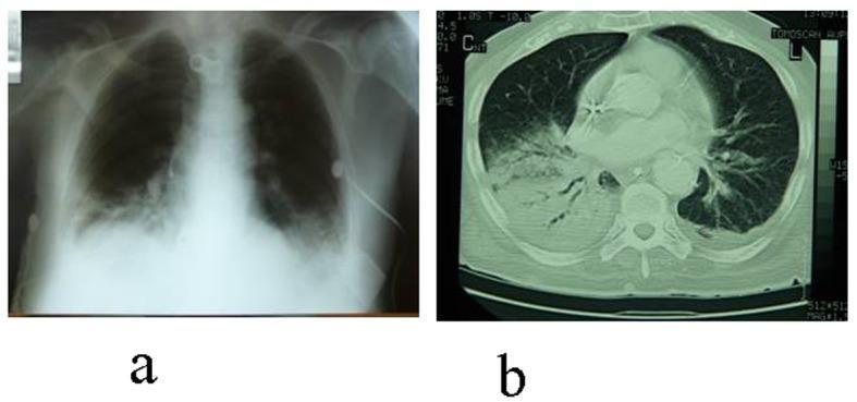
CASE 2 — Left Upper Lobe Collapse (Luftsichel Sign)
CASE 3 — Right Upper Lobe Collapse (Golden S Sign)
CASE 4 — Left Lower Lobe Collapse
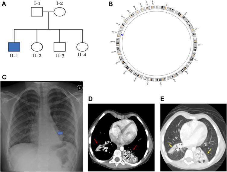
CASE 5 — Right Middle Lobe Collapse
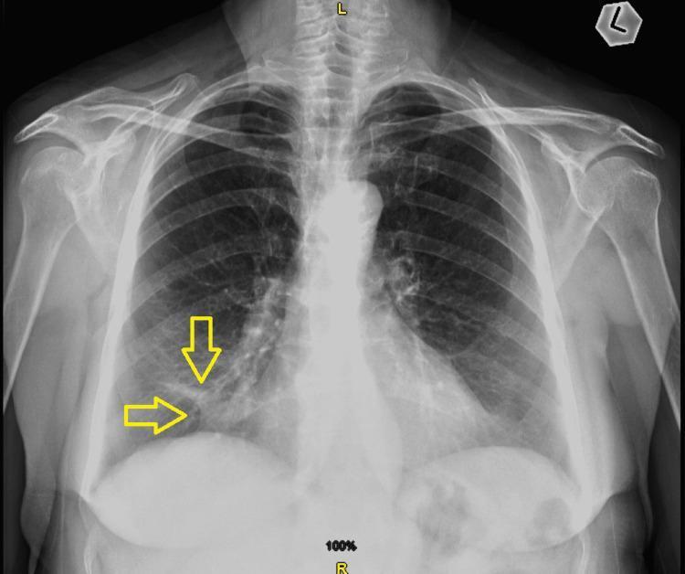
CASE 6 — COVID-19 Bilateral Pneumonia
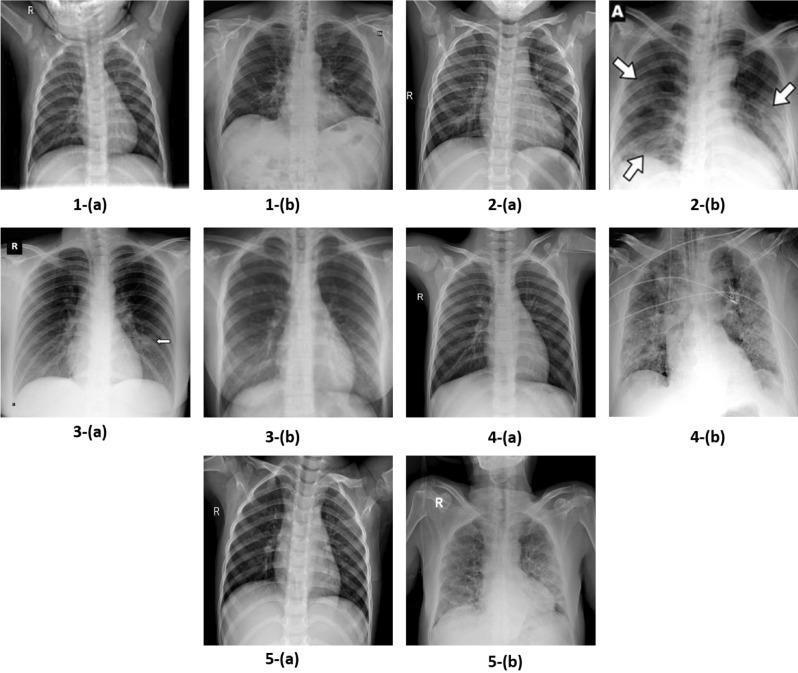
CASE 7 — Aspiration Pneumonia
CASE 8 — Left Lower Lobe Pneumonia
CASE 9 — Bronchopneumonia
CASE 10 — Pulmonary Oedema (Cardiogenic)
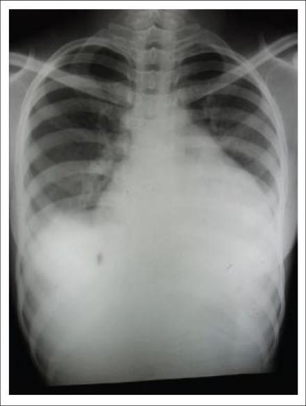
CASE 11 — Cardiogenic vs Non-Cardiogenic Oedema
| Feature | Cardiogenic | ARDS (Non-cardiogenic) |
|---|---|---|
| Heart size | Enlarged | Normal |
| Effusions | Bilateral | Absent |
| Kerley lines | Present | Absent |
| Distribution | Perihilar | Diffuse/peripheral |
| Context | IHD, CCF | Sepsis, trauma |
CASE 12 — Interstitial Pneumonia / Pulmonary Fibrosis

CASE 13 — Miliary Tuberculosis
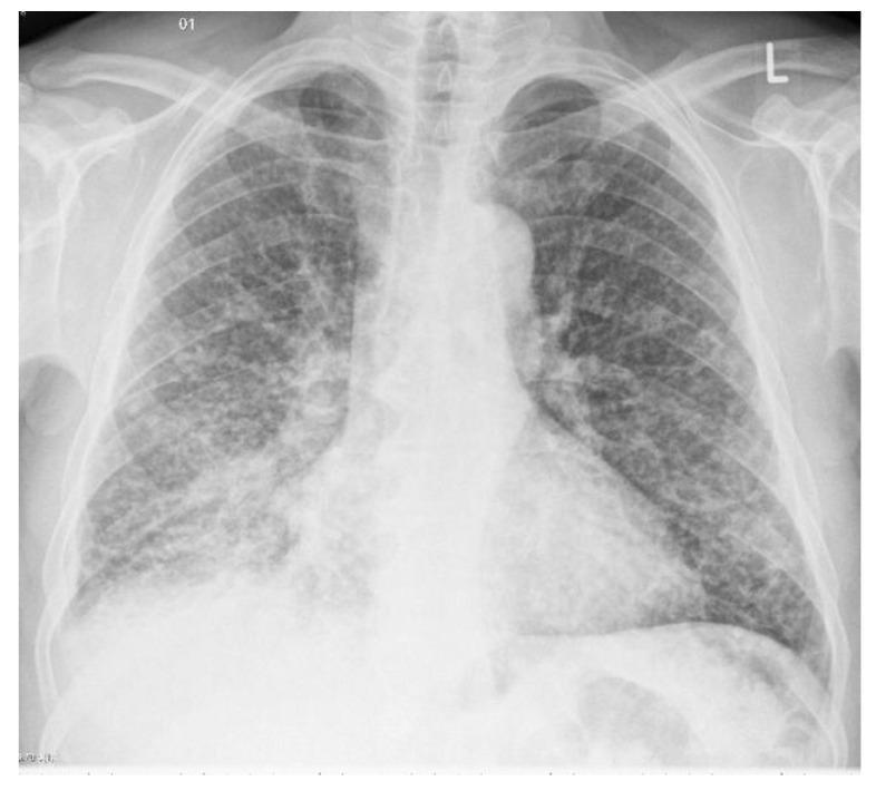
CASE 14 — Discoid/Linear Atelectasis
CASE 15 — Right Middle Lobe Syndrome
CASE 16 — Round Pneumonia (Child)
CASE 17 — Lipoid Pneumonia
CASE 18 — PCP (Pneumocystis Pneumonia)
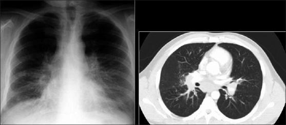
CASE 19 — Organising Pneumonia (COP)
CASE 20 — Eosinophilic Pneumonia
DAY 1 COMPLETE ✅
theres two station on xray posioting
TOACS STATION — X-RAY POSITIONING
Two Stations You Will Face
STATION 1 — IDENTIFY THE PROJECTION/POSITION
CHEST X-RAY PROJECTIONS
1. PA (Posteroanterior) — Standard
- Patient stands facing the detector
- X-ray beam travels back to front
- Features:
- Scapulae projected OUTSIDE lung fields
- Heart size accurate (heart magnified < 5%)
- Sharp, well-defined image
- Clavicles horizontal
- Full inspiration — 6 anterior ribs or 10 posterior ribs visible
2. AP (Anteroposterior) — Portable/Sick Patient
- Patient supine or sitting, detector behind them
- X-ray beam travels front to back
- Features:
- Scapulae projected OVER lung fields ← key difference
- Heart appears larger (magnified — don't comment on cardiomegaly)
- Less sharp image
- Usually labelled "AP" or "PORTABLE"
- Clavicles appear more horizontal or angled
TOACS tip: Always say "This is an AP film — I cannot accurately assess heart size on this projection."
3. Lateral CXR
- Patient stands with left side against detector (usually)
- Features:
- Spine visible posteriorly
- Sternum anteriorly
- Retrosternal and retrocardiac spaces visible
- Diaphragms: left higher (stomach bubble beneath), right lower
- Hilum: left hilum higher than right
4. Lateral Decubitus
- Patient lying on their side
- Used to confirm: free-flowing pleural effusion (layers out) vs loculated (stays in place)
- Also shows: small pneumothorax in non-ambulant patients
5. Expiratory CXR
- Used to accentuate a pneumothorax
- Lungs smaller → pneumothorax relatively larger → easier to see
6. Lordotic View (AP Lordotic)
- Patient leans backward
- Clavicles projected ABOVE lung apices
- Used to assess: right middle lobe, apical lesions, confirm RML collapse
ABDOMEN X-RAY PROJECTIONS
1. Supine AXR — Standard
- Most common
- Shows bowel gas pattern, calcifications, soft tissue masses
- Both psoas shadows visible normally
2. Erect AXR
- Patient standing
- Shows: air-fluid levels in bowel obstruction (step-ladder pattern)
- Shows: free air under diaphragm (pneumoperitoneum)
3. Erect CXR (for pneumoperitoneum)
- Better than erect AXR for detecting free air
- As little as 1 mL of free air detectable
4. Left Lateral Decubitus AXR
- Patient lying on LEFT side, beam horizontal
- Used when patient CANNOT stand
- Free air collects between liver and right lateral abdominal wall
SKELETAL X-RAY PROJECTIONS — Common Examples
| Body Part | Standard Views | Special Views |
|---|---|---|
| Shoulder | AP + Axillary lateral | Y-view (scapular Y), Stryker notch (Hill-Sachs) |
| Elbow | AP + Lateral | Radial head-capitellum view |
| Wrist | PA + Lateral | Scaphoid views (4 views), Carpal tunnel |
| Hip | AP pelvis + Lateral hip | Judet views (acetabulum) |
| Knee | AP + Lateral | Skyline/Merchant (patella), Tunnel/Notch view |
| Ankle | AP + Lateral + Mortise | Stress views |
| Foot | AP + Lateral + Oblique | Weight-bearing views |
| Spine (C) | AP + Lateral | Peg view (C1/C2 odontoid), Obliques (foramina) |
| Spine (L) | AP + Lateral | Obliques (pars — "Scottie dog") |
| Skull | PA + Lateral | Towne's view (occiput), SMV (base) |
STATION 2 — PATIENT POSITIONING / TECHNIQUE ASSESSMENT
HOW TO ASSESS CXR TECHNICAL QUALITY — RIPE
R — Rotation
- Check: Spinous processes should be equidistant from medial ends of both clavicles
- If rotated → mediastinum appears widened falsely → don't overcall pathology
- Say: "The film is rotated to the [left/right] as the spinous process is displaced toward the [left/right] clavicular head"
I — Inspiration
- Adequate = 6 anterior ribs or 10 posterior ribs above the diaphragm
- Poor inspiration → lung bases appear congested/hazy → mimics pulmonary oedema
- Say: "This is a poorly inspired film with only [X] anterior ribs visible"
P — Projection
- PA vs AP (as above)
- State which projection and consequences for interpretation
E — Exposure
- Underexposed (too white/light): Intervertebral disc spaces NOT visible through cardiac shadow → overestimates opacification
- Overexposed (too dark): Normal lung markings may be obscured
- Ideal: Vertebral bodies just visible behind cardiac shadow
COMMON POSITIONING ERRORS TO IDENTIFY
| Error | What You See | What to Say |
|---|---|---|
| Patient rotated | Spinous process off-centre, one clavicle higher | "Rotated film — mediastinum may appear falsely widened" |
| Poor inspiration | < 6 anterior ribs, elevated diaphragms | "Inadequate inspiration — basal opacities may be artefactual" |
| AP instead of PA | Scapulae over lung fields, magnified heart | "AP projection — cardiac size cannot be reliably assessed" |
| Underexposed | White film, disc spaces invisible | "Underexposed film — subtle opacities may be missed" |
| Overexposed | Very dark, lung markings washed out | "Overexposed — pneumothorax may be missed" |
| Off-centring | Ribs asymmetric, one lung appears smaller | "Film not centred — one hemithorax appears smaller artefactually" |
TOACS MARKING POINTS — What Examiners Want to Hear
"This is a [PA/AP] chest X-ray of an [adult/child]. Before interpreting, I will assess technical quality using the RIPE criteria..."
- Rotation: "The spinous process is [central/deviated] — the film is [adequately positioned/rotated]"
- Inspiration: "[X] anterior ribs are visible — this is [adequate/inadequate] inspiration"
- Projection: "This is a [PA/AP] projection based on [scapular position/labelling]"
- Exposure: "The film is [adequately exposed/under/overexposed] as vertebral bodies are [visible/not visible] behind the heart"
QUICK MEMORY CARD
PA CXR = Scapulae OUTSIDE lungs = accurate heart size
AP CXR = Scapulae OVER lungs = magnified heart
RIPE = Rotation, Inspiration, Projection, Exposure
Rotation check = spinous process vs clavicular heads
Adequate inspiration = 6 anterior ribs / 10 posterior ribs