
Open lip schizencephaly content for my paper presentation
Reading File
Finding Sources
Finding Sources
Searching Images
open-lip schizencephaly MRI axial coronal image
Searching PubMed
schizencephaly AND imaging
Searching the Web
open lip schizencephaly MRI review diagnosis management
Reading File
Reading File
Reading File
query_images_db
Reading Web Page
https://www.ncbi.nlm.nih.gov/sites/books/NBK560913
Open-Lip Schizencephaly: Presentation Content
1. Introduction
- Schizencephaly is a rare congenital malformation of cortical development.
- It is defined by a full-thickness cerebral cleft lined by dysplastic gray matter, extending from the pial surface/subarachnoid space to the lateral ventricle.
- It may be unilateral or bilateral.
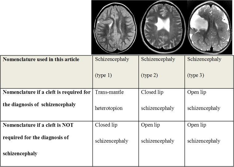
- The two traditional forms are:
- Closed-lip: cleft walls are apposed or fused.
- Open-lip: cleft walls are widely separated, with a CSF-filled channel communicating between the ventricle and subarachnoid space.
2. Definition of Open-Lip Schizencephaly
- In open-lip schizencephaly, the margins of the cleft are separated by CSF.
- The cleft is lined on both sides by gray matter, often dysplastic polymicrogyric cortex.
- This gray-matter lining is the key feature distinguishing it from porencephaly.
- Clefts commonly occur near the perisylvian, frontoparietal, and central regions.
3. Etiology and Pathogenesis
The precise cause is often uncertain and is probably heterogeneous.
Proposed mechanisms include:
- Disordered neuronal migration and cortical organization during fetal brain development.
- Early fetal ischemic or vascular insult.
- Intrauterine infection, especially congenital cytomegalovirus in some cases.
- Genetic and sporadic developmental mechanisms.
- Associated developmental malformations.
Do not describe EMX2 mutation as an established cause. Earlier reports suggested an association, but this has not been consistently confirmed. Bradley and Daroff's Neurology in Clinical Practice, Schizencephaly section.
4. Clinical Manifestations
The clinical severity depends mainly on:
- Unilateral versus bilateral involvement
- Size and location of the cleft
- Associated malformations
- Presence of hydrocephalus and epilepsy
Common features
- Global developmental delay
- Motor delay
- Speech and language delay
- Seizures, often focal or generalized
- Spastic hemiparesis in unilateral lesions
- Spastic quadriparesis in extensive bilateral lesions
- Intellectual disability
- Feeding difficulty and visual impairment in severe cases
Clinical correlation
- Unilateral lesions often cause contralateral hemiparesis and may have milder cognitive effects.
- Bilateral wide open-lip clefts are more often associated with severe developmental impairment, quadriparesis, and difficult-to-control epilepsy.
5. Associated Anomalies
Open-lip schizencephaly may occur with:
- Polymicrogyria around the cleft
- Septum pellucidum agenesis
- Corpus callosum dysgenesis or agenesis
- Optic nerve hypoplasia and septo-optic dysplasia
- Gray-matter heterotopia
- Ventriculomegaly or hydrocephalus
- Other cortical malformations
6. Neuroimaging: MRI Findings
MRI is the investigation of choice.
Typical MRI findings:
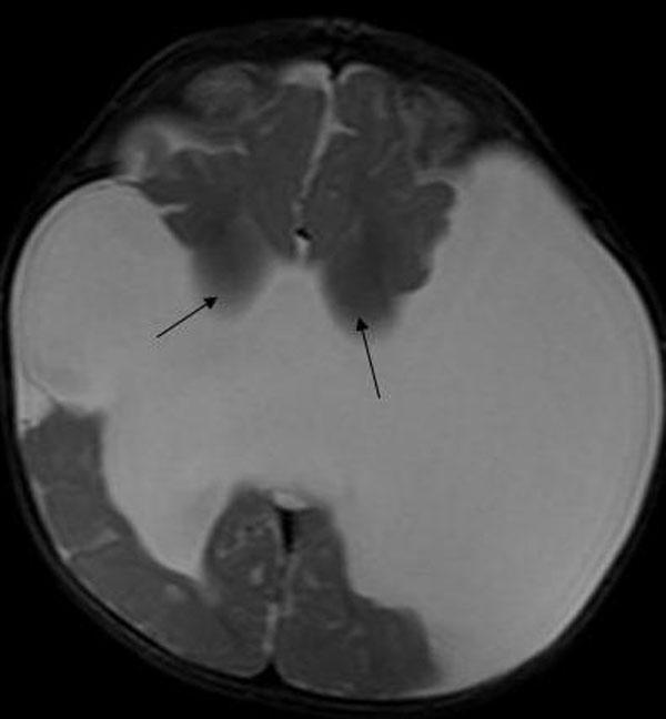
- CSF-signal cleft extending from the cortical surface to the lateral ventricle.
- Cleft margins lined by gray matter.
- Widely separated walls in the open-lip form.
- Dysplastic cortex and polymicrogyria adjacent to the cleft.
- Evaluation for associated callosal, septal, optic, and cortical abnormalities.
T1- and T2-weighted MRI, especially high-resolution coronal and 3D sequences, are useful for defining the gray-matter-lined cleft and associated cortical malformations. CT can show large clefts or calcification but is less reliable for identifying gray-matter lining. Grainger & Allison's Diagnostic Radiology, Schizencephaly section.
7. Differential Diagnosis
| Condition | Distinguishing feature |
|---|---|
| Porencephaly | CSF cavity due to destructive lesion, usually lined by white matter/gliosis rather than dysplastic gray matter |
| Arachnoid cyst | Extra-axial CSF collection; does not form a gray-matter-lined transmantle cleft |
| Holoprosencephaly | Failure of forebrain division with midline structural abnormalities |
| Focal cortical dysplasia | Cortical thickening and blurring of gray-white junction, without a complete ventricular-to-pial cleft |
| Hydranencephaly | Extensive destruction/absence of cerebral hemispheres rather than a discrete gray-matter-lined cleft |
High-yield point: A cleft lined by gray matter strongly supports schizencephaly; a cavity with a gliotic or white-matter margin supports porencephaly.
8. Management
There is no treatment that repairs the congenital cleft. Management is individualized and multidisciplinary.
- Anti-seizure medication for epilepsy.
- Physical therapy for spasticity, gait, posture, and motor function.
- Occupational therapy to improve activities of daily living.
- Speech and language therapy for communication and feeding issues.
- Developmental and educational support.
- Ophthalmologic evaluation if septo-optic dysplasia is suspected.
- Endocrine evaluation when midline abnormalities suggest pituitary dysfunction.
- Neurosurgical CSF diversion, such as ventriculoperitoneal shunting, if hydrocephalus or raised intracranial pressure develops.
- Genetic counseling may be considered, particularly where there are syndromic features or family history.
9. Prognosis
- Prognosis is variable.
- Better outcomes are generally seen in small unilateral clefts with preserved surrounding cortex.
- Poorer outcomes are associated with:
- Bilateral lesions
- Large open clefts
- Severe epilepsy
- Hydrocephalus
- Major associated malformations
- Early rehabilitation and seizure control improve functional outcomes, although the structural brain anomaly persists.
10. Take-Home Messages
- Open-lip schizencephaly is a congenital gray-matter-lined cleft from the ventricle to the pial surface.
- Its hallmark is a widely separated, CSF-filled cleft.
- MRI is the best test because it demonstrates the gray-matter lining and associated malformations.
- Differentiate it from porencephaly by identifying the gray-matter lining.
- Management focuses on seizures, hydrocephalus when present, and early multidisciplinary rehabilitation.
- Lesion laterality, size, associated anomalies, and epilepsy determine outcome.
Suggested references for your final slide
- Grainger & Allison's Diagnostic Radiology, “Schizencephaly” section.
- Bradley and Daroff's Neurology in Clinical Practice, “Schizencephaly” section.
- The NCBI clinical overview summarizes presentation, MRI diagnosis, and supportive management.
- A recent open-lip case report illustrates MRI findings, hemiparesis, and rehabilitation-focused care.
Recent-evidence note: A targeted search of systematic reviews from the last two years did not identify a relevant review specifically addressing open-lip schizencephaly, reflecting the rarity of the condition and reliance on imaging reviews and case-based literature.
This is a shared conversation. Sign in to Orris to start your own chat.