
Give detailed notes on the main topic "Introduction to neurological disorder (CVA, UMNL, LMNL)" based on the following topic learning outcomes: 1. Discuss the anatomy of vascular supply to brain 2. Explain the types of stroke, predisposing factors and causes 3. Discuss the clinical presentation and complication of CVA 4. Outline the management of CVA 5. Discuss the clinical approach to UMNL and LMNL. Use the following references: Davidson's principles and Practice of Medicine 24th Edition, Kumar and Clark Clinical Medicine 10th Edition, MacLeod's Clinical Examination 15th Edition, Harrison's principles of internal medicine 21st Edition, Nicholas J Talley O' Conner Clinical Examination 9th Edition, Malaysia Ministry of Heath Websites, and Malaysian Clinical Practice Guidelines.
Malaysia Clinical Practice Guidelines stroke management MOH 2023 2024
circle of Willis cerebral vascular anatomy brain arteries diagram
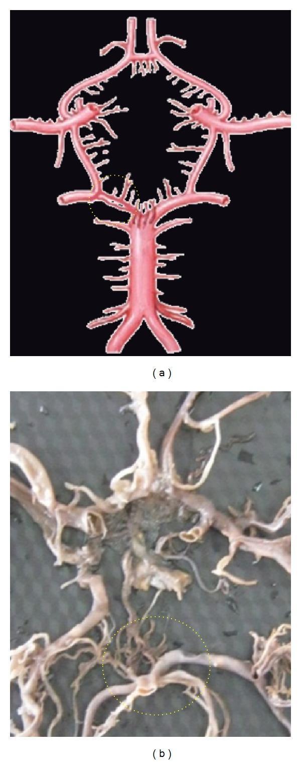
The image consists of two panels (a and b) illustrating the anatomy of the Circle of Willis at the base of the brain. Panel (a) is an anatomical diagram showing a standard arterial configuration with a dotted yellow circle highlighting the junction of the basilar artery and the posterior cerebral artery (PCA). Panel (b) is a clinical photograph of a post-mortem anatomical specimen showing the cerebral vasculature. A dotted yellow circle in panel (b) highlights a vascular anomaly: a duplication of the P1 segment of the posterior cerebral artery. In this region, the arterial pathway consists of two parallel, distinct vascular channels rather than a single vessel. The broader anatomical context displays the basilar artery, internal carotid arteries (ICA), anterior cerebral arteries (ACA), and communicating arteries (ACoA and PCoA). The image is intended for medical education in neuroanatomy and neuroradiology to demonstrate normal anatomical variants and congenital anomalies of the cerebral collateral circulation.
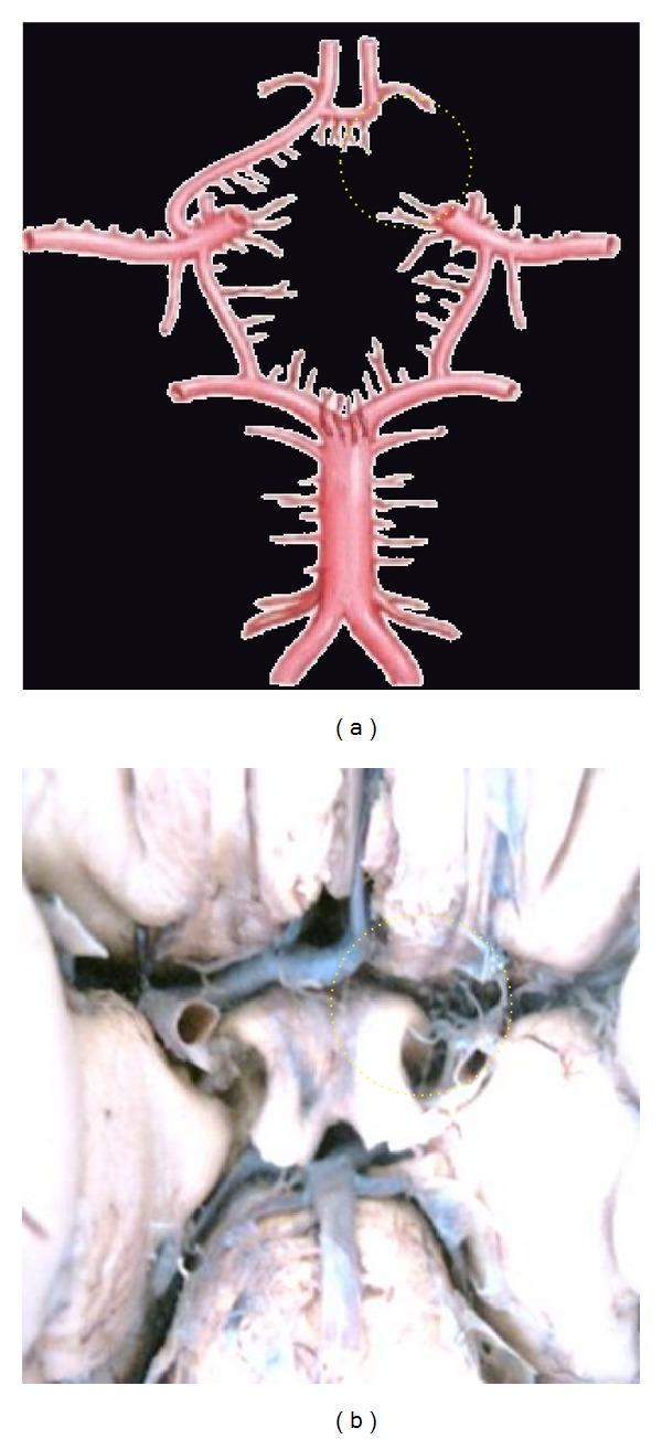
This medical visual content consists of an anatomical diagram (a) and a corresponding clinical photograph of a cadaveric dissection (b) illustrating neurovascular anatomy. The focus is on the Circle of Willis, specifically demonstrating a congenital vascular variation known as aplasia of the A1 segment of the anterior cerebral artery (ACA). In both panels, a yellow dotted circle highlights the area where the right A1 segment should connect the internal carotid artery to the anterior communicating artery (ACoA). The absence of this segment results in an incomplete Circle of Willis, which has clinical significance for collateral cerebral blood flow. The diagram (a) clearly depicts the major components of the cerebral arterial circle, including the basilar artery, posterior cerebral arteries, internal carotid arteries, and the intact left A1 and A2 segments. The clinical photograph (b) provides a real-world view of the same anatomical deficiency at the base of the brain. This material is designed for advanced medical education in neuroanatomy and radiology, emphasizing common vascular anomalies.
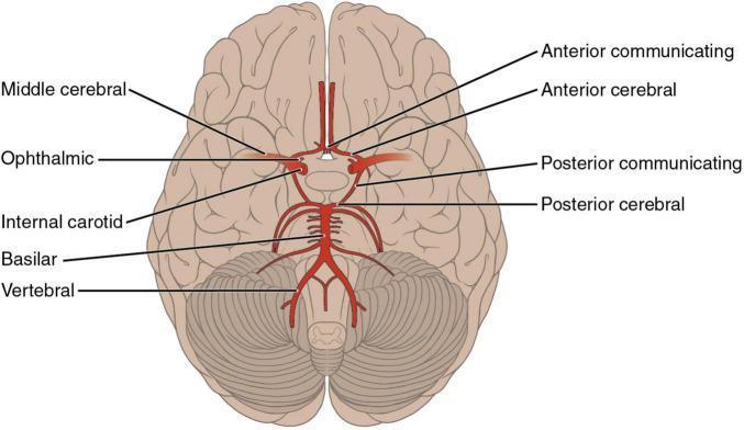
An anatomical diagram illustrating the arterial supply of the brain from an inferior (basal) perspective, specifically highlighting the Circle of Willis and its contributing vessels. The diagram shows the bilateral internal carotid arteries branching into the middle cerebral (MCA) and ophthalmic arteries. Centrally, the anterior cerebral arteries (ACA) are seen originating and coursing medially into the longitudinal fissure, connected by the anterior communicating artery (ACoA). The posterior circulation is depicted with the vertebral arteries merging into the basilar artery, which subsequently divides into the posterior cerebral arteries (PCA). Posterior communicating arteries (PCoA) are shown forming the critical anastomotic link between the internal carotid and posterior cerebral systems. The diagram is superimposed on the ventral surface of the brain, demonstrating the spatial relationship of these vessels to the frontal, temporal, and occipital lobes, as well as the cerebellum and brainstem. This illustration is an educational resource for understanding neurovascular anatomy and the territories susceptible to ischemic stroke.
upper motor neuron lower motor neuron lesion comparison diagram
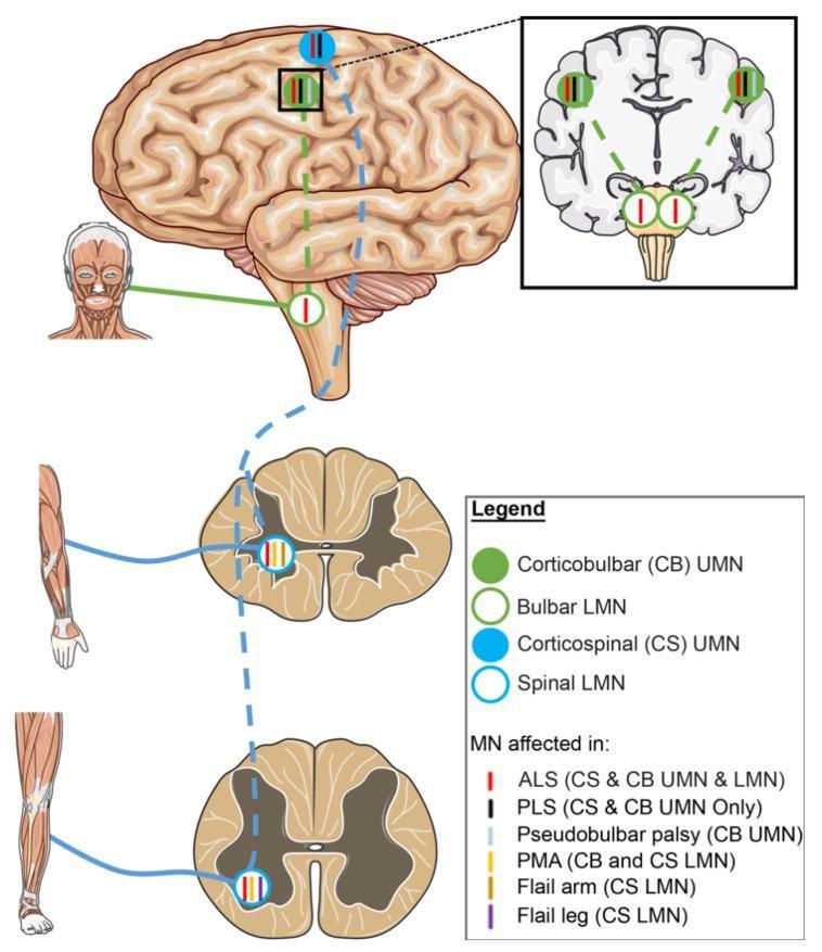
A medical schematic diagram illustrating the neuroanatomical pathways of upper motor neurons (UMN) and lower motor neurons (LMN) and their selective involvement in Amyotrophic Lateral Sclerosis (ALS) variants. The diagram features a lateral view of the human brain, a coronal section of the brain, and transverse sections of the spinal cord (cervical and lumbar levels). Two primary tracts are highlighted: the corticobulbar (CB) tract, represented by green solid circles (UMN) and open circles (LMN), and the corticospinal (CS) tract, represented by blue solid circles (UMN) and open circles (LMN). Solid circles represent UMN cell bodies in the motor cortex, while open circles denote LMNs in the brainstem (bulbar) and spinal cord. Colored vertical tick marks inside these circles indicate specific disease phenotypes: red for classic ALS (CS & CB UMN and LMN), black for Primary Lateral Sclerosis (PLS, UMN only), and yellow for Progressive Muscular Atrophy (PMA, LMN only). Other variants shown include Pseudobulbar palsy (CB UMN), Flail arm, and Flail leg (specific CS LMN segments). This illustration serves as an educational tool for differentiating motor neuron disease phenotypes based on the anatomical site of degeneration.
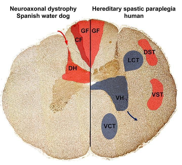
This Comparison Diagram presents a cross-sectional illustration of the spinal cord, contrasting the localization of axonal spheroids and neuronal loss in Neuroaxonal Dystrophy (NAD) and Hereditary Spastic Paraplegia (HSP). The diagram is split into two halves: the left represents a Spanish Water Dog with NAD, and the right represents human HSP. Ascending sensory pathways are highlighted in red, including the Gracile Fasciculus (GF), Cuneate Fasciculus (CF), and Dorsal Horn (DH) on the NAD side, and GF, Dorsal Spinocerebellar Tract (DST), and Ventral Spinocerebellar Tract (VST) on the HSP side. Descending motor pathways, affected only in the HSP model, are highlighted in blue, depicting the Lateral Corticospinal Tract (LCT), Ventral Corticospinal Tract (VCT), and Ventral Horn (VH). The visual demonstrates that in NAD, pathology is restricted to sensory, ascending tracts in the dorsal funiculus and dorsal horn. In contrast, human HSP shows involvement of both sensory pathways and descending motor pathways (pyramidal tracts and ventral horns). This diagram illustrates the pathophysiology of upper motor neuron and sensory system degeneration relevant to neurology and neurohistology.
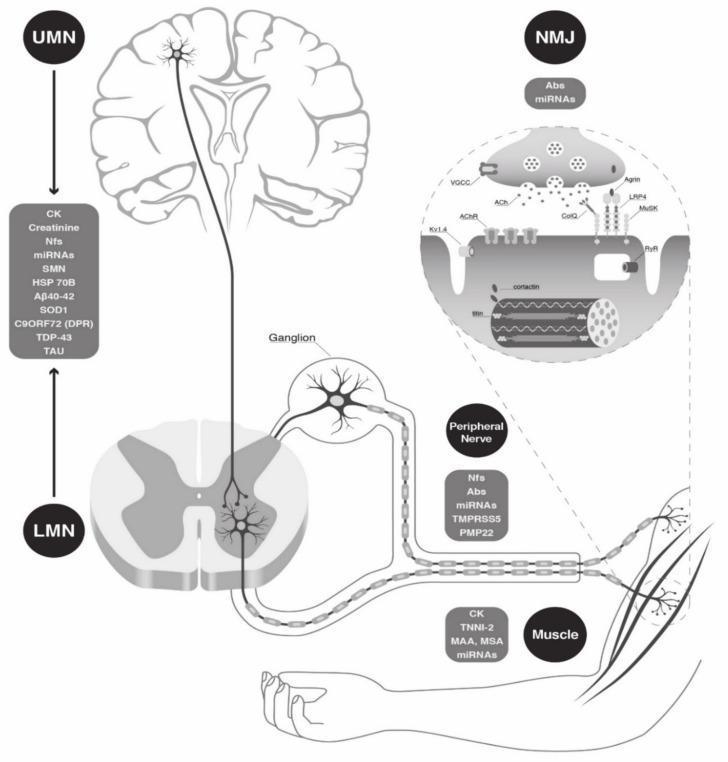
This medical illustration is a pathophysiology diagram detailing the anatomical components of the human neuromuscular unit and their associated circulating biomarkers. The schematic follows a top-down approach: 1) Upper Motor Neuron (UMN), shown within a coronal section of the brain with listed biomarkers including CK, Nfs, miRNAs, and neurodegenerative markers like TDP-43 and A̠40-42. 2) Lower Motor Neuron (LMN), depicted within a cross-section of the spinal cord, showing axons exiting via a ganglion into the peripheral nerve (associated with PMP22 and TMPRSS5). 3) Neuromuscular Junction (NMJ), magnified to show synaptic vesicles, ACh receptors, and muscle endplates with related antibodies (Abs) and miRNAs. 4) Skeletal Muscle, illustrated at the periphery with biomarkers such as CK, TNNI-2, and myositis-specific antibodies (MSA). The diagram serves as an educational tool for neurology and neuromuscular diseases, highlighting the potential for diagnostic and prognostic monitoring across the motor pathway from the central nervous system to the peripheral effector organ.
ischemic stroke CT scan MRI brain infarction hemorrhagic stroke
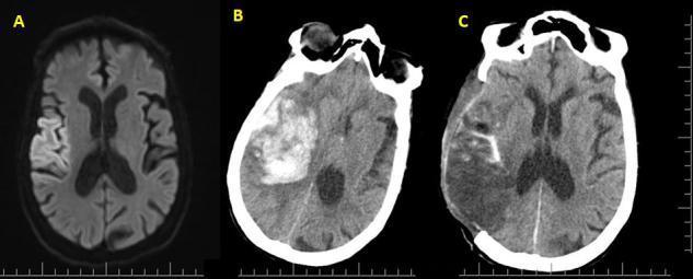
This Comparison Chart consists of three axial brain scans (MRI and CT) documenting the clinical progression and surgical management of a right middle cerebral artery (MCA) infarction. Image A is an axial T2-FLAIR MRI showing hyperintense signals in the right cortical region, consistent with early cytotoxic edema and acute ischemic stroke. Image B is a non-contrast CT scan taken 24 hours later, revealing a massive, hyperdense intracerebral hematoma within the infarcted area. This hemorrhagic transformation demonstrates significant mass effect, leading to subfalcine herniation and a distinct leftward midline shift. Image C is a post-operative CT scan following a decompressive hemicraniectomy (DHC) and hematoma evacuation. Key features include a large right-sided skull defect, reduced hematoma volume, and improvement in the midline shift, though residual parenchymal distortion and encephalomalacia persist. This sequence serves as an educational tool for neurosurgery and neuroradiology, illustrating the complications of large-vessel ischemia and the radiological appearance of life-saving surgical decompression.
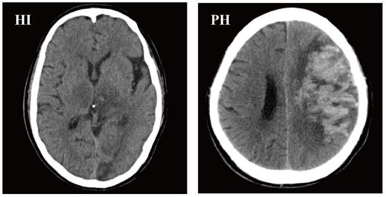
This diagnostic image consists of two axial non-contrast CT scans of the brain demonstrating hemorrhagic transformation (HT) in acute ischemic stroke, categorized according to the ECASS II classification. The first scan, labeled 'HI' (Hemorrhagic Infarction), shows subtle, ill-defined areas of hypodensity within the cerebral parenchyma, particularly in the right hemisphere, suggesting petechial hemorrhage or small areas of bleeding within an infarcted zone without a significant space-occupying effect. The second scan, labeled 'PH' (Parenchymal Hemorrhage), displays a prominent, heterogeneous hyperdense region in the left cerebral hemisphere indicative of acute blood. This lesion involves multiple lobes (frontal and parietal) and exhibits significant mass effect, including compression of the ipsilateral lateral ventricle and a rightward midline shift. These images serve as educational examples for differentiating types of hemorrhagic complications after stroke, highlighting the visual distinction between scattered petechiae (HI) and confluent hematoma (PH).
Introduction to Neurological Disorders: CVA, UMNL, and LMNL
Comprehensive Study Notes
LEARNING OUTCOME 1: Anatomy of Vascular Supply to the Brain
1.1 Overview of Cerebral Blood Supply
- The common carotid arteries arise from the aorta (left side directly; right side from the brachiocephalic artery) and bifurcate at the neck into the internal and external carotid arteries
- The ICA enters the skull through the carotid canal and gives rise to the Anterior Cerebral Artery (ACA) and the Middle Cerebral Artery (MCA)
- Arise from the subclavian arteries, ascend through the transverse foramina (foramina transversaria) of cervical vertebrae, enter the skull through the foramen magnum, and unite to form the Basilar Artery
- The basilar artery gives rise to the Posterior Cerebral Arteries (PCA), anterior inferior cerebellar arteries (AICA), superior cerebellar arteries, and posterior inferior cerebellar arteries (PICA)
1.2 The Circle of Willis
| Vessel | Origin | Connection |
|---|---|---|
| Anterior Cerebral Arteries (ACA) | Terminal branches of ICA | Connected by Anterior Communicating Artery (AComm) |
| Middle Cerebral Arteries (MCA) | Terminal branches of ICA | Largest branch |
| Posterior Communicating Arteries (PComm) | From ICA | Links anterior to posterior circulation |
| Posterior Cerebral Arteries (PCA) | Top of basilar artery | Receives flow from PComm |
1.3 Territories of the Three Main Cerebral Arteries
Middle Cerebral Artery (MCA)
- Largest and most clinically important cerebral artery
- Supplies the lateral surface of the cerebral hemisphere including: primary motor cortex (face, arm, leg laterally), primary sensory cortex, Broca's area (left), Wernicke's area (left), visual radiation, and insula
- Deep branches (lenticulostriate arteries) supply the basal ganglia and internal capsule
- The recurrent artery of Heubner (from ACA) also supplies parts of the basal ganglia
Anterior Cerebral Artery (ACA)
- Supplies the medial surface of the frontal and parietal lobes
- The medial motor cortex (leg area predominantly) is ACA territory
- ACA infarcts produce contralateral leg weakness > arm weakness (inverse of MCA pattern)
Posterior Cerebral Artery (PCA)
- Supplies: occipital lobe (primary visual cortex), inferior temporal lobe, thalamus, midbrain
- PCA infarcts cause contralateral homonymous hemianopia with macular sparing (in cortical infarcts)
- Thalamic perforating branches (thalamoperforators) supply the thalamus
Vertebrobasilar System
- Basilar artery: supplies the pons, cerebellum via AICA and superior cerebellar arteries
- PICA (from vertebral artery): supplies lateral medulla - occlusion causes Wallenberg syndrome (lateral medullary syndrome)
1.4 Deep Perforating Arteries
- Lenticulostriate arteries (from proximal MCA): supply putamen, globus pallidus, posterior limb of internal capsule
- Thalamoperforators (from PCA/PComm): supply thalamus
- Anterior choroidal artery (from ICA): supplies posterior limb of internal capsule, optic tract, hippocampus
LEARNING OUTCOME 2: Types of Stroke, Predisposing Factors, and Causes
2.1 Definition
2.2 Classification / Types of Stroke
A. Ischaemic Stroke (~85%)
- Caused by in situ thrombosis on an atherosclerotic plaque (large vessel) or lipohyalinosis (small vessel)
- Large vessel atherosclerosis: involves ICA, MCA stem, vertebral/basilar arteries
- Small vessel disease (lacunar infarcts): involves deep perforating arteries; associated with hypertension and diabetes
- Deficits often progress gradually (stuttering onset) or occur during sleep
- Thrombus or material from a proximal source travels to occlude a cerebral artery
- Sources include:
- Cardiac (most common embolic source): atrial fibrillation, mechanical valves, left ventricular thrombus (post-MI), infective endocarditis, dilated cardiomyopathy, patent foramen ovale (PFO)
- Artery-to-artery: from carotid plaque
- Usually sudden onset, maximal at onset; may involve cortical branches
- Emboli tend to lodge in MCA territory (largest flow)
- Small (<1.5 cm) deep infarcts from occlusion of a single perforating artery
- Classic syndromes: pure motor hemiparesis, pure sensory stroke, sensorimotor stroke, ataxic hemiparesis, dysarthria-clumsy hand syndrome
- Strongly associated with hypertension and diabetes
B. Haemorrhagic Stroke (~15%)
- Spontaneous bleeding into brain parenchyma
- Most common cause: chronic hypertension causing Charcot-Bouchard microaneurysms in deep perforating vessels
- Common sites: putamen/basal ganglia (most common), thalamus, pons, cerebellum, subcortical white matter
- Also caused by: cerebral amyloid angiopathy (lobar bleeds in elderly), AVM, anticoagulation, cocaine
- Bleeding into the subarachnoid space
- Most commonly from rupture of a saccular (berry) aneurysm at the Circle of Willis
- Classic presentation: sudden-onset "thunderclap" headache ("worst headache of my life")
- Other causes: AVM, trauma
2.3 Predisposing Factors and Risk Factors
Non-Modifiable Risk Factors
- Age (risk doubles every decade after age 55)
- Sex (male > female; though women have higher lifetime risk)
- Race (higher incidence in Black/Asian populations)
- Family history / genetics
- Prior stroke or TIA (strongest predictor of future stroke)
Modifiable Risk Factors
| Risk Factor | Mechanism |
|---|---|
| Hypertension | Most important modifiable risk factor; damages vessel walls, promotes atherosclerosis and lipohyalinosis; ~5x increase in stroke risk |
| Atrial fibrillation | Leads to intracardiac thrombus formation; ~5-6x increase in stroke risk |
| Diabetes mellitus | Promotes atherosclerosis and lacunar disease |
| Hypercholesterolaemia | Accelerates atherogenesis |
| Cigarette smoking | Promotes atherosclerosis, hypercoagulable state |
- Obesity, sedentary lifestyle
- Excess alcohol
- Cardiac disease: valvular disease, low ejection fraction, patent foramen ovale
- Substance abuse (cocaine causes vasospasm and ICH)
2.4 Causes / Aetiology
TOAST Classification (Trial of Org 10172 in Acute Stroke Treatment):
- Large-artery atherosclerosis
- Cardioembolism
- Small-vessel occlusion (lacunar)
- Stroke of other determined aetiology (e.g., vasculitis, dissection, hypercoagulable states)
- Stroke of undetermined aetiology (cryptogenic)
Hypercoagulable States (rarer causes, important in young patients):
- Protein S deficiency, Protein C deficiency, Antithrombin III deficiency
- Factor V Leiden mutation, Prothrombin gene mutation
- Antiphospholipid antibody syndrome
- Sickle cell disease, Polycythaemia, Hyperhomocysteinaemia
- Vasculitis (temporal arteritis, primary CNS vasculitis, SLE-related)
- Disseminated intravascular coagulation (DIC)
Arterial Dissection
- Carotid or vertebral artery dissection - important cause in young patients
- Presents with ipsilateral neck/head pain, partial Horner's syndrome, then ischaemic deficits
LEARNING OUTCOME 3: Clinical Presentation and Complications of CVA
3.1 General Clinical Presentation
- F - Face drooping
- A - Arm weakness
- S - Speech difficulty
- T - Time to call emergency
3.2 Syndrome-Based Clinical Presentations by Territory
Middle Cerebral Artery (MCA) Syndrome
| MCA Division | Key Deficits |
|---|---|
| Left MCA superior division | Right face + arm weakness (UMN type), Broca's (non-fluent) aphasia |
| Left MCA inferior division | Wernicke's (fluent) aphasia, right visual field defect; minimal motor deficit; patient appears confused |
| Left MCA deep territory | Right pure motor hemiparesis (UMN type); aphasia possible with larger infarcts |
| Left MCA stem (complete) | Right hemiplegia + hemisensory loss + right homonymous hemianopia + global aphasia + left gaze preference |
| Right MCA superior | Left face + arm weakness, left hemineglect |
| Right MCA inferior | Profound left hemineglect, left visual field defect, somatosensory deficits |
| Right MCA stem | Left hemiplegia + hemisensory loss + left hemianopia + hemispatial neglect + right gaze preference |
Anterior Cerebral Artery (ACA) Syndrome
- Contralateral leg weakness > arm (medial motor cortex - leg area)
- Frontal lobe signs: urinary incontinence, grasp reflex, abulia (lack of motivation)
- Possible contralateral leg sensory loss
Posterior Cerebral Artery (PCA) Syndrome
- Contralateral homonymous hemianopia (often with macular sparing)
- Thalamic infarcts: contralateral hemisensory loss (all modalities)
- Midbrain involvement: CN III palsy, vertical gaze palsy (top-of-basilar syndrome)
- Memory impairment (if mesial temporal lobe/hippocampus involved)
Vertebrobasilar Syndromes
- Basilar artery occlusion: "locked-in" syndrome (quadriplegia, aphonia, preserved vertical eye movement); high mortality
- Lateral medullary (Wallenberg) syndrome (PICA/vertebral artery): ipsilateral facial sensory loss + contralateral body sensory loss (crossed sensory loss), ipsilateral Horner's syndrome, dysphagia, dysarthria, ipsilateral limb ataxia, vertigo/nausea
Lacunar Syndromes (from small perforating vessel occlusion)
- Pure motor hemiparesis: face + arm + leg weakness; posterior limb of internal capsule (lenticulostriate arteries) or ventral pons
- Pure sensory stroke: thalamic infarct (VPL/VPM nuclei)
- Sensorimotor stroke: thalamocapsular infarct
- Ataxic hemiparesis: hemiparesis + ipsilateral limb ataxia
- Dysarthria-clumsy hand syndrome: dysarthria + hand clumsiness
3.3 Haemorrhagic Stroke - Clinical Features
- ICH: Sudden-onset focal deficit + headache + vomiting + reduced consciousness (mass effect from haematoma)
- Basal ganglia/capsular: contralateral hemiplegia, eyes deviate toward haemorrhage side
- Pontine: quadriplegia, pinpoint pupils, coma
- Cerebellar: ataxia, ipsilateral gaze palsy, no hemiplegia
- SAH: Thunderclap headache (sudden explosive onset), neck stiffness, photophobia, reduced consciousness, positive Kernig's/Brudzinski's signs
3.4 Complications of CVA
Acute Complications
- Haemorrhagic transformation: Secondary haemorrhage into ischaemic territory; common with large embolic infarcts; risk increased by tPA
- Cerebral oedema and raised ICP: Peaks at 3-4 days in large MCA infarcts; can cause herniation and death
- Seizures: 3-10% of stroke patients; usually after stroke but occasionally at onset
- Aspiration pneumonia: From dysphagia (most common infection complication)
- Hydrocephalus: From cerebellar infarct/haemorrhage compressing 4th ventricle, or SAH blocking CSF drainage
Subacute Complications
- Deep vein thrombosis (DVT) and pulmonary embolism: Immobility; major cause of morbidity
- Pressure sores / decubitus ulcers: Immobility
- Urinary tract infection: Indwelling catheters, neurogenic bladder
- Constipation / bowel dysfunction
- Contractures and spasticity: From UMN lesion and immobility
- Shoulder pain / subluxation: In hemiplegic limb
- Depression: Occurs in 30-50% of stroke survivors; greatly affects rehabilitation
Long-term Complications
- Post-stroke dementia / vascular cognitive impairment
- Epilepsy: Late-onset seizure disorder
- Recurrent stroke: ~10% per year without secondary prevention
- Spasticity and contractures
- Dysphagia and nutritional deficits
LEARNING OUTCOME 4: Management of CVA
4.1 Emergency/Acute Management: "Time is Brain"
Initial Assessment (on arrival)
- ABCs - Airway, Breathing, Circulation; oxygen if SpO2 <94%
- Blood glucose (correct hypo/hyperglycaemia immediately)
- IV access + bloods: FBC, U&E, LFT, glucose, coagulation (PT/APTT/INR), lipids, cardiac enzymes, ECG
- Non-contrast CT head - within 20 minutes of arrival - to rule out haemorrhage before any thrombolysis
- Clinical stroke scoring: NIH Stroke Scale (NIHSS)
- Vital signs: BP, temperature, oxygen saturation
Reperfusion Therapies (Ischaemic Stroke only)
- Indication: Acute ischaemic stroke within 4.5 hours of onset
- No evidence of intracranial haemorrhage on CT
- "Time is Brain" - earlier treatment = better outcomes
- Dose: 0.9 mg/kg IV (max 90 mg); 10% as bolus, 90% over 60 min
- Contraindications include: history of intracranial haemorrhage, known AVM/aneurysm, active bleeding, platelet count <100,000, INR >1.7, BP >185/110 mmHg (uncontrolled), recent major surgery, pregnancy
- For large vessel occlusion (MCA, ICA, basilar artery)
- Time window: up to 6 hours (standard); up to 24 hours with advanced imaging (CT perfusion/MR diffusion-perfusion) if penumbra is salvageable
- Combined with IV tPA or alone ("drip and ship")
- Catheter-based device removes clot from occluded artery
- Malaysia CPG for Ischaemic Stroke (3rd Edition, 2020) - recommends EVT at stroke-capable centres with neurointerventional services
4.2 General Supportive Care
| Aspect | Recommendation |
|---|---|
| Blood pressure | Allow permissive hypertension in acute ischaemic stroke (up to 220/120 mmHg if not thrombolysing); treat if BP >185/110 for tPA eligibility |
| Blood glucose | Target 7.7-10 mmol/L; aggressively correct hypoglycaemia; treat hyperglycaemia |
| Temperature | Treat fever aggressively (worsens infarction); target normothermia |
| IV fluids | Maintain hydration; avoid hypotonic fluids (worsen cerebral oedema) |
| Oxygen | Only if hypoxic (SpO2 <94%); routine O2 not beneficial |
| Positioning | Head elevated 30° for raised ICP; flat position may optimise perfusion in acute phase |
| Swallowing assessment | Before oral intake; use bedside swallow test; NGT if needed |
| DVT prophylaxis | Low molecular weight heparin + compression stockings when haemorrhage excluded |
4.3 Antithrombotic Therapy (Non-tPA patients / Secondary Prevention)
- Aspirin 300 mg loading dose orally within 48 hours of ischaemic stroke (if not thrombolysed, wait 24h post-tPA)
- Long-term: Aspirin 75-100 mg daily, or Clopidogrel 75 mg daily
- Dual antiplatelet (aspirin + clopidogrel) for 21-90 days in minor stroke/high-risk TIA (POINT/CHANCE trials)
- Atrial fibrillation-related stroke: Start anticoagulation (DOAC preferred - apixaban, rivaroxaban, dabigatran) after 2 weeks for moderate stroke, 2 days for TIA/minor stroke
- IV heparin: limited role; use in progressing cardioembolic stroke, carotid/basilar dissection
- Warfarin still used for mechanical heart valves
- High-intensity statin (Atorvastatin 40-80 mg) for all ischaemic stroke/TIA - reduces recurrence beyond lipid-lowering effects
- Target LDL <1.8 mmol/L (or <1.4 mmol/L in very high risk)
4.4 Management of Complications
- Cerebral oedema/raised ICP: Head elevation, osmotherapy (mannitol 20%), hyperventilation (temporary); hemicraniectomy for malignant MCA infarction (young patients, dominant hemisphere with patient consent)
- Cerebellar infarct with mass effect: Surgical decompression
- Post-stroke seizures: Anticonvulsants (levetiracetam or carbamazepine); no prophylactic anticonvulsants
- ICH: Reverse anticoagulation immediately; control BP (systolic target <140 mmHg); surgical evacuation in cerebellar haematoma >3 cm or accessible cortical haematoma with deterioration
- SAH: Nimodipine (calcium channel blocker) to prevent vasospasm; secure aneurysm (coiling preferred over clipping)
4.5 Stroke Unit Care
- Dedicated stroke unit care reduces mortality and disability vs general ward (Level A evidence)
- Multidisciplinary team: neurologist, nurse, physiotherapist, occupational therapist, speech therapist, dietitian, social worker
- Malaysia MOH recommends all stroke patients be managed in a dedicated stroke unit or stroke bay
4.6 Secondary Prevention / Long-term Management
| Intervention | Target |
|---|---|
| Antihypertensives (ACE inhibitor + diuretic) | BP <130/80 mmHg |
| Statin therapy | LDL <1.8 mmol/L |
| Antiplatelet/anticoagulation | Lifelong |
| Diabetic control | HbA1c <7% |
| Smoking cessation | Complete cessation |
| Carotid endarterectomy/stenting | If symptomatic carotid stenosis >50-70% |
| Cardiac monitoring | Detect paroxysmal AF (prolonged ambulatory ECG) |
| PFO closure | Consider in young cryptogenic stroke |
- Begin early mobilisation within 24-48 hours if medically stable
- Physiotherapy, occupational therapy, speech-language therapy, neuropsychology
- Long-term recovery possible up to ~1 year; neuroplasticity allows unaffected brain areas to compensate
LEARNING OUTCOME 5: Clinical Approach to UMNL and LMNL
5.1 Anatomical Framework
- Cell body in the motor cortex (precentral gyrus)
- Axon travels via the corticospinal tract (corona radiata → internal capsule → cerebral peduncle → pyramidal decussation → lateral corticospinal tract in spinal cord)
- For cranial nerve nuclei: corticobulbar tract (cortex → brainstem motor nuclei)
- Synapse with LMN in the anterior horn of the spinal cord (or cranial nerve nuclei in brainstem)
- Cell body in the anterior horn of the spinal cord or cranial nerve motor nuclei
- Axon travels via peripheral nerves to skeletal muscle
- Acts as the "final common pathway"
5.2 Comparison: UMN vs LMN Signs
| Clinical Feature | UMN Lesion | LMN Lesion |
|---|---|---|
| Weakness | Yes (contralateral to lesion if above decussation) | Yes (ipsilateral, in distribution of affected nerve/root) |
| Muscle atrophy | Absent (or mild from disuse) | Prominent (denervation atrophy) |
| Fasciculations | Absent | Present (spontaneous motor unit firing) |
| Muscle tone | Increased (spasticity) | Decreased (flaccidity) |
| Deep tendon reflexes | Increased (hyperreflexia) | Decreased/absent (hyporeflexia/areflexia) |
| Plantar response | Extensor (Babinski's sign positive) | Flexor (normal) or absent |
| Distribution | Usually pyramidal pattern (extensors > flexors in arm; flexors > extensors in leg) | Distribution of affected nerve, root, or plexus |
| Clonus | May be present | Absent |
| Hoffmann's sign | May be positive | Absent |
5.3 Mechanism of UMN Signs - Spasticity
5.4 Clinical Approach to UMN Lesion
History
- Sudden onset (stroke) vs gradual (tumour, MS, spondylosis)
- Associated features: headache (SAH, ICH), fever (abscess, encephalitis), prior episodes (MS, TIA)
- Risk factors: vascular risk factors, malignancy, autoimmune disease
Examination - Motor System
- Pyramidal posture: arm flexed + pronated; leg extended
- Wasting: absent (or mild disuse atrophy late)
- Fasciculations: absent
- Spasticity: velocity-dependent resistance ("clasp-knife" phenomenon - initial high resistance that suddenly releases)
- Distinguish from rigidity (Parkinson's - "lead pipe", "cogwheel") and from normal
- Pyramidal pattern: UL - shoulder abductors, elbow extensors, wrist extensors, finger extensors weakest; LL - hip flexors, knee flexors, ankle dorsiflexors weakest
- Grade using MRC scale (0-5)
- Deep tendon reflexes: brisk/hyperreflexia; test biceps (C5,6), triceps (C7), supinator (C6), knee (L3,4), ankle (S1)
- Babinski's sign (extensor plantar): dorsiflexion of big toe + fanning of other toes on stroking the lateral sole - hallmark of UMN lesion above L1
- Hoffmann's sign (UL equivalent of Babinski): flicking terminal phalanx of middle finger causes thumb flexion
- Clonus: rhythmic involuntary muscular contractions at ankle or knee
- Assess for associated hemisensory loss, visual field defect
- Coordination may be impaired if cerebellum or its tracts affected
Localisation of UMN Lesions
| Location | Key Features |
|---|---|
| Motor cortex | Contralateral monoparesis (face, arm, or leg only - somatotopic) |
| Internal capsule | Contralateral hemiplegia (face + arm + leg); often with sensory loss (posterior limb) |
| Brainstem (above decussation) | Contralateral hemiplegia + ipsilateral cranial nerve palsy ("crossed signs") |
| Spinal cord | Bilateral weakness + sensory level + bladder/bowel dysfunction (paraplegia/quadriplegia) |
| Cervical cord | Quadriparesis (arms + legs); UMN signs in legs, possibly UMN or LMN in arms at level of lesion |
5.5 Clinical Approach to LMN Lesion
History
- Onset, progression, distribution of weakness
- Pain (nerve root compression), sensory symptoms
- Autonomic features: sweating, sphincter control
Examination - Motor System
- Wasting/atrophy (prominent - present within 3 weeks of denervation)
- Fasciculations (visible twitches of muscle groups - pathological)
- Decreased (flaccidity) - hypotonia
- Distribution follows affected nerve, nerve root, or plexus
- Know dermatomes and myotomes for localisation
- Decreased or absent (hyporeflexia/areflexia) in distribution of affected nerve
- Plantar response: flexor (normal) or absent
- Sensory loss in distribution of affected nerve/root
Localisation of LMN Lesions
| Location | Pattern | Examples |
|---|---|---|
| Anterior horn | Weakness without sensory loss | Poliomyelitis, motor neuron disease (PMA) |
| Nerve root | Segmental weakness + dermatomal sensory loss + reflex loss | Disc prolapse (L4/5, L5/S1, C5/6, C6/7) |
| Plexus | Weakness in territory of multiple roots/nerves | Brachial neuritis, lumbosacral plexopathy |
| Peripheral nerve (mononeuropathy) | Weakness + sensory loss in single nerve territory | Carpal tunnel (median), common peroneal palsy |
| Polyneuropathy | Distal, symmetrical, "stocking-glove" sensory + motor | Diabetic neuropathy, GBS |
| Neuromuscular junction | Weakness, fatiguability, no sensory loss | Myasthenia gravis |
| Muscle | Proximal > distal weakness, no sensory loss | Myopathy, muscular dystrophy |
5.6 FAST Neurological Examination Approach (MacLeod's / Talley O'Connor Framework)
- Inspect: posture, gait (hemiplegia = circumduction gait; LMN foot drop = steppage gait), wasting, fasciculations
- Tone: passive movement at each joint - note spasticity vs flaccidity vs rigidity
- Power: MRC grading; test against resistance; pyramidal vs non-pyramidal distribution
- Reflexes: deep tendon reflexes + pathological reflexes (Babinski, Hoffmann, clonus)
- Coordination: finger-nose test, heel-shin test, rapid alternating movements (assess cerebellum)
- Sensation: light touch, pain, vibration, proprioception; determine dermatomal/peripheral nerve pattern
- Special tests:
- Pronator drift (arms extended, palms up - affected arm pronates and drifts down in UMN)
- Romberg's test (posterior column / vestibular)
5.7 Combined UMN + LMN Signs
| Condition | UMN Features | LMN Features |
|---|---|---|
| Motor Neuron Disease (ALS) | Spasticity, hyperreflexia, Babinski | Wasting, fasciculations, hyporeflexia |
| Cervical myelopathy with radiculopathy | Spastic paraparesis (legs) | Wasting + areflexia at level of lesion (arms) |
| Subacute combined degeneration (B12) | Spastic paraparesis | Peripheral neuropathy |
| Conus medullaris lesion | Variable UMN | LMN at conus level |
Summary Comparison Table: CVA by Type
| Feature | Ischaemic Stroke | Intracerebral Haemorrhage | Subarachnoid Haemorrhage |
|---|---|---|---|
| Onset | Sudden, maximal | Sudden, progressive | Sudden explosive |
| Headache | Mild/absent | Present (raised ICP) | Severe "thunderclap" |
| Vomiting | Uncommon | Common | Common |
| Consciousness | Variable | Often impaired | Often impaired |
| Focal deficit | Yes | Yes | Initially may be absent |
| Meningism | No | No | Yes (neck stiffness, photophobia) |
| CT finding | Hypodense (after 24-48h) / normal early | Hyperdense (white) | Hyperdense in subarachnoid space |
| Treatment | tPA + EVT | BP control, reverse anticoag | Nimodipine, secure aneurysm |
Key References
- Neuroanatomy through Clinical Cases, 3rd Edition (Blumenfeld) - primary source for vascular anatomy, stroke syndromes, UMN/LMN framework
- Davidson's Principles and Practice of Medicine, 24th Edition - stroke management, complications
- Kumar and Clark Clinical Medicine, 10th Edition - stroke classification, risk factors
- Harrison's Principles of Internal Medicine, 21st Edition - stroke pathophysiology, hypercoagulable states
- MacLeod's Clinical Examination, 15th Edition - systematic neurological examination approach
- Talley O'Connor Clinical Examination, 9th Edition - motor examination framework, UMNL/LMNL clinical approach
- Fuster and Hurst's The Heart, 15th Edition - cardioembolic stroke, atrial fibrillation and stroke risk
- Malaysian CPG: Management of Ischaemic Stroke, 3rd Edition (2020) - Malaysian Society of Neurosciences/MOH - local stroke management recommendations
- Malaysian CPG: Management of Spontaneous Intracerebral Haemorrhage (2025, 1st Edition) - MOH Malaysia
- Malaysia National Stroke Registry 2017-2024 - epidemiology and real-world stroke data in Malaysia
Clinical Exam Tip: In any patient presenting with acute neurological deficit, always (1) establish the time of onset, (2) localise the lesion (hemisphere/brainstem/cord), (3) classify as UMN/LMN, and (4) determine the likely vascular territory or aetiology. The combination of these steps guides both immediate management (tPA eligibility) and long-term secondary prevention.