
How to check tibia length
- Direct measurement - from the medial knee joint line to the medial malleolus with a tape measure, knee slightly flexed.
- Scanogram/orthoroentgenogram - the gold standard X-ray method for precise bone length.
- Ultrasonography - used especially in fetal/prenatal assessment.
- CT scanogram - most accurate for pre-surgical planning.
how to measure tibia length clinical methods
tibia length measurement clinical technique
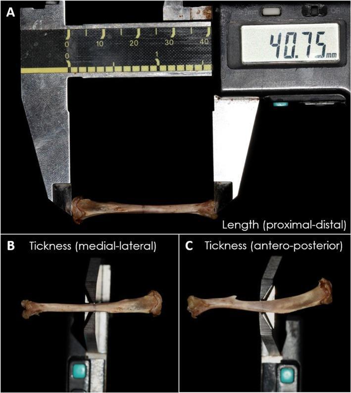
A series of three macroscopic clinical photographs (A, B, C) documenting the morphometric analysis of a tibia specimen using a digital pachymeter (caliper). Panel A demonstrates the measurement of the total longitudinal length (proximal-distal), with the digital display indicating a value of 40.75 mm. Panel B shows the measurement of diaphyseal thickness in the medial-lateral plane. Panel C illustrates the measurement of diaphyseal thickness in the antero-posterior plane. The images highlight standardized techniques for quantifying gross anatomical dimensions in orthopedic or osteological research, specifically focusing on bone morphology and physical characteristics. This type of macroscopic analysis is critical for assessing bone development, structural integrity, or the effects of experimental interventions on skeletal tissue.
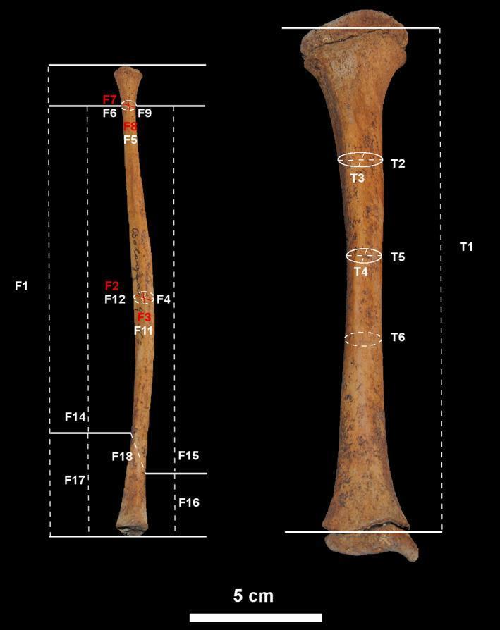
This clinical imaging figure displays human subadult skeletal remains of the right tibia and fibula, positioned vertically against a black background for osteometric analysis. The image serves as an anatomical and anthropological guide for bone measurement. The tibia on the right shows a broad proximal end with developing condylar morphology and a distal end exhibiting post-mortem damage. Labeled measurements (T1–T6) indicate longitudinal length and cross-sectional diameters at the proximal, midshaft, and distal thirds. The fibula on the left is slender and elongated, with labels (F1–F18) marking anthropometric points including the fibular neck, midshaft circumference, and the distal malleolar region. Dotted lines and arrows illustrate the specific planes for metric assessment. A 5 cm scale bar is provided at the bottom. This visual is designed for orthopedic research, forensic anthropology, and pediatric osteology to study growth patterns and developmental changes in the lower limb bones of children.
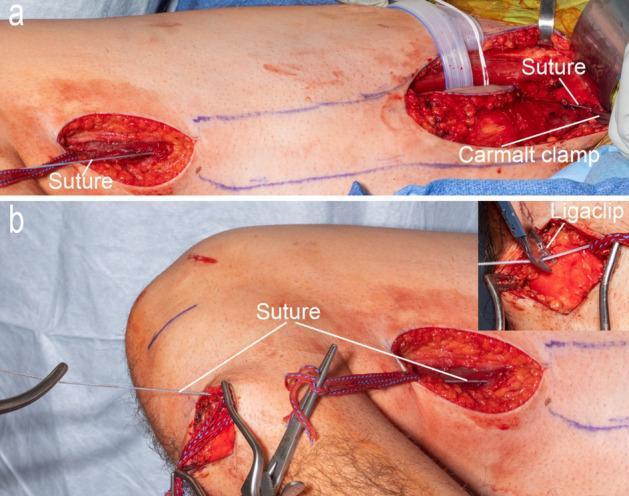
This clinical photograph consists of two panels (a and b) illustrating a surgical technique for musculotendinous unit (MTU) length measurement during a gracilis muscle harvest. Panel (a) shows a proximal thigh incision where a white #2 fiber wire suture is secured at the gracilis origin on the pubic ramus using a Carmalt clamp. The suture is threaded subcutaneously toward a distal thigh incision. Panel (b) depicts the suture continuing from the distal thigh incision through to a third incision at the pes anserine on the medial proximal tibia. An inset in panel (b) shows the use of a titanium Ligaclip to mark the specific length of the MTU at the level of the tibial tubercle. The surgical field includes visible longitudinal incisions, anatomical blue skin markings for surgical planning, umbilical tape for tendon manipulation, and standard retractors. This procedure is critical for intraoperative muscle force measurements and functional transfers, ensuring precise length-tension relationships are maintained.
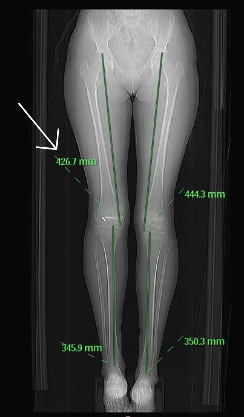
This diagnostic image is a CT scanogram of the lower limbs, a clinical imaging modality used for precise orthometric assessment. The coronal view visualizes the entire lower body skeletal anatomy, including the pelvis, femurs, tibias, fibulas, and feet. Anatomical landmarks for measurement are identified, with digital green measurement lines extending from the femoral heads to the ankle joints. Numerical annotations in millimeters quantify the length of individual long bones: the right femur measures 426.7 mm compared to the left femur at 444.3 mm, and the right tibia measures 345.9 mm compared to the left tibia at 350.3 mm. Visible surgical hardware (orthopedic screws) is noted in the distal right femur, consistent with a history of fracture fixation. The primary educational focus is the quantification of limb length discrepancy (LLD) following pediatric trauma and physeal injury. The image demonstrates how scanography provides an objective basis for surgical planning, such as epiphysiodesis, by comparing corresponding segments on the contralateral side.
How to Check / Measure Tibia Length
1. Clinical (Tape Measure) Methods
True Leg Length (Segmental - Tibial Component)
- Patient lies supine with limbs in a neutral position (identical alignment for both sides).
- Measure from the medial knee joint line (medial tibial plateau) to the medial malleolus using a tape measure.
- Compare both sides. A difference >1 cm is clinically significant.
Other Tape Landmarks Used in Practice
- ASIS to medial malleolus (for total true limb length, includes femur + tibia)
- ASIS to lateral malleolus (alternative landmark)
2. Anatomical / Osteological Landmarks (Dry Bone or Imaging)
From the lateral condyle (proximal) to the tip of the medial malleolus (distal)
-
The tibia accounts for ~22% of body height - useful for stature estimation.
-
When measuring a dry bone, subtract 5 mm from the measurement before applying stature formulae (to account for cartilage loss).
-
The Essentials of Forensic Medicine and Toxicology, 36th ed., p. 136
3. Radiographic Methods
| Method | Landmarks | Notes |
|---|---|---|
| Orthoroentgenogram (long leg X-ray) | Medial tibial plateau to tibial plafond | Traditional gold standard |
| CT Scanogram | Cursor at medial tibial plateau to tibial plafond | Most accurate; 80% less radiation than orthoroentgenogram |
| Standing full-length radiograph | Center of medial tibial plateau (MP) to tibial plafond (TP) | Normative values available |
4. Tibial Tuberosity to Medial Malleolus (Surgical Nail Sizing)
-
Measure from the tibial tuberosity to the medial malleolus
-
This can be done directly with a ruler or indirectly via X-ray
-
Comminuted fractures require measuring the contralateral tibia radiographically
-
Campbell's Operative Orthopaedics, 15th Ed 2026
5. Ultrasonography
Visual Reference
Quick Summary
| Setting | Method | Landmark |
|---|---|---|
| Bedside/clinic | Tape measure | Medial tibial plateau → medial malleolus |
| Osteology/forensics | Dry bone calipers | Lateral condyle → tip of medial malleolus |
| Surgical planning | CT scanogram | Medial tibial plateau → tibial plafond |
| Nail sizing | Tape/X-ray | Tibial tuberosity → medial malleolus |
| Prenatal | Ultrasound | Proximal → distal epiphysis |