
Varicose vein explain with treatment
Reading File
Finding Sources
Searching Images
varicose veins legs clinical photo
Finding Sources
Finding Sources
Reading File
Reading File
Searching Content
"Management of varicose veins" in /textbooks/9780367548117
Reading File
Varicose Veins
What they are
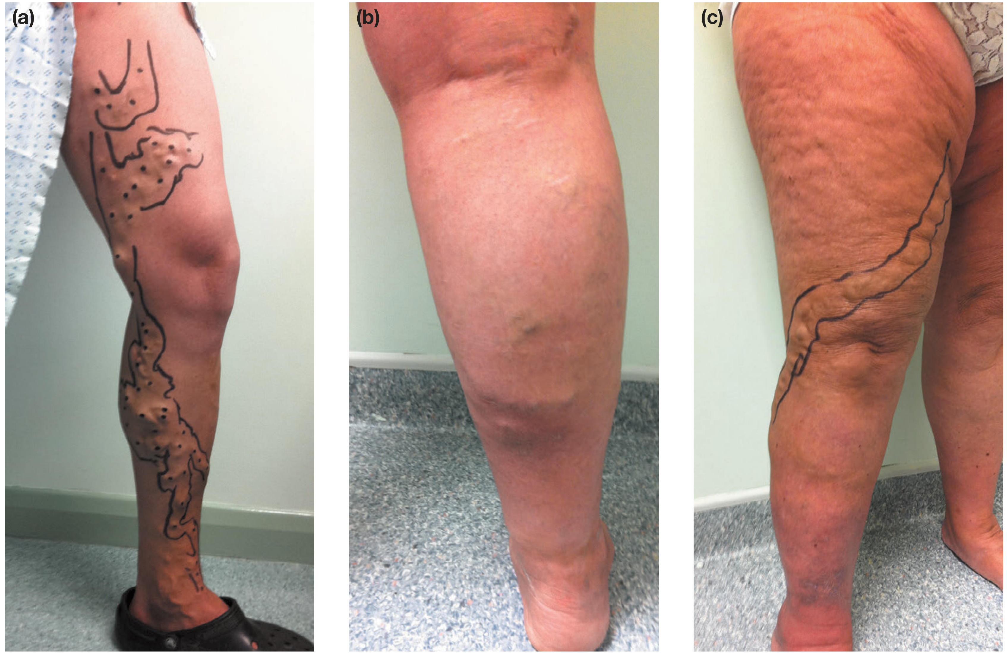
Varicose veins are dilated, tortuous, elongated superficial veins in the legs, caused by valvular incompetence that allows blood to flow backward (reflux) under gravity instead of being pushed up toward the heart. The great saphenous vein (GSV) and small saphenous vein (SSV) systems are most commonly affected.
Pathophysiology
The vein wall itself is the primary problem in most cases, not a primary mechanical valve failure. Changes include inflammatory cell infiltration, dysfunctional smooth muscle proliferation, collagen deposition, reduced elastin, and increased matrix metalloproteinase activity. This leads to loss of vein wall compliance, dilatation, elongation/tortuosity, and secondary valve dysfunction. This process can start anywhere in the venous tree - secondary varicose veins arise from post-thrombotic damage (DVT) or congenital venous abnormalities, while primary varicose veins have no identifiable underlying cause (- Bailey & Love's Short Practice of Surgery).
Epidemiology and risk factors
- Prevalence of visible varicose veins in adults: 30-50%
- More common in women; rises steeply with age (11.5% at 18-24 years to 55.7% at 55-64 years, Edinburgh Vein Study)
- Pregnancy, family history, higher BMI/height increase risk
- Prolonged standing, smoking, and constipation are suggested but not conclusively proven risk factors
Symptoms
Aching, heaviness, throbbing, burning, or a bursting sensation that worsens through the day or with standing, and improves with leg elevation or compression stockings. Itching and ankle swelling can occur, more so with complications. Symptom severity does not always correlate with the visible extent of varicosity.
Complications
- Chronic: skin pigmentation (haemosiderin deposition), lipodermatosclerosis, eczema, venous ulceration
- Acute: superficial vein thrombosis (thrombophlebitis), bleeding from a varix
Diagnosis
Clinical examination plus duplex ultrasound to map reflux and identify the source (saphenofemoral or saphenopopliteal junction incompetence, perforator or deep vein involvement) - this guides which vein(s) need treatment.
Treatment
1. Conservative management
- Graduated compression stockings - reduce symptoms and venous hypertension but do not cure the underlying reflux
- Weight loss, leg elevation, regular exercise (calf muscle pump)
- First-line when intervention is not indicated, not desired, or contraindicated
2. Sclerotherapy
- Injection of a sclerosant (e.g., sodium tetradecyl sulfate) into the vein, causing endothelial damage and fibrotic closure
- Liquid sclerotherapy is used for smaller/reticular and spider veins; foam sclerotherapy (ultrasound-guided) can treat larger truncal veins
- Simple, outpatient, but higher recurrence than thermal ablation for large truncal veins
3. Endovenous thermal ablation (now first-line for truncal reflux in most guidelines)
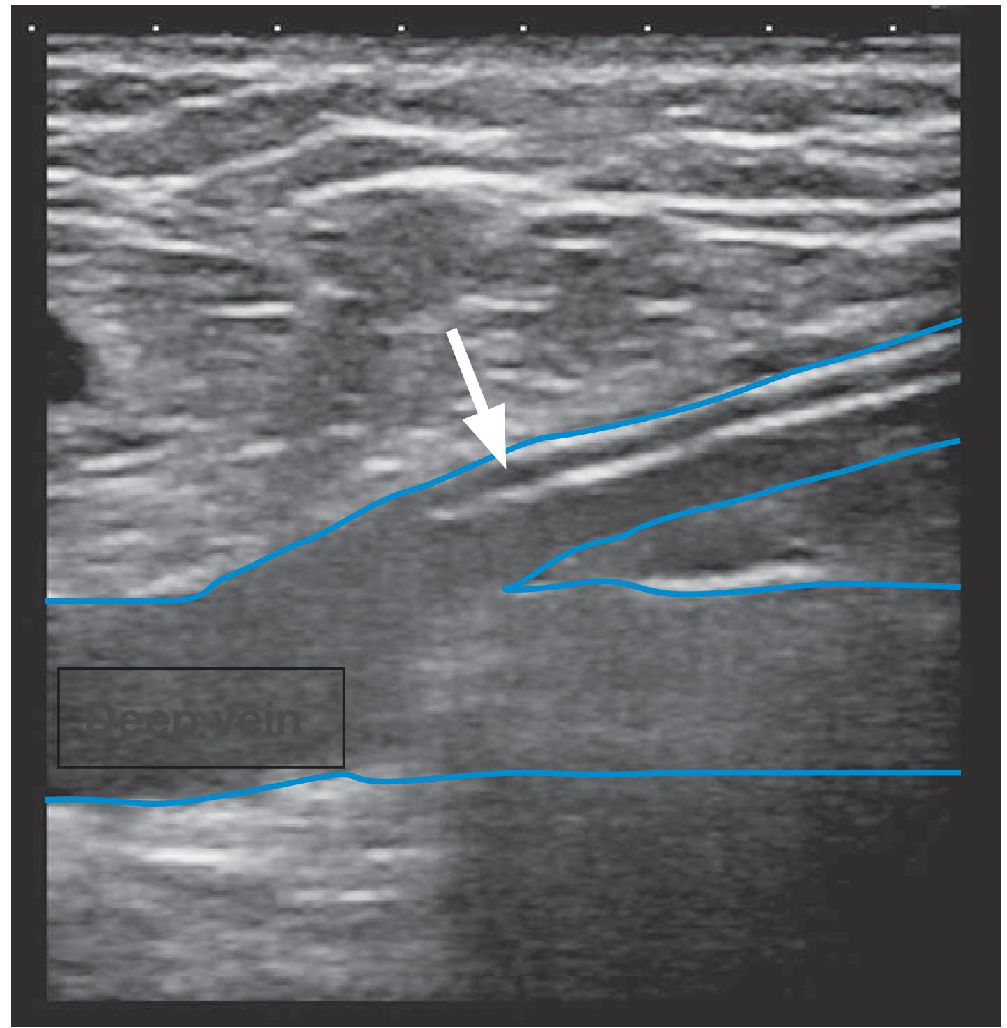
- Endovenous laser ablation (EVLA) and radiofrequency ablation (RFA): an ultrasound-guided catheter/fibre is passed into the incompetent vein (GSV or SSV), tumescent local anaesthesia is infiltrated around the vein, and thermal energy is delivered as the fibre is withdrawn, causing the vein wall to collapse and fibrose shut
- Performed under local anaesthetic, day-case, faster recovery than open surgery, and comparable or better long-term closure rates
4. Non-thermal, non-tumescent techniques
- Mechanochemical ablation (MOCA) and cyanoacrylate glue closure - avoid the need for tumescent anaesthesia, useful when thermal ablation is unsuitable (e.g., near skin/nerves)
5. Surgery
- High tie and stripping of the GSV/SSV with or without multiple stab avulsions (phlebectomy) of tributary varicosities
- Reserved now mainly for cases unsuitable for endovenous techniques (very tortuous veins, patient/anatomical factors) since thermal ablation has largely replaced open surgery as first-line
- CPT coding example: stab phlebectomy for 10-20 incisions vs. more than 20 (- Pfenninger and Fowler's Procedures for Primary Care)
6. Concomitant/adjunct phlebectomy or foam sclerotherapy of residual tributary varicosities is often done at the same time as truncal ablation.
Treatment choice depends on venous anatomy (superficial vs. perforator vs. deep, per the CEAP/anatomical classification), vein size and tortuosity, patient comorbidities, and local expertise. In patients with uncomplicated but symptomatic varicose veins, ablation/removal of the refluxing vein gives the greatest quality-of-life benefit; once skin changes or ulceration have developed, some morbidity may be irreversible even after successful treatment.
Sources: Bailey and Love's Short Practice of Surgery, 28th Edition (Chapter 62, "Dysfunction of the venous system" / "Varicose Veins"); Pfenninger and Fowler's Procedures for Primary Care, 3rd Edition; S Das, A Manual on Clinical Surgery, 13th Edition.
This is a shared conversation. Sign in to Orris to start your own chat.