
For md dermatology exam make notes on difference between Lymphangioma circumscriptum vs lymohangiectasia
lymphangioma circumscriptum vs lymphangiectasia difference dermatology
lymphangioma circumscriptum frog spawn vesicles skin
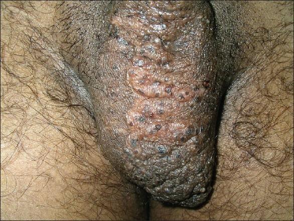
This clinical photograph shows a close-up view of a male scrotum on the first postoperative day following radiofrequency current coagulation for lymphangioma circumscriptum. The scrotal skin is hyperpigmented (consistent with Fitzpatrick skin type V) and displays an irregular, cobblestone texture. Multiple grouped vesicles are distributed across the surface, particularly concentrated on the left side. Some vesicles appear whitish and translucent, containing clear lymphatic fluid, while others are dark-red to blackish, indicating the presence of blood-tinged fluid or hemorrhagic crusting. The overall appearance is characteristic of superficial lymphangioma (lymphangioma circumscriptum), which often presents as persistent clusters of small, fluid-filled vesicles described as 'frog spawn.' Areas of hyperpigmented skin and slight erythema around the lesions are consistent with early postoperative changes and chronic skin irritation from lymphorrhoea.
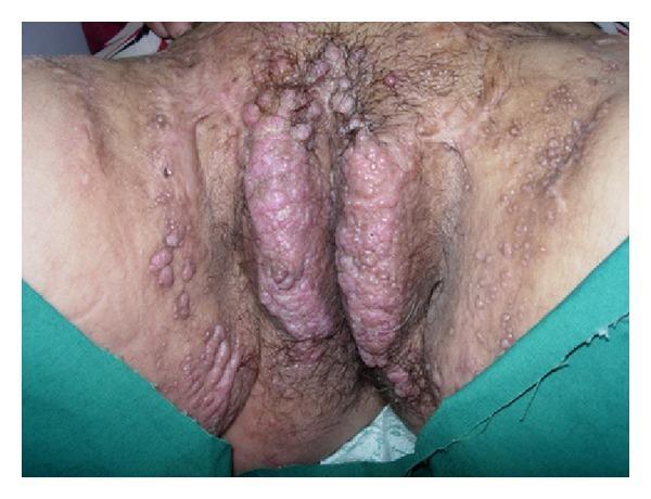
This clinical photograph displays a primary content type of dermatological clinical imaging, specifically focused on the female external genitalia and surrounding inguinal regions. The image demonstrates significant bilateral vulvar lymphedema characterized by marked enlargement and thickening of the labia majora. The affected skin surface is densely covered with multiple, translucent, flesh-colored to erythematous papules and vesicles, ranging in size from a few millimeters to larger confluent clusters. These lesions exhibit a characteristic 'frog-spawn' or 'cobblestone' morphology, typical of lymphangioma circumscriptum. The distribution extends beyond the vulva to involve the mons pubis and the medial inguinal folds. The underlying skin shows signs of inflammation and erythema. This visual evidence illustrates secondary lymphangioma circumscriptum, which often occurs as a late-stage complication of chronic lymphatic obstruction or pelvic surgery, representing the superficial manifestation of deeper lymphatic dilatation.
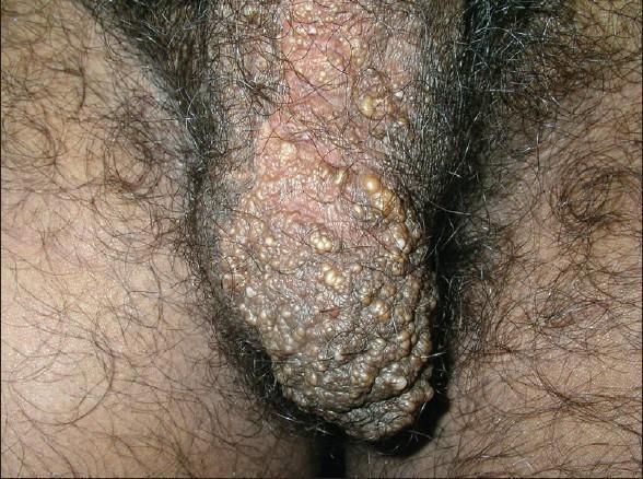
Clinical photograph of the male genital region, specifically showing the scrotum. The image depicts a localized cluster of numerous small, translucent to whitish-yellow vesicles on the left side of the scrotum. These lesions, characteristic of lymphangioma circumscriptum (LC), appear as multiple, thin-walled, fluid-filled papules resembling 'frog spawn.' The vesicles vary in size and are set against a background of skin that exhibits secondary changes, including hyperpigmentation and a thickened, pebbly, or verrucous (wart-like) texture. Some vesicles appear slightly more prominent and pearly, while the surrounding scrotal skin shows normal hair distribution. The clinical presentation is consistent with superficial lymphatic malformation, demonstrating clustered lymphangiectasias that may occasionally ooze clear or blood-tinged serous fluid (lymphorrhea).
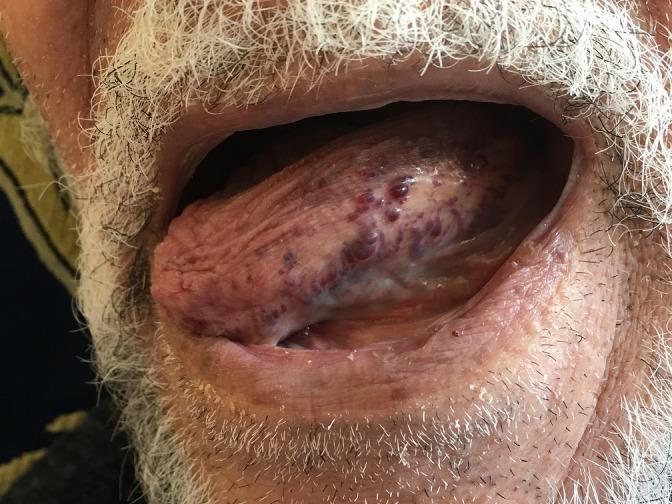
A clinical photograph of a human oral cavity showing the tongue protruding from the mouth. The tongue exhibits macroglossia (enlargement) and a highly irregular surface texture characterized by numerous clustered, small, dome-shaped papules or vesicles. These lesions vary in color from translucent to deep red and purple-blue, representing a 'frog-spawn' appearance typical of lymphangioma circumscriptum. This superficial lymphatic malformation displays intralesional hemorrhage, causing the darkened, hemorrhagic vesicles visible on the dorsal and lateral surfaces of the tongue. The surrounding lips and perioral skin, including the beard of an elderly male patient, appear unremarkable except for slight retraction to facilitate visualization of the lingual pathology. This image is an educational example of oral lymphatic malformations in a geriatric patient, highlighting the classic morphology of superficial circumscribed lymphangioma.
"lymphangioma circumscriptum" treatment
Lymphangioma Circumscriptum vs Lymphangiectasia (Acquired Lymphangioma)
Quick Comparison Table
| Feature | Lymphangioma Circumscriptum (LC) | Lymphangiectasia (Acquired Lymphangioma) |
|---|---|---|
| Nature | True congenital lymphatic malformation (microcystic LM) | Acquired dilation of previously normal superficial lymphatics |
| Cause | Developmental malformation of the lymphatic system (no prior damage needed) | Secondary to damage/obstruction of deep lymphatics - surgery, radiotherapy, malignancy/lymph node dissection, infection (filariasis, TB), Crohn disease, scleroderma, chronic lymphedema |
| Onset/Age | Present at birth or appears in early childhood | Adults, typically 5th-7th decade; appears months to years after the inciting insult (e.g., post-mastectomy/pelvic surgery/radiation) |
| Pathophysiology | Sequestered lymphatic cisterns in the deep dermis/subcutis communicate with dilated superficial dermal lymphatics that bulge the epidermis | Increased hydrostatic pressure in obstructed deep lymphatics causes backward dilation of normal superficial channels - not a new malformation |
| Common sites | Proximal limbs, trunk/chest, axilla, oral cavity (tongue, buccal mucosa), can occur anywhere | Site of prior surgery/radiation/trauma - e.g., vulva/mons pubis (post-pelvic surgery or radiotherapy), chest wall (post-mastectomy/axillary dissection), abdomen |
| Clinical appearance | Plaques with clusters of clear or hemorrhagic vesicles ("frog spawn"), lesions often far more extensive subcutaneously than the visible vesicles suggest; may weep lymph, bleed, or recur after minor trauma | Same "frog spawn" vesicular morphology, but confined to the area of prior lymphatic injury; often with associated lymphedema of the region |
| Depth of defect | Deep dermal/subcutaneous malformed lymphatic cisterns (deep component) with secondary superficial ectasia | Only superficial dermal lymphatics are ectatic; no deep malformed cisterns |
| Complications | Cellulitis/erysipelas-like flares after minor trauma, lymphorrhea, rarely lymphangiosarcoma | Recurrent cellulitis, lymphorrhea; rarely lymphangiosarcoma (classically in chronic post-mastectomy lymphedema = Stewart-Treves syndrome) |
| Histology | Dilated, thin, endothelium-lined lymphatic channels in the dermis, often with deeper communicating cisterns; channels may show papillary projections into lumen | Dilated superficial dermal lymphatic channels - histologically can be indistinguishable from LC on a superficial biopsy |
| Imaging | Deep component often demonstrable on MRI/ultrasound (helps confirm malformation) | No deep malformed cisterns on imaging - only surface ectasia at the site of prior insult |
| Key distinguishing clue | No history of preceding surgery/radiation/infection; congenital | History of trauma, surgery, irradiation, or chronic obstruction at the same site |
| Treatment | Surgical excision (needs to include the deep cisterns to prevent recurrence), sclerotherapy, laser (CO2/Nd:YAG), electrocautery/radiofrequency | Treat/avoid the underlying cause where possible; CO2 laser, excision, sclerotherapy for symptomatic lesions - recurrence common if the underlying lymphatic damage persists |
Exam Pearls
- Mnemonic: LC = "born with it" (congenital malformation); Lymphangiectasia = "acquired after it" (surgery/radiation/infection damages deep lymphatics first).
- Some authors (e.g., Medscape/older literature) use "acquired lymphangioma" and "lymphangiectasia" as synonyms, and use them interchangeably with LC - but modern texts (Bolognia, Andrews) treat LC as a true microcystic lymphatic malformation, and lymphangiectasia as a secondary/acquired ectasia of normal channels following lymphatic injury.
- Both share the "frog-spawn" vesicle sign and can look identical under the dermatoscope/on biopsy - clinical history of surgery/radiotherapy/trauma before the eruption is often the only way to separate acquired lymphangiectasia from congenital LC.
- Vulvar/scrotal/thigh lesions after pelvic malignancy surgery + radiotherapy = classic acquired lymphangiectasia vignette.
- Differential for both: cutaneous metastasis (carcinoma telangiectoides), hemangioma, viral warts, molluscum (if vesicles are mistaken for papules).
- Malignant transformation (lymphangiosarcoma) is a rare but testable complication of long-standing lesions, especially in chronic lymphedema (Stewart-Treves syndrome after mastectomy).
Representative Clinical Images
Sources
- Dermatology, 2-Volume Set, 5th ed. (Bolognia) - "Microcystic lymphatic malformation (lymphangioma circumscriptum)," p. 2179-2180
- Andrews' Diseases of the Skin, Clinical Dermatology - "Acquired Progressive Lymphangioma," p. 695
- Lymphangiectasia overview - Medscape, on nomenclature distinguishing acquired lymphangioma (lymphangiectasia) from lymphangioma circumscriptum
- Huilgol SC, et al. CO2 laser therapy of vulval lymphangiectasia and lymphangioma circumscriptum (PMID: 12135508)
Based on information in this PDF make notes on lymphoedema for md dermatology theory exam
cd /tmp/workspace/attachments/9312e611-7a5c-4848-9af2-f00dd9b979b6 && python3 -c " import pdfplumber with pdfplumber.open('Lymphedema JAAD.pdf') as pdf: print('pages:', len(pdf.pages)) "
cd /tmp/workspace/attachments/9312e611-7a5c-4848-9af2-f00dd9b979b6 && python3 -c " import pdfplumber with pdfplumber.open('Lymphedema JAAD.pdf') as pdf: full = [] for i, page in enumerate(pdf.pages): t = page.extract_text() or '' full.append(f'--- PAGE {i+1} ---\n' + t) text = '\n'.join(full) print(len(text)) with open('/tmp/workspace/attachments/9312e611-7a5c-4848-9af2-f00dd9b979b6/extracted.txt','w') as f: f.write(text) "
wc -l /tmp/workspace/attachments/9312e611-7a5c-4848-9af2-f00dd9b979b6/extracted.txt
Read the file /tmp/workspace/attachments/9312e611-7a5c-4848-9af2-f00dd9b979b6/extracted.txt (1433 lines, extracted text of a JAAD CME article "Lymphedema: Pathophysiology and clinical manifestations" by Grada and Phillips, J Am Acad Dermatol 2017;77:1009-20). This is text extracted from a two-column PDF via pdfplumber, so some lines (especially table content) may appear garbled, reversed, or out of order (e.g. "emordnyslianwolleY" = "Yellow nail syndrome" reversed) - use your judgment to reconstruct the intended meaning from context, cross-referencing nearby text. Read the ENTIRE file in sequential chunks using read_file with offset/limit (e.g. limit=200 per call, advancing offset each time) until you have covered all 1433 lines. Do not skip any part. Then extract and organize ALL the clinically/exam-relevant content into a detailed, well-structured set of notes covering: 1. Definition and pathophysiology of lymphedema (key mechanisms, chronic inflammation, fibrosis cycle) 2. Classification: Primary lymphedema (genetic causes/syndromes - Milroy disease, Meige disease, lymphedema-distichiasis/FOXC2, hypotrichosis-lymphedema-telangiectasia/SOX18, Hennekam syndrome/CCNE1, Turner syndrome, other genes - with a table of gene, syndrome, clinical features if present in text) and its subtypes by age of onset (congenital, praecox, tarda) 3. Secondary lymphedema: all causes mentioned (filariasis, malignancy-related treatment/surgery/radiation, podoconiosis, morbid obesity, recurrent cellulitis/erysipelas, herpesvirus, lymphogranuloma venereum, tuberculosis/scrofula, etc.) with details/statistics given in the text 4. Clinical manifestations and cutaneous features/complications (e.g. skin changes, elephantiasis nostras verrucosa, lymphangiectasia, papillomatosis, verrucous changes, cellulitis/erysipelas risk, lymphangiosarcoma/Stewart-Treves, yellow nail syndrome, staging system if mentioned - Stage 0/1/2/3, Brunner or ISL staging) 5. Diagnostic workup: clinical exam, lymphoscintigraphy, MRI, ultrasonography, bioimpedance spectroscopy, laboratory tests, genetic testing (which genes are clinically testable) 6. Management: compression therapy, complete decongestive therapy, pneumatic compression devices (APCD), surgical options, skin care, treatment of complications - list all key points and specific details/statistics given 7. Any tables, staging systems, or key numeric facts (percentages, prevalence numbers, sensitivity/specificity values) present in the text - reproduce them accurately Return your final answer as complete, well-organized markdown notes (using headers, bullet points, and tables) suitable for a postgraduate (MD) dermatology theory exam, staying strictly faithful to the content of the article (do not invent facts not in the text, but you may supplement very briefly with widely-accepted standard knowledge if something is clearly incomplete in the extracted text, and mark such additions clearly as "[general knowledge, not from article]"). Include page/section references where helpful (e.g., "p. 1010" based on the --- PAGE X --- markers, noting the original journal page numbers 1009-1020 that appear in the text near each page break).
/large_tool_results
Read the file /tmp/workspace/attachments/9312e611-7a5c-4848-9af2-f00dd9b979b6/extracted.txt (1433 lines). It is text extracted from a two-column PDF (a JAAD CME article "Lymphedema: Pathophysiology and clinical manifestations" by Grada and Phillips, J Am Acad Dermatol 2017;77:1009-20). Some lines, especially table content, may be garbled/reversed due to PDF column extraction issues (e.g. "emordnyslianwolleY" reversed reads "Yellow nail syndrome") - use context to reconstruct meaning. Read the ENTIRE file sequentially using read_file with limit=200 per call, advancing offset (0, 200, 400, 600, 800, 1000, 1200) until all 1433 lines are covered. Do not skip content. After reading everything, write a detailed, well-organized set of exam notes in markdown covering: 1) definition & pathophysiology, 2) classification of primary lymphedema (genes/syndromes: Milroy/FLT4, Meige disease, lymphedema-distichiasis/FOXC2, hypotrichosis-lymphedema-telangiectasia/SOX18, Hennekam syndrome/CCNE1, GJC2, Turner syndrome, congenital/praecox/tarda subtypes), 3) causes of secondary lymphedema (filariasis, cancer treatment/surgery/radiation, podoconiosis, obesity, infection, etc.) with any stats given, 4) clinical/cutaneous manifestations and complications (elephantiasis nostras verrucosa, papillomatosis, cellulitis/erysipelas, lymphangiosarcoma/Stewart-Treves, yellow nail syndrome, staging system), 5) diagnostic workup (lymphoscintigraphy, MRI, ultrasound, bioimpedance spectroscopy, labs, genetic testing), 6) management (compression therapy, complete decongestive therapy, pneumatic compression devices, surgery, skin care). Return ONLY the final complete markdown notes as your answer message (this is the only thing that will be seen - do not omit it). Stay strictly faithful to the article content; you may note "[general knowledge]" for any minor supplementation. Include original journal page numbers where visible (article spans pages 1009-1020).
Lymphedema - Notes for MD Dermatology Theory Exam
1. Definition and Pathophysiology
- Lymphedema = localized tissue swelling from excessive retention of lymphatic fluid in the interstitial compartment, caused by impaired lymphatic drainage in the presence of normal capillary function (this distinguishes it from venous/capillary edema) (p. 1009).
- Classified as primary (developmental lymphatic vascular anomalies) or secondary (acquired, due to underlying systemic disease, trauma, or surgery).
- Core pathophysiologic cycle: lymphatic congestion → chronic inflammation → fibrosis → further lymphatic damage (a self-perpetuating cycle that makes the disease progressive) (p. 1009).
- Chronic disease with no definitive cure; goal of management is to limit progression and complications.
- High protein content of stagnant lymph (compared to venous edema) predisposes to recurrent skin infection.
- Chronic venous insufficiency can itself cause secondary lymphatic malfunction ("phlebolymphedema").
2. Classification
A. Primary Lymphedema
| Gene | Associated Syndrome |
|---|---|
| FLT4 (VEGFR3) | Milroy disease (congenital hereditary lymphedema, AD) |
| SOX18 | Hypotrichosis-lymphedema-telangiectasia syndrome |
| FOXC2 | Lymphedema-distichiasis syndrome |
| GJC2 | Hereditary lymphedema type IC |
| CCBE1 (printed as CCNE1 in article) | Hennekam lymphangiectasia-lymphedema syndrome-1 |
- Turner syndrome: infants/children with lymphedema should undergo chromosomal karyotyping or targeted testing.
- Classification by age of onset:
- Congenital - present at/soon after birth
- Lymphedema praecox - onset from puberty up to 35 years (most common form of primary lymphedema)
- Lymphedema tarda - hereditary onset after 35 years, often triggered by trauma or infection
B. Secondary (Acquired) Lymphedema
- Filariasis - most common cause of secondary lymphedema worldwide. Caused by the roundworm Wuchereria bancrofti; larvae deposited on skin by mosquitoes migrate to lymphatics and block flow. WHO (March 2017): >120 million infected, ~40 million disfigured/incapacitated, mainly sub-Saharan Africa and India.
- Malignancy-related therapeutic interventions - most common cause of secondary lymphedema in the United States. Surgical lymph node dissection/excision (mastectomy for breast cancer, melanoma surgery) impairs drainage; subsequent radiation causes loss of dermal lymphatics and nodal fibrosis, making lymphatic regeneration unlikely. Usually chronic unilateral extremity swelling, but prostate or cervical cancer surgery may cause bilateral swelling.
- Podoconiosis - nonfilarial elephantiasis from chronic barefoot exposure to irritant clay soil containing silica microparticles; 2nd most common cause of tropical lymphedema worldwide. Causes subendothelial lymphatic inflammation/edema and luminal blockade. Endemic in highland tropical Africa, North India, Central America.
- Morbid obesity - BMI >60 kg/m² linked to impaired lymphatic flow; adipose tissue obstructs lymphatics; decreased physical activity worsens it; large abdominal pannus can obstruct lower-limb lymphatic drainage. Associated with massive localized lymphedema (benign overgrowth of lymphoproliferative tissue).
- Infections: recurrent cellulitis/erysipelas (damages cutaneous lymphatics, causes unilateral lymphedema), herpesvirus infection (rare - e.g., herpetic whitlow), lymphogranuloma venereum (Chlamydia trachomatis - genital lymphedema), scrofula (tuberculous cervical lymphadenitis - much less common).
- Drug-induced tissue swelling/fluid retention (may mimic or worsen lymphedema) - via sodium overload, renal dysfunction, vascular hyperpermeability:
- Corticosteroids
- Antihypertensives (calcium channel blockers, minoxidil, methyldopa, hydralazine, clonidine, beta-blockers) - CCBs specifically impair pumping of collecting lymphatics
- NSAIDs
- Estrogens/progesterone
- Thiazolidinediones
- Cytokines (GM-CSF, G-CSF, IL-2, IL-4, interferon alfa-2a)
- Chemotherapy agents (cyclophosphamide, cyclosporine, cytosine arabinoside)
- Antivirals (acyclovir)
- Antidepressants (phenothiazines, trazodone)
3. Important Differential Diagnoses (Mimics of Lymphedema)
| Condition | Key Features |
|---|---|
| Hypoalbuminemia (nephrotic syndrome, glomerulonephritis, burns) | Decreased oncotic pressure → impaired reabsorption → usually bilateral edema |
| Myxedema | Dermal mucin deposition promotes fluid retention; pretibial myxedema can show warty nodules mimicking elephantiasis nostras verrucosa; associated with Graves' disease |
| Lipedema | Chronic disorder of adipose tissue, often misdiagnosed as primary lymphedema |
| Feature | Lipedema | Lymphedema |
|---|---|---|
| Sex | Almost always female | Both sexes |
| Foot | Spared | Involved |
| Laterality | Usually bilateral | Usually unilateral |
| Kaposi-Stemmer sign | Negative | Positive |
| Consistency | Nonpitting, soft, tender | Nonpitting (when chronic), firm, usually nontender |
4. Clinical Manifestations and Complications
- Kaposi-Stemmer sign (inability to pinch/tent the skin at the base of the second toe/finger) - pathognomonic of chronic lymphedema.
- Characteristic "squared-off" appearance of digits.
- Chronic lymphedema does not improve with overnight elevation (unlike venous edema).
- Heaviness and discomfort
- Decreased range of motion
- Recurrent skin infection (cellulitis/erysipelas) - interdigital maceration predisposes to fungal/bacterial infection
- Elephantiasis nostras verrucosa (ENV) - verrucous, cobblestone/warty hyperkeratotic skin changes from chronic lymphedema
- Recurrent skin ulcers
- Cutaneous angiosarcoma (rare but serious)
- Depression
- Anxiety
- Negative body image
Cutaneous Angiosarcoma / Stewart-Treves Syndrome (STS)
- Rare but life-threatening complication of long-standing chronic lymphedema.
- Classically associated with radical mastectomy, but the term is now applied broadly to angiosarcoma arising in any long-standing primary or secondary lymphedema.
- Incidence of post-mastectomy STS of the arm: 0.07%-0.45%.
- Presents as painless, reddish-purple skin nodules that increase in size/number, form satellite lesions and telangiectasia.
- Predilection for local recurrence and pulmonary metastasis.
Elephantiasis Nostras Verrucosa - Treatment
5. Diagnostic Workup
- Lymphedema is primarily a clinical diagnosis based on history and physical exam; imaging is generally reserved to rule out other causes or when surgery is being considered.
- Lymphoscintigraphy = criterion standard imaging test to confirm the diagnosis. Sensitivity ~73%, specificity ~100%.
- MRI superior to CT - detects free water and the characteristic "honeycomb" pattern in subcutaneous tissue (specific for lymphedema).
- Duplex ultrasonography - excludes concomitant DVT/venous reflux disease, detects obstructing neoplasms causing secondary lymphedema, can identify adult worms in scrotal filariasis.
- Bioimpedance spectroscopy - painless, cost-effective; measures extracellular fluid via tissue resistance to electrical current; validated for established breast-cancer-related lymphedema (BCRL) and increasingly for early/subclinical detection.
- Laboratory tests: liver function tests, urinalysis, albumin, urea, creatinine (rule out hepatic/renal causes); full blood count if infection suspected; blood smear microscopy for microfilaria in patients from/traveling to endemic areas; ELISA for circulating filarial antigen.
- Genetic testing: karyotyping/targeted testing for Turner syndrome in infants/children; consider in patients with family history. Panels available for FLT4, SOX18, FOXC2, GJC2, CCBE1/CCNE1 (see table above).
6. Management
Combined Decongestive Therapy (CDT) - standard of care
| Phase | Components | Frequency | Duration | Notes |
|---|---|---|---|---|
| Reductive (intensive) | MLD, compression, multilayer low-stretch bandaging, intermittent pneumatic compression, skin/wound care, lymph-reducing exercise (in bandages) | 5 days/week | 4-8 weeks | Usually hospital/outpatient setting |
| Maintenance | Self-lymph drainage (as needed), compression, elastic bandages (night)/garments (day), intermittent pneumatic compression | Daily | Lifelong | Starts right after intensive phase; home-based, individualized |
- Manual lymphatic drainage (MLD): Cochrane review - safe and beneficial when combined with compression bandaging, more pronounced benefit in mild-moderate disease. Session: 40-60 min by trained physical therapist. Low pressure (30-40 mmHg), circular/spiral technique to enhance lymphatic contractility and redirect flow through non-obstructed channels. Nonelastic, high-grade (40-80 mmHg) wraps applied after MLD/worn during exercise.
- Compression therapy: supported by RCTs; achieves improved lymphatic/venous flow, reduces protein debris, shapes limb, sustains volume control, maintains skin integrity, protects from trauma.
Pharmacologic Therapy
- Benzopyrones (coumarin): increase local proteolysis; mean annual reduction in edema volume ~55% reported; oral coumarin can cause hepatotoxicity; topical coumarin reduces edema without hepatotoxicity. Cochrane review: insufficient quality evidence overall for efficacy.
- Diuretics: NOT recommended - lymphedema results from excess protein-rich interstitial fluid, not systemic vascular fluid overload.
Filariasis-specific Treatment
- Diethylcarbamazine (DEC) - treatment of choice for active lymphatic filariasis; active against microfilariae, limited effect on adult worms. Dose: 6 mg/kg/day orally in 3 divided doses for 12 days.
- Ivermectin: single dose 150 µg/kg for 1 day.
- Albendazole 400 mg orally, single dose.
- Doxycycline 200 mg/day for 4-8 weeks - kills intracellular symbiont Wolbachia, can also help eliminate microfilariae.
- Filariasis is a reportable disease in Hawaii (USA).
Exam-Ready Summary Points
- Lymphedema = impaired lymphatic drainage + normal capillary function → chronic inflammation → fibrosis (irreversible cycle).
- Worldwide, filariasis is the #1 cause of secondary lymphedema; in the US, cancer treatment (surgery + radiation) is #1.
- Kaposi-Stemmer sign = pathognomonic clinical sign; lymphoscintigraphy = gold-standard imaging.
- Compression + CDT = backbone of management; diuretics are not indicated.
- Know the feared complication: Stewart-Treves syndrome (angiosarcoma in chronic lymphedema, classically post-mastectomy arm, incidence 0.07-0.45%).
- Differentiate from lipedema (bilateral, spares foot, negative Stemmer sign, tender) and drug-induced/hypoalbuminemic/myxedematous edema.