
Use of steroid in aloppecia
"alopecia areata"[MeSH Terms] AND "corticosteroids"[MeSH Terms]
alopecia areata intralesional steroid injection scalp
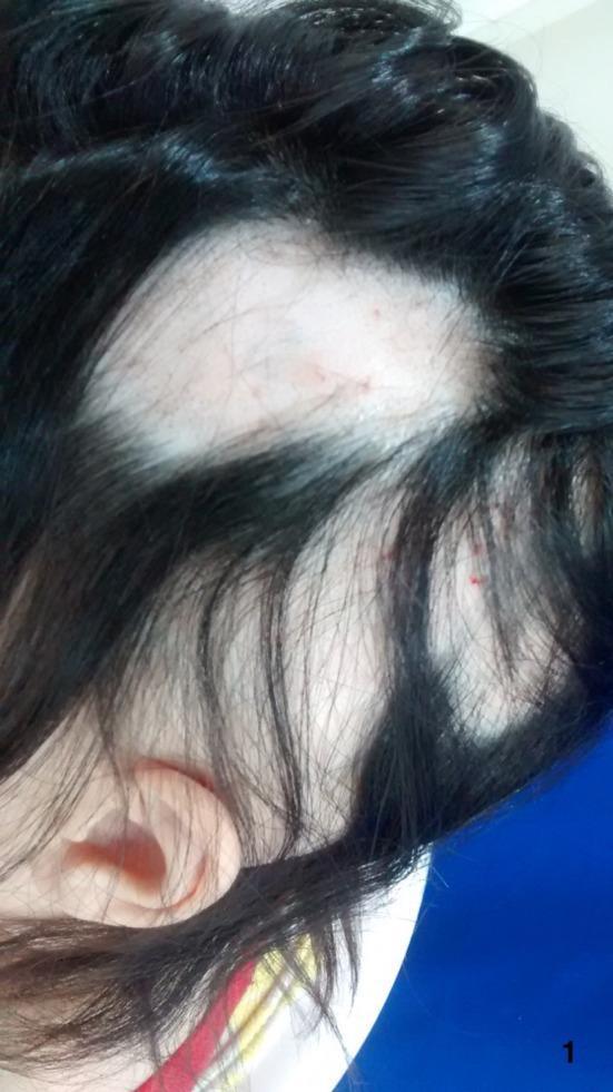
This clinical photograph shows a close-up, superior-lateral view of the scalp of a patient with severe alopecia areata. The image reveals multiple large, well-demarcated, non-scarring patches of alopecia (hair loss). The exposed scalp skin appears smooth, pale, and lacks follicular ostia plugging, which is characteristic of non-cicatricial alopecia. Within the bald patches, a few vellus hairs and some short, broken hairs are visible. Minor erythematous punctate lesions are present on the bare scalp surface, possibly related to recent intralesional treatments. The remaining hair is dark, terminal, and demonstrates normal density and length in unaffected regions. This visual presentation is typical of autoimmune hair loss, where the disease progresses from focal patches toward more extensive involvement such as alopecia totalis. The photograph serves as a clinical reference for diagnosing patchy hair loss and monitoring disease activity and treatment response in dermatological settings.
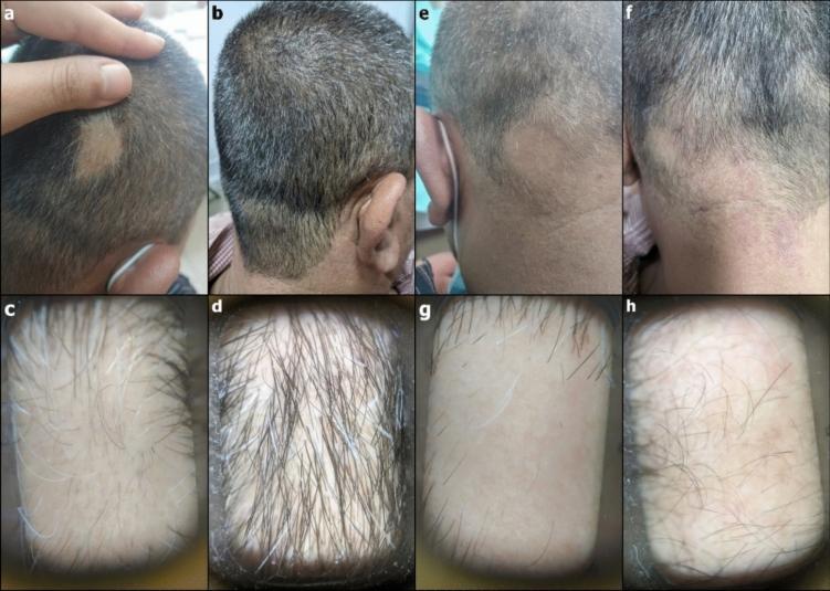
This composite educational graphic presents clinical and trichoscopic comparisons of alopecia areata treatment outcomes over a three-month period. The image is divided into two main comparison groups: a treated representative patch and an untreated control patch. Panels (a) and (b) show the clinical progression of a well-defined, smooth, non-scarring bald patch on the scalp. Panel (a) illustrates the baseline lesion, while panel (b) demonstrates significant hair regrowth and closure of the patch following topical steroid treatment. Corresponding trichoscopic images (c) and (d) provide high-magnification views; panel (c) shows the baseline with sparse terminal hairs and visible follicular openings, while (d) reveals a marked increase in terminal hair density and thickness. Panels (e) through (h) represent the control patch. Clinical images (e) and (f) show a persistent alopecic area with no significant clinical change. Trichoscopic views (g) and (h) confirm minimal hair regrowth, maintaining low hair density. This visual material serves to illustrate diagnostic trichoscopic features of alopecia areata and the quantifiable visual markers used to assess therapeutic efficacy in dermatology.
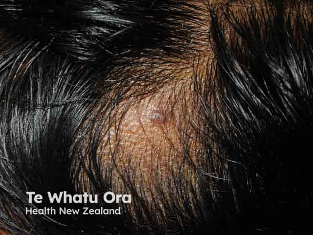
This image depicts a dermatologic clinical photograph of alopecia areata involving the lateral scalp. Imaging modality: clinical photography with macro close-up view of scalp skin and hair follicles, captured without illumination modifiers or contrast. The primary subject is a localized alopecic patch on the lateral scalp, best described as a round to oval, well-demarcated area of hair loss. The overlying skin appears normal in texture, with preserved epidermis and minor surface desquamation or perifollicular subtle erythema not readily observable in non-dermoscopic photography. Hair density within the patch is markedly reduced with visible expansion of follicular openings at the border and intact surrounding hair. The lesion is non-scarring, with no obvious scarring plaques or scarring tracts; there is no evidence of ornamentation or scaly scale. This presentation is characteristic of alopecia areata, a non-scarring autoimmune condition associated with peribulbar lymphocytic infiltrates histologically. Clinically significant for differential diagnosis against tinea capitis, traction alopecia, androgenetic alopecia, and lichen planopilaris. This image is valuable for education, tele-dermatology, and case repositories, enabling recognition of patchy alopecia patterns, conferral of prognosis, and assessment of treatment response to topical or intralesional corticosteroids and immunomodulatory therapies. Useful in training, patient counseling, and multi-disciplinary discussion.
Use of Steroids in Alopecia
1. Pathophysiologic Basis for Steroid Use
- Andrews' Diseases of the Skin, p. 869
- Dermatology 2-Volume Set 5e, p. 3328-3330
2. Topical Corticosteroids
- Ultrapotent (Class I) topical corticosteroids are preferred - e.g., clobetasol propionate 0.05% cream/lotion
- Can be applied under occlusion for acute or more severe patches
- Less reliable than intralesional injections but safer and non-invasive
- Dermatology 5e, p. 3383
3. Intralesional Corticosteroids (ILC)
| Parameter | Detail |
|---|---|
| Concentration | 2.5-5 mg/mL (up to 10 mg/mL) |
| Technique | Injected into mid-to-deep dermis, or superficial subcutaneous tissue |
| Volume per site | ~0.1 mL per injection site |
| Spacing | Injection sites 1 cm apart |
| Max dose per session | 10-20 mg in an average adult |
| Frequency | Every 3-4 weeks |
- Skin atrophy - more likely with large volumes and higher concentrations
- Retinal artery embolization (rare but serious) - risk increased with injection under high pressure or via small-bore syringe
- Subcutaneous fat atrophy (lipodystrophy)
- Andrews' Diseases of the Skin, p. 869
- Dermatology 5e, p. 3385
4. Systemic Corticosteroids
4a. Oral Daily Steroids
- Prednisolone: Initial dose 0.4-0.6 mg/kg/day
- Gradual taper over at least 12 weeks
- Possible transition to a pulse regimen after stabilization
- Used for rapidly progressive disease
4b. Pulse Systemic Steroids
| Regimen | Dose | Notes |
|---|---|---|
| Methylprednisolone pulse | 500 mg/day IV for 3 days, repeated monthly | In adults; 5 mg/kg twice daily over 3 days in children |
| Dexamethasone pulse | 0.1 mg/kg orally twice weekly | Oral pulse option |
| Prednisolone weekly pulse | 200 mg orally once weekly | Oral pulse option |
| Prednisolone monthly pulse | 5 mg/kg (up to 300 mg) once monthly | Used in children and adults |
-
60% of patients with widespread patchy AA responded
- ~50% with alopecia totalis had a good response
- ~25% with alopecia universalis responded
- Ophiasic AA did not respond
-
Disease duration 6 months or less
-
Age of onset younger than 10 years
-
Multifocal disease pattern
-
Goldman-Cecil Medicine, p. 1417
-
Andrews' Diseases of the Skin, p. 869
-
Dermatology 5e, p. 3390-3391
5. Steroid-Sparing Agents (used alongside or after steroids)
-
Cyclosporine: 3-5 mg/kg/day (first choice steroid-sparing agent)
-
Methotrexate: 15-20 mg weekly (adults); ~0.4 mg/kg/week in children
-
Mycophenolate mofetil, azathioprine, dapsone, sulfasalazine - limited data
-
Dermatology 5e, p. 3391
6. Summary: Steroid Selection by Disease Extent
| Disease Extent | Preferred Steroid Approach |
|---|---|
| Single/few patches | Intralesional triamcinolone acetonide 2.5-5 mg/mL every 3-4 weeks |
| Moderate patchy AA | Ultrapotent topical corticosteroid +/- intralesional TA |
| Rapidly progressive/widespread | Pulse prednisolone or methylprednisolone; consider daily prednisolone 0.4-0.6 mg/kg/day |
| Alopecia totalis (acute) | Pulse corticosteroids - partial response in ~50% |
| Alopecia universalis | Pulse corticosteroids - limited response (~25%); prefer JAK inhibitors |
| Ophiasis | Steroids are NOT effective; use contact immunotherapy or JAK inhibitors |
| Eyebrow AA | Intralesional TA is preferred |
7. Important Cautions
- Long-term systemic steroid use carries risks of HPA axis suppression, osteoporosis, weight gain, hypertension, and infection - risks must be carefully weighed against benefits
- Hair regrowth achieved with steroids may not be sustained after discontinuation; AA is a relapsing disease
- JAK inhibitors (baricitinib, ritlecitinib) are now FDA-approved for severe AA in adults and adolescents aged 12+ and are increasingly preferred over prolonged systemic steroids for extensive disease
- Spontaneous remission occurs in 34-50% of patients within 1 year, making anecdotal reports of treatment success difficult to interpret
- Andrews' Diseases of the Skin - Clinical Dermatology, Chapter 33
- Dermatology 2-Volume Set 5e, Chapter 69
- Goldman-Cecil Medicine, Chapter 409
- Fitzpatrick's Dermatology, Volumes 1 & 2
Effect on zinc on hair loss
"zinc" AND "hair loss" AND "alopecia"
PMID: 39440586
zinc deficiency alopecia acrodermatitis enteropathica skin hair loss
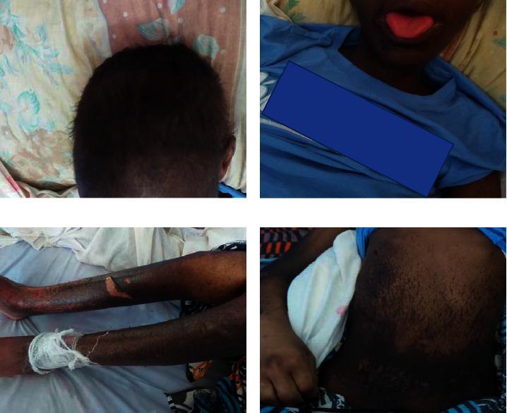
A composite of four clinical photographs demonstrating the systemic dermatological and mucosal manifestations of Acrodermatitis Enteropathica (AE) in a pediatric patient. The top-left panel shows the scalp with diffuse alopecia and dyspigmented, lightened hair. The top-right panel displays the orofacial region, highlighting beefy red glossitis of the tongue and perioral dermatitis. The bottom-left panel illustrates the lower extremities, featuring widespread hyperpigmented patches, skin erosions, and a prominent flaccid bulla on the shin, with one limb partially bandaged. The bottom-right panel depicts the abdomen, characterized by extensive hyperpigmentation and scattered erosive lesions. Collectively, these images illustrate the classic clinical triad of zinc deficiency: periorificial dermatitis, alopecia, and diarrhea-associated systemic skin breakdown. The presentation is essential for medical students and clinicians for the diagnostic recognition of nutritional deficiencies and zinc malabsorption syndromes.
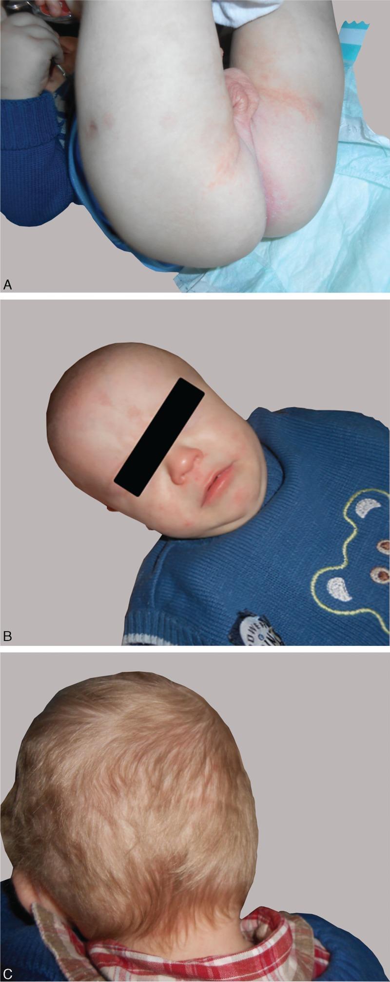
A composite of three clinical photographs (A, B, C) showing a pediatric patient following two months of treatment for acrodermatitis enteropathica. Panel A illustrates the diaper area (buttocks and perianal region), showing significant resolution of previous erythematous, scaly, and pustular lesions; the skin appears mostly clear with minimal residual faint pink patches within the gluteal folds. Panel B displays the infant's face, demonstrating remission of perioral and periorificial dermatitis, with a clear facial complexion and absence of acute inflammation around the mouth and nose. Panel C provides a posterior view of the scalp, showing successful hair regrowth following previous diffuse alopecia. The hair is light-colored, fine, and demonstrates uniform density without visible thinning or patchy hair loss. Collectively, these images document clinical improvement and therapeutic response to zinc supplementation, highlighting the remission of characteristic periorificial skin manifestations and hair loss associated with metabolic zinc deficiency.
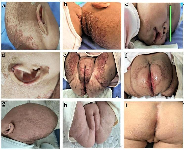
A series of nine clinical photographs (a-i) illustrating the cutaneous manifestations and therapeutic response of acrodermatitis enteropathica-like lesions in an infant. Images a-f depict the pre-treatment state, characterized by well-demarcated, erythematous, and erosive plaques with overlying scaly crusts and scabs. The distribution is classic for zinc deficiency, primarily affecting the acral and periorificial regions: occipital scalp (a, b), perioral area (c), auricle (d), and the perineal and perianal zones (e, f). Diffuse alopecia and sparse hair growth are also visible on the scalp. Images g-i demonstrate the clinical status after 6 days of oral zinc supplementation, showing rapid and significant improvement. The inflammatory erythema and erosions on the scalp (g) and anogenital region (h, i) have largely resolved, leaving behind smooth skin with minimal residual post-inflammatory changes. This comparison serves as a pedagogical tool for recognizing the diagnostic distribution of zinc deficiency-related dermatitis and its dramatic response to targeted replacement therapy.
Effect of Zinc on Hair Loss
1. Role of Zinc in Hair Follicle Biology
-
Enzymatic function: Zinc is a cofactor for over 300 enzymes, including RNA polymerases and alkaline phosphatase - both essential for the rapid cell division required in the hair matrix. Low alkaline phosphatase levels are in fact a useful marker of zinc deficiency.
-
Protein synthesis: Hair is primarily keratin; zinc supports keratinocyte proliferation and protein synthesis in the follicular bulb.
-
5-alpha-reductase inhibition: Zinc inhibits the enzyme 5-alpha-reductase (which converts testosterone to DHT), offering a potential role in limiting androgenetic alopecia.
-
Immune modulation: Zinc is required for normal T-cell function and immune homeostasis; deficiency impairs immune defense, which can dysregulate follicular cycling.
-
Antioxidant protection: Zinc has antioxidant properties that may protect the skin and follicles from UV-induced and oxidative damage.
-
Yamada's Textbook of Gastroenterology, 7e
-
Dermatology 2-Volume Set 5e, p. 218
2. Zinc Deficiency and Hair Loss
A. Clinical Presentation
Triad of zinc deficiency: Dermatitis + Diarrhea + Alopecia (complete triad seen in only ~20% of patients)
-
Periorificial and acral erosive/pustular/crusting dermatitis
-
Paronychia and onychodystrophy (nail changes)
-
Angular cheilitis and stomatitis
-
Blepharitis and conjunctivitis
-
Superimposed Candida and staphylococcal skin infections
-
Dermatology 5e, p. 220-222
-
Andrews' Diseases of the Skin, p. 2916
B. Inherited Zinc Deficiency: Acrodermatitis Enteropathica (AE)
| Feature | Detail |
|---|---|
| Inheritance | Autosomal recessive |
| Gene | SLC39A4 (encodes zinc transporter ZIP4) |
| Presentation | Onset within 1-2 weeks after weaning from breast milk |
| Hair findings | Diffuse alopecia of scalp, eyebrows, and eyelashes |
| Associated | Periorificial dermatitis, diarrhea, growth retardation, irritability |
| Treatment | Lifelong zinc supplementation: 3 mg/kg/day |
C. Acquired Zinc Deficiency
- Alcoholism (poor intake + increased urinary excretion)
- Malabsorption syndromes (IBD, celiac disease, short bowel)
- Parenteral nutrition without adequate zinc
- Anorexia nervosa
- Pregnancy (increased zinc requirement)
- Vegan/phytate-rich diets (phytate binds zinc, reducing bioavailability - endemic in Middle East/North Africa)
- Chronic renal failure, HIV disease, sickle cell disease
- Drugs: penicillamine, diuretics, antacids, quinolones (reduce zinc absorption)
- Andrews' Diseases of the Skin, p. 2906-2913
- Dermatology 5e, p. 224-230
D. Hair Regrowth After Zinc Supplementation
3. Zinc in Alopecia Areata
4. Zinc and Androgenetic Alopecia (AGA)
"Deficiencies or imbalances in specific vitamins and minerals, especially vitamin B, vitamin D, Fe, Se, and Zn are involved in the pathogenesis of AGA and may represent modifiable risk factors for treatment and prevention."
- Zinc deficiency has been associated with increased risk of AGA
- Zinc supplementation shows potential benefit in improving hair growth and preventing hair loss in AGA
- However, the evidence is not entirely consistent - some studies report no significant associations
- Zinc's mechanism in AGA includes 5-alpha-reductase inhibition (reducing DHT) and anti-inflammatory effects
5. Diagnosis of Zinc Deficiency in Hair Loss
| Test | Notes |
|---|---|
| Serum zinc | Low (<40 μg/mL confirms deficiency) |
| Serum alkaline phosphatase | Low level (zinc-dependent enzyme) - useful adjunctive marker; results may be available faster |
| Therapeutic trial | If serum zinc is borderline but clinical picture is suggestive, a zinc supplementation trial is reasonable |
6. Treatment: Zinc Supplementation Dosing
| Indication | Dose |
|---|---|
| Acquired zinc deficiency (adults) | Zinc sulfate 50 mg elemental zinc daily (220 mg zinc sulfate tablet) = 1-2 mg/kg/day |
| Acrodermatitis enteropathica (genetic) | 3 mg/kg/day - lifelong |
| Transient neonatal/acquired (correctable cause) | Supplement until replete; address underlying cause |
Caution: Excessive zinc supplementation causes copper deficiency (zinc upregulates metallothionein, which sequesters copper). Overzealous supplementation should be avoided. Fatal copper deficiency from zinc-based denture adhesive overuse has been reported.
- Andrews' Diseases of the Skin, p. 2931-2933
7. Summary
| Type of Hair Loss | Zinc's Role |
|---|---|
| Zinc deficiency-associated alopecia | Direct cause; reverses completely with supplementation |
| Acrodermatitis enteropathica | Classic triad includes alopecia; lifelong Zn replacement required |
| Alopecia areata | Low zinc levels found; immunomodulatory role; adjunctive supplementation considered |
| Androgenetic alopecia | Deficiency is a modifiable risk factor; 5-alpha-reductase inhibition is mechanism; evidence moderately supportive |
| Telogen effluvium | Zinc deficiency is one of many nutritional triggers |
- Andrews' Diseases of the Skin - Clinical Dermatology, Chapter 22
- Dermatology 2-Volume Set 5e, Chapter 51
- Goldman-Cecil Medicine, Chapter 2
- Yamada's Textbook of Gastroenterology, 7e
- Wang R et al. Micronutrients and Androgenetic Alopecia: A Systematic Review. Mol Nutr Food Res 2024 [PMID: 39440586]
Lupus erythemayous of hair
"lupus erythematosus" AND "alopecia"
discoid lupus erythematosus scalp scarring alopecia

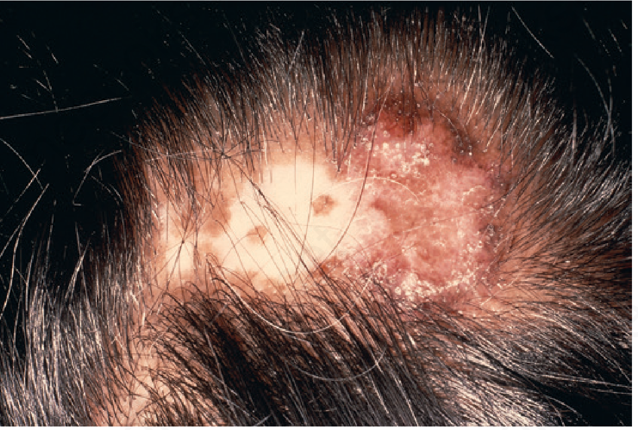
This is a clinical scalp photograph documenting discoid lupus erythematosus (DLE)–associated scarring alopecia. The image shows a localized left-parietal scalp patch with hair loss persisting in the focal area, featuring an atrophic center and a clearly defined, active-appearing border. Surrounding skin may display mild erythema and scaling; follicular openings are diminished or absent within the scar, consistent with cicatricial alopecia. Visual cues—well-demarcated plaque with scarring—help distinguish DLE from non-scarring alopecias such as androgenetic alopecia or alopecia areata in this clinical setting. This photograph supports dermatologic assessment, documentation of disease extent, and monitoring of progression or response to therapy. Clinically, DLE on the scalp necessitates biopsy confirmation when uncertain, photoprotection, and consideration of antimalarials or immunosuppressants depending on activity and systemic involvement risk. The image is suitable for education, differential diagnosis exercises, and research into scarring mechanisms, disease activity, and treatment outcomes. When correlated with histopathology and serologic data, it aids in comprehensive lupus evaluation and longitudinal patient management.
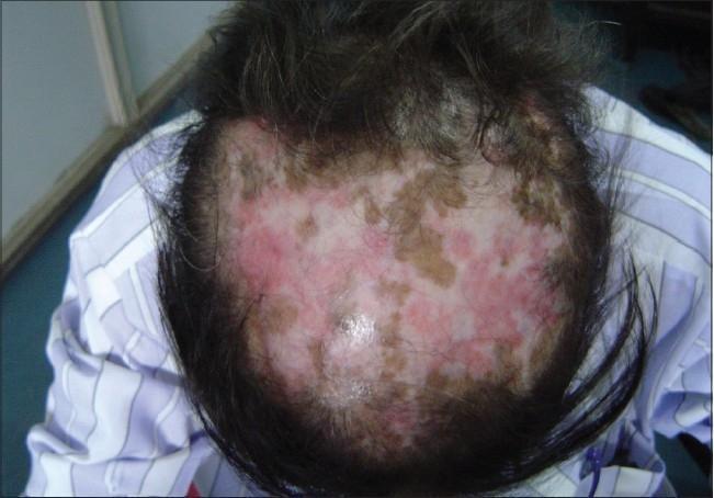
This clinical photograph displays a superior view of the scalp demonstrating classic features of Discoid Lupus Erythematosus (DLE). The image reveals extensive, patchy scarring alopecia (cicatricial alopecia) characterized by a significant reduction in hair density and visible destruction of hair follicles. The skin lesions exhibit a tri-colored, variegated appearance: erythematous (pink-red) areas indicating active inflammation, hyperpigmented (brown-tan) macules and patches, and focal regions of porcelain-white hypopigmentation suggestive of established dermal fibrosis and scarring. The distribution is irregular across the vertex and crown, showing the progressive nature of the follicular damage. This visual serves as a primary educational example of the cutaneous manifestations of chronic cutaneous lupus erythematosus on the scalp, emphasizing the transition from active inflammatory plaques to permanent, irreversible hair loss.
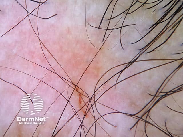
Clinical photography of the scalp depicts discoid lupus erythematosus-associated cicatricial (scarring) alopecia. The frontal to vertex scalp shows patchy erythematous and atrophic plaques with loss of normal follicular openings and hair shafts. Dermal sclerosis and peripilar telangiectasias are evident within affected areas, producing a smooth, scarred surface with diminished skin appendages. The remaining hair is sparse at the lesion margins, emphasizing the irregular scarring process. The vascular pattern includes visible dilated vessels, consistent with telangiectasia, and surrounding mild erythema. Overall morphology is characteristic of chronic inflammatory scarring alopecia with angular plaque margins and hypo- or hyperpigmented zones. The locoregional involvement on the scalp supports a diagnosis of discoid lupus erythematosus, a cutaneous manifestation of lupus erythematosus. The image captures essential features used for differential diagnosis from non-scarring alopecias and other cicatricial conditions, including lichen planopilaris and frontal fibrosing alopecia. The finding has clinical significance for patient management, as DLE may respond to antimalarial therapy and sun protection, and chronic inflammation risks progression to deeper dermal damage. This image is valuable for dermatology education, cicatricial alopecia research, and documented cutaneous lupus case repositories; it informs pattern recognition, staging, and treatment planning. Educational use includes pattern recognition, differential diagnosis, and treatment planning principles.
Lupus Erythematosus and Hair Loss
1. Classification of LE-Related Alopecia
| Type | Form of Lupus | Reversibility | Mechanism |
|---|---|---|---|
| Non-scarring diffuse alopecia ("lupus hairs") | SLE (systemic) | Reversible with disease control | Inflammatory telogen effluvium |
| Frontal "lupus hairs" | SLE during flares | Reversible | Fragile short hairs, frontal area |
| Scarring (cicatricial) alopecia | Discoid LE (DLE) | Permanent | Follicular destruction by chronic inflammation |
- Harrison's Principles of Internal Medicine 22e, p. 2078-2080
2. Non-Scarring Alopecia in Systemic Lupus Erythematosus (SLE)
Clinical Features
- Occurs during flares of systemic disease
- Can involve the entire scalp or preferentially the frontal scalp
- Characteristic appearance: multiple short, fragile, broken hairs along the frontal hairline - the so-called "lupus hairs" - which represent initial regrowth after shedding
- Pattern may resemble telogen effluvium - diffuse thinning without a defined patch
- Hair regrowth occurs as the systemic disease is controlled
Mechanism
Key Point
3. Discoid Lupus Erythematosus (DLE) - Scarring Alopecia
Clinical Features on Scalp
- Active (peripheral) zone: erythema, edema, scaling, follicular plugging (keratin plugs in follicular ostia), and perifollicular erythema
- Established (central) zone: atrophy, loss of follicular ostia (the hallmark of scarring alopecia), telangiectasia
- Dyspigmentation: Central hypopigmentation (white/ivory) surrounded by a rim of hyperpigmentation - a key diagnostic feature
- Permanent hair loss in the central atrophic zone as follicles are destroyed
Histopathology of DLE Scalp Biopsy
- Hyperkeratosis with follicular plugging
- Interface dermatitis (vacuolar degeneration at dermo-epidermal junction)
- Basement membrane zone thickening (PAS-positive)
- Dense perifollicular and periadnexal lymphocytic infiltrate
- Dermal mucin deposition (a helpful diagnostic clue)
- Late: loss of follicles replaced by fibrosis/scarring
- Andrews' Diseases of the Skin, p. 3663-3667
Epidemiology
- More common in African American women
- 5-10% of adults with DLE develop systemic lupus erythematosus (SLE), especially those with widespread lesions - monitoring is essential
4. Trichoscopy (Dermoscopy of the Scalp) in DLE
- Large yellow dots (dilated follicular infundibula with keratinous material)
- Red-white structureless areas (fibrosis)
- Absence of follicular ostia (in scarred zones)
- Arborizing telangiectasia
- Perifollicular white halos
5. Differential Diagnosis
| Condition | Key Distinguishing Feature |
|---|---|
| Lichen planopilaris | Violet perifollicular macules; no dyspigmentation; oral LP common |
| Frontal fibrosing alopecia | Band-like recession at hairline; eyebrow loss; postmenopausal women |
| Alopecia areata | Non-scarring; exclamation-point hairs; follicular ostia preserved |
| Tinea capitis | Fungal infection; can mimic inflammatory alopecia; culture + KOH positive |
| Folliculitis decalvans | Neutrophilic cicatricial; follicular pustules; tufted hairs |
| Secondary syphilis | "Moth-eaten" non-scarring alopecia; serology positive |
- Harrison's 22e, p. 2080; Andrews', p. 3657
6. Treatment
General Measures (All Forms of LE)
- Strict photoprotection - LE is photosensitive; UV light triggers both skin flares and hair loss
- Broad-spectrum high-SPF sunscreen daily
- Smoking cessation (smoking reduces antimalarial efficacy)
- Avoid trauma to scalp lesions; biopsies/scar revision can flare disease
Local (Topical/Intralesional) Treatment
| Treatment | Details |
|---|---|
| High-potency topical corticosteroids | First-line for localized DLE; apply under occlusion if needed; superpotent class I agents |
| Intralesional triamcinolone acetonide | Single most effective local treatment; 2.5-10 mg/mL injected every 4-6 weeks; max 40 mg per session |
| Topical calcineurin inhibitors (tacrolimus, pimecrolimus) | Second-line topical; useful on face/sensitive areas |
| Topical retinoids | Helpful for hypertrophic lesions |
Systemic Treatment
| Drug | Dose / Notes |
|---|---|
| Hydroxychloroquine (Plaquenil) | 5 mg/kg/day or less - First-line systemic; 2/3 of patients respond; if no response in 3 months, switch |
| Chloroquine | 250 mg/day; higher ocular toxicity; second choice antimalarial |
| Quinacrine | 100 mg/day; add to HCQ (no added retinal toxicity); causes yellowish pigmentation |
| Acitretin (retinoid) | Similar efficacy to HCQ in RCTs; more side effects |
| Methotrexate | Often next agent after antimalarials; for recalcitrant cases |
| Dapsone | Drug of choice in bullous SLE; also useful in some DLE/SCLE |
| Thalidomide | Effective but limited by teratogenicity and peripheral neuropathy |
| Oral prednisone | Reserved for acute flares only; not for long-term scarring alopecia management |
| Belimumab, MMF, azathioprine | For refractory/systemic disease |
- Andrews' Diseases of the Skin, p. 3853-3862
- Goldman-Cecil Medicine, p. 1469
7. Prognosis
- Non-scarring SLE alopecia: Completely reversible when SLE is controlled
- DLE scarring alopecia: Hair loss in fibrotic zones is permanent and irreversible - treatment only arrests progression
- Early diagnosis and aggressive treatment of active DLE on the scalp are essential to preserve hair-bearing scalp
- Fibrosis leaves a permanent "footprint" on elastic fiber staining (Verhoeff-van Gieson) even after inflammation resolves
- Harrison's Principles of Internal Medicine 22e, Chapter 61
- Andrews' Diseases of the Skin - Clinical Dermatology, Chapters 2, 9, and SLE treatment section
- Goldman-Cecil Medicine International Edition, Chapter 409
- Dermatology 2-Volume Set 5e (Dermatology 5e), Chapter 69