
Hi
Hi! How can I help you today?
Liver ? From basic to MBBS level
Reading File
Finding Sources
Finding Sources
Finding Sources
Reading File
Reading File
Finding Sources
Finding Sources
Finding Sources
Finding Sources
Reading File
Reading File
Searching Images
liver anatomy lobule hepatocyte sinusoid diagram
Finding Sources
Searching Images
liver gross anatomy lobes segments surface
Here is a comprehensive, MBBS-level guide to the Liver — from basic anatomy to physiology and clinical relevance.
🫀 THE LIVER — Basic to MBBS Level
1. INTRODUCTION & GROSS ANATOMY
The liver is the largest solid organ in the body, weighing ~1.5 kg (about 2% of body weight) in adults. It lies in the right hypochondrium and epigastrium, under the diaphragm.
Surfaces
| Surface | Relation |
|---|---|
| Diaphragmatic (superior/anterior) | Faces diaphragm; smooth and convex |
| Visceral (inferior) | Faces abdominal organs; bears impressions and the porta hepatis |
Lobes (Anatomical/Morphological)
- Right lobe — largest
- Left lobe — smaller
- Caudate lobe — on posterior visceral surface, between IVC and fissure for ligamentum venosum
- Quadrate lobe — on inferior visceral surface, between gallbladder fossa and fissure for ligamentum teres
The H-shaped arrangement of fossae and fissures on the visceral surface defines these four lobes.
Couinaud Segments (Functional/Surgical Anatomy)
The liver is divided into 8 functional segments (Couinaud's classification), each with its own:
- Portal venous branch
- Hepatic arterial branch
- Bile duct
- Hepatic venous drainage
| Segment | Location |
|---|---|
| I | Caudate lobe |
| II–III | Left lateral |
| IV | Left medial (quadrate) |
| V–VIII | Right lobe |
This classification allows segmental hepatic resections — removing one segment without affecting others.
2. BLOOD SUPPLY (Dual Blood Supply — Key Feature!)
The liver has a dual blood supply — unique among abdominal organs.
| Vessel | Volume | Oxygen contribution |
|---|---|---|
| Portal vein | ~1050 mL/min (75%) | 50–60% |
| Hepatic artery | ~300 mL/min (25%) | 40–50% |
| Total | ~1350 mL/min (27% of cardiac output) | — |
- Portal vein pressure: ~9 mmHg; Hepatic vein pressure: ~0 mmHg
- Low pressure gradient → very low resistance through sinusoids
- Blood drains via hepatic veins → IVC
Portal Hypertension
When sinusoidal resistance increases (e.g., cirrhosis), portal pressure rises → portal hypertension → varices, ascites, splenomegaly.
3. MICROSTRUCTURE / HISTOLOGY
The Classic Liver Lobule
The functional unit of the liver is the liver lobule — a hexagonal prism:
- Central vein at the center
- Portal triads (hepatic artery, portal vein, bile duct) at each of the 6 corners
- Hepatocyte plates radiate outward like spokes of a wheel, 2 cells thick
- Between plates: hepatic sinusoids carry blood from portal triads → central vein
The human liver contains 50,000–100,000 lobules.

Hepatic Acinus (Functional Unit by Rappaport)
- Based on blood flow direction (portal triad → central vein)
- Zone 1 (periportal) — richest O₂ and nutrients → gluconeogenesis, urea synthesis, β-oxidation
- Zone 2 (midzone)
- Zone 3 (pericentral/centrilobular) — lowest O₂ → glycolysis, lipogenesis, CYP450 metabolism; most vulnerable to ischemia and drugs
Cells of the Liver
| Cell | Function |
|---|---|
| Hepatocytes | ~80% of volume; metabolic workhorses |
| Kupffer cells | Resident macrophages lining sinusoids; phagocytose bacteria, old RBCs, foreign material |
| Stellate cells (Ito cells) | Store vitamin A; when activated → produce collagen → fibrosis/cirrhosis |
| Sinusoidal endothelial cells (LSECs) | Fenestrated (pores ~1 µm); allow free exchange of plasma proteins |
| Bile duct epithelial cells (cholangiocytes) | Line bile ducts; add HCO₃⁻ to bile |
Space of Disse
- Narrow perisinusoidal space between endothelial cells and hepatocytes
- Connects to lymphatics → ~50% of all body lymph originates in the liver
- Fluid transudation here → ascites when hepatic venous pressure rises
Bile Canaliculi
- Formed by apical membranes of adjacent hepatocytes
- Run in opposite direction to blood flow (centrifugal)
- Bile flows: canaliculi → canals of Hering → bile ductules → bile ducts → hepatic duct → common bile duct
4. BILE SECRETION
The liver secretes 600–1000 mL of bile per day.
Functions of Bile
- Fat digestion — bile acids emulsify fats and enable lipase action; form micelles for fat-soluble vitamin absorption (A, D, E, K)
- Excretion — excretes bilirubin (hemoglobin breakdown product), cholesterol, drugs, toxins
Composition of Bile
| Component | Role |
|---|---|
| Bile salts | Emulsification of fats (primary: cholic & chenodeoxycholic acid; secondary: deoxycholic & lithocholic) |
| Bilirubin | Excretory product of heme metabolism |
| Cholesterol | Excreted; gallstones form when excess |
| Lecithin (phospholipids) | Solubilizes cholesterol in bile |
| Water + electrolytes | Vehicle |
Bile Secretion in Two Stages
- Hepatocytes secrete primary bile (bile acids, cholesterol, bilirubin) into canaliculi
- Ductular epithelial cells add Na⁺ + HCO₃⁻ (stimulated by secretin)
Gallbladder
- Stores and concentrates bile 5–20× by absorbing water/electrolytes
- Maximum capacity: 30–60 mL (holds ~12 hours of secretion)
- Bile ejection triggered by CCK (cholecystokinin)
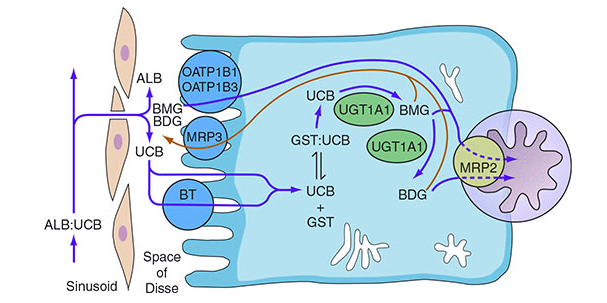
5. BILIRUBIN METABOLISM (High-Yield!)
| Step | Details |
|---|---|
| Production | Heme (from old RBCs) → biliverdin → unconjugated bilirubin (UCB) = indirect bilirubin |
| Transport | UCB bound to albumin in blood (insoluble in water) |
| Hepatic uptake | Via OATP1B1/1B3 transporters |
| Conjugation | UCB + glucuronic acid → conjugated bilirubin (CB) = direct bilirubin, by UGT1A1 enzyme |
| Excretion into bile | Via MRP2 transporter into bile canaliculus |
| Intestinal fate | CB → urobilinogen by gut bacteria → urobilin (yellow in urine) or stercobilin (brown in stool) |
Jaundice (Icterus)
Normal bilirubin: < 1 mg/dL | Jaundice visible: > 2–3 mg/dL
| Type | Cause | Predominant bilirubin |
|---|---|---|
| Pre-hepatic (hemolytic) | Excess RBC breakdown (malaria, hemolytic anemia) | Indirect ↑↑ |
| Hepatic (hepatocellular) | Hepatitis, cirrhosis — uptake/conjugation/excretion all impaired | Both ↑ |
| Post-hepatic (obstructive) | Gallstones, cholangiocarcinoma, pancreatic CA | Direct ↑↑ |
6. METABOLIC FUNCTIONS OF THE LIVER
The liver is the central metabolic organ of the body.
Carbohydrate Metabolism
- Glycogenesis — converts glucose → glycogen (stores ~100 g)
- Glycogenolysis — breaks down glycogen → glucose (fasting)
- Gluconeogenesis — synthesizes glucose from lactate, amino acids, glycerol
- Maintains blood glucose between meals
Protein Metabolism
- Synthesis of plasma proteins: albumin, fibrinogen, clotting factors (I, II, V, VII, IX, X, XI), transferrin, haptoglobin, complement proteins
- Transamination & deamination of amino acids
- Urea cycle — detoxifies ammonia → urea (excreted in urine)
Tip: Liver disease → low albumin (edema), low clotting factors (bleeding), high ammonia (encephalopathy)
Fat Metabolism
- β-oxidation of fatty acids for energy
- Ketogenesis — produces ketone bodies during starvation
- Synthesis of cholesterol, triglycerides, VLDLs, HDLs
- Lipogenesis from excess carbohydrates
Vitamin and Mineral Storage
| Stored | Amount |
|---|---|
| Vitamin A | ~10-month supply |
| Vitamin D | ~3–4 months |
| Vitamin B₁₂ | 1–3 years |
| Iron (as ferritin) | Significant stores |
| Copper | Released by ceruloplasmin |
7. DETOXIFICATION & DRUG METABOLISM
- CYP450 enzymes (mainly in Zone 3) → phase I metabolism (oxidation, reduction, hydroxylation)
- Conjugation (phase II) → glucuronidation, sulfation, acetylation → water-soluble products for excretion
- First-pass metabolism — orally absorbed drugs pass through portal circulation → liver before reaching systemic circulation
- Detoxifies ammonia, hormones (estrogen, aldosterone), drugs, alcohol, toxins
8. OTHER IMPORTANT FUNCTIONS
| Function | Details |
|---|---|
| Blood reservoir | Stores ~450 mL blood (~10% of total volume); can release in hypovolemia |
| Immune function | Kupffer cells phagocytose gut bacteria from portal blood |
| Hormonal | Produces IGF-1, thrombopoietin, angiotensinogen, hepcidin |
| Fetal hematopoiesis | Major site of blood cell production in fetus (weeks 6–30) |
| Vitamin K activation | Needed for synthesis of factors II, VII, IX, X (coagulation) |
| Regeneration | Can regenerate after 70% removal; driven by HGF and TNF-α |
9. LYMPHATIC DRAINAGE & NERVE SUPPLY
- Lymphatics: Drain to hepatic nodes → celiac nodes → cisterna chyli. Liver produces ~50% of total body lymph.
- Nerve supply: Sympathetic (T7–T10), Parasympathetic (vagus), Phrenic nerve (pain referred to right shoulder)
10. CLINICAL CORRELATIONS (MBBS Must-Know)
| Condition | Key Mechanism |
|---|---|
| Cirrhosis | Repeated injury → stellate cell activation → fibrosis → nodular regeneration; causes portal hypertension, ascites, hepatic encephalopathy |
| Hepatic encephalopathy | Liver fails to convert NH₃ → urea; ammonia crosses BBB |
| Ascites | ↓ albumin (↓ oncotic pressure) + portal hypertension + ↑ aldosterone |
| Portal hypertension | ↑ resistance in sinusoids → esophageal varices, splenomegaly, caput medusae |
| Hepatitis | Inflammation → ↑ ALT/AST (hepatocellular enzymes) |
| Cholestasis | ↑ ALP, GGT; obstructive → ↑ direct bilirubin, pale stools, dark urine |
| NAFLD/NASH | Fat accumulation in hepatocytes (steatosis) → inflammation → fibrosis |
| Wilson's disease | Copper accumulation (ceruloplasmin mutation) |
| Hemochromatosis | Iron overload → cirrhosis, diabetes, bronze skin |
11. LIVER FUNCTION TESTS (LFTs) — Quick Reference
| Test | Normal | Significance |
|---|---|---|
| ALT (SGPT) | 7–56 U/L | Hepatocellular damage (specific to liver) |
| AST (SGOT) | 10–40 U/L | Hepatocellular damage (also heart, muscle) |
| ALP | 40–150 U/L | Cholestasis, bone disease |
| GGT | 8–61 U/L | Cholestasis, alcohol use |
| Albumin | 3.5–5.0 g/dL | Synthetic function |
| PT/INR | PT 11–13s | Synthetic function (clotting factors) |
| Total bilirubin | < 1 mg/dL | Excretory function |
| Direct bilirubin | < 0.3 mg/dL | Conjugated (obstructive) |
Summary Mind-Map
LIVER
├── Anatomy: Right > Left > Caudate > Quadrate; Couinaud 8 segments
├── Blood Supply: Portal vein (75%) + Hepatic artery (25%)
├── Histology: Lobule → Hepatocyte plates → Sinusoids → Central vein
│              Acinus Zones 1-2-3 (periportal to centrilobular)
│              Cells: Hepatocytes, Kupffer, Stellate, LSEC
├── Bile: 600-1000 mL/day → fat digestion + excretion
├── Bilirubin: Heme → UCB → CB (UGT1A1) → bile → stercobilin/urobilin
├── Metabolism: Glucose (glycogen), Proteins (albumin, urea), Fats (lipoproteins)
├── Detox: CYP450 (Zone 3), Phase I & II, first-pass
└── Clinical: Cirrhosis, Jaundice, Portal HTN, Hepatic encephalopathy
Sources: Guyton & Hall Textbook of Medical Physiology | Medical Physiology (Boron & Boulpaep) | Yamada's Textbook of Gastroenterology | Harrison's Principles of Internal MedicineThat covers the full scope of the liver from basics to MBBS level. Here's a quick orientation of what's included:
- Gross anatomy — lobes, surfaces, Couinaud's 8 segments
- Blood supply — dual supply (portal vein + hepatic artery), flow volumes, portal hypertension
- Histology — liver lobule, hepatic acinus zones, all cell types, Space of Disse, bile canaliculi
- Bile secretion — composition, two-stage secretion, gallbladder concentration
- Bilirubin metabolism — full pathway with types of jaundice
- Metabolic functions — carbohydrates, proteins, fats, vitamins/minerals
- Detoxification — CYP450, phase I & II, first-pass metabolism
- Other functions — blood reservoir, immunity, hematopoiesis, regeneration
- Clinical correlates — cirrhosis, ascites, portal hypertension, hepatic encephalopathy, NAFLD, Wilson's, hemochromatosis
- LFTs — all key tests with normal values
Let me know if you want any topic expanded — e.g., portal hypertension in detail, bile acid synthesis, hepatic drug metabolism, or liver pathology (hepatitis, cirrhosis histology, etc.).
This is a shared conversation. Sign in to Orris to start your own chat.