
Explain the pathophysiology of AVNRT
Reading File
Finding Sources
Finding Sources
Searching PubMed
"atrioventricular nodal reentry tachycardia"[MeSH Terms]
Reading File
Reading File
Reading File
Reading File
Reading File
Searching Images
AV nodal reentry tachycardia fast and slow pathway diagram
Pathophysiology of AVNRT
AVNRT is a re-entrant supraventricular tachycardia. The circuit sits in and around the AV node and uses two functionally different conduction routes, called dual AV nodal pathways.
1. Substrate: dual AV nodal physiology
Dual pathways were proposed in the 1950s to explain AVNRT. The AV node is thought to split functionally into two pathways:
-
Fast pathway: conducts quickly and has a longer refractory period.
-
Slow pathway: conducts slowly and has a shorter refractory period (it repolarizes sooner).
-
Fuster and Hurst's The Heart, 15th Ed, p. 1100
Anatomically, the slow pathway is usually located anterior to the coronary sinus ostium. This is why slow pathway ablation there works without damaging normal AV conduction through the fast pathway. The fast pathway lies more superiorly, near the apex of Koch's triangle (my addition from general knowledge).
2. Three conditions needed for re-entry
Braunwald describes these as the key determinants:
- Two conducting pathways that are separated longitudinally but connected at each end.
- Different refractory periods in the two pathways, so a premature beat can be blocked in one but not the other.
- A circuit time longer than the longest refractory period in the circuit.
- Braunwald's Heart Disease, Ch. 62 (Anatomic Reentry), Fig. 62.18
3. Initiation (typical slow-fast AVNRT)
- A premature atrial beat arrives while the fast pathway is still refractory.
- The impulse is blocked in the fast pathway and conducts down the slow pathway, which has already recovered. This causes a long PR interval on the premature beat.
- The impulse reaches the His bundle and activates the ventricles through the His-Purkinje system.
- By now the fast pathway has had time to recover. The impulse travels retrogradely up the fast pathway to the atrium.
- If the circuit time is longer than the slow pathway's refractory period, the slow pathway is re-excited antegradely. The loop then repeats.
- Fuster and Hurst's The Heart, Table 35-1 legend, and Braunwald's Heart Disease, Ch. 62
The circuit is therefore: antegrade down the slow pathway, then retrograde up the fast pathway.
4. Why the ECG looks the way it does
-
Retrograde atrial activation via the fast pathway happens at the same time as antegrade ventricular activation. The atria and ventricles are activated almost simultaneously.
-
In about 50% of cases the P wave is buried inside the QRS.
-
In about 45% of cases the P wave falls at the end of the QRS. This gives a pseudo-R' in V1 and a pseudo-S wave in the inferior leads.
-
Occasionally there is 2:1 conduction to the ventricles. The block is usually within or below the His bundle, which shows the atria and ventricles are not required parts of the circuit.
-
Braunwald's Heart Disease, Ch. 65, Typical AVNRT
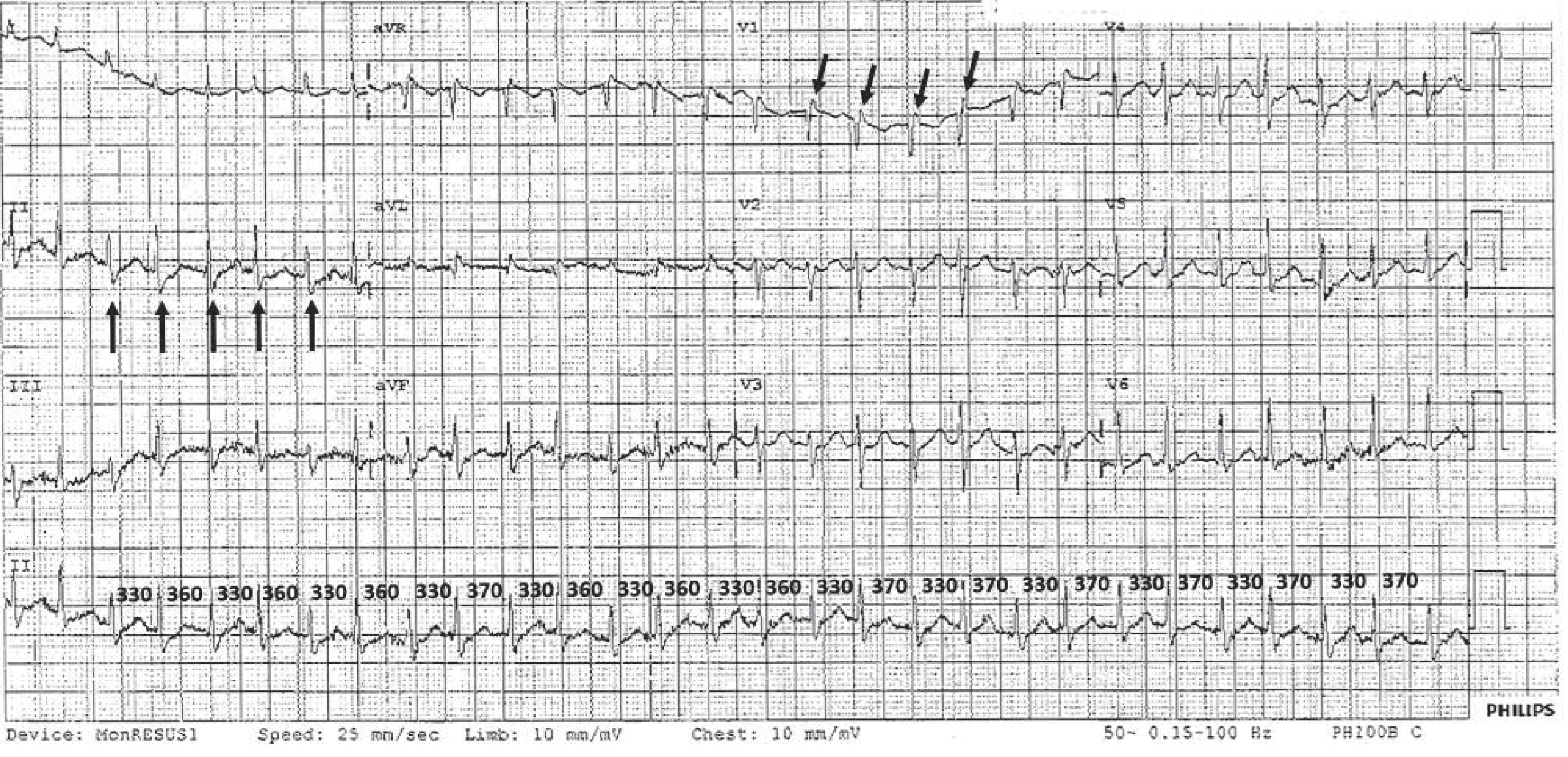
5. Types
- Typical (slow-fast): 85-90% of cases (Fuster and Hurst's The Heart, p. 1100).
- Atypical: the remaining 10-15% (same source). There are two subtypes:
- Fast-slow: antegrade conduction down the fast pathway and retrograde conduction up the slow pathway.
- Slow-slow: antegrade conduction down one slow pathway and retrograde conduction up a second slow pathway. Braunwald describes this as a rare variety.
- In both, the retrograde P wave falls later, so the RP interval is longer than in typical AVNRT.
6. Clinical correlates of the mechanism
- The tachycardia is paroxysmal, with abrupt onset and termination. It is typically triggered by premature atrial beats, and sometimes premature ventricular beats (my addition).
- Because the AV node is part of the circuit, it ends with AV nodal blockade (vagal maneuvers, adenosine, and so on). This is general knowledge and not taken from the sections I read.
- Ablating the slow pathway cures the arrhythmia. The older approach of ablating the fast pathway carried a small but definite risk of AV block.
Recent literature check
A PubMed search of the last 2 years of systematic reviews and meta-analyses found nothing that contradicts these mechanisms. The results were about ablation catheter types (PMID 38212906), supraventricular arrhythmia genetics (PMID 38804128), and supraventricular arrhythmias in arrhythmogenic cardiomyopathy and Brugada syndrome (PMID 42597178).
This is a shared conversation. Sign in to Orris to start your own chat.