

What is happened to it
cellulitis lower leg redness swelling erythema
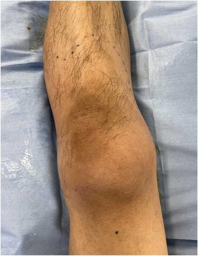
This clinical photograph shows the anterior view of a patient's left leg, including the distal thigh, knee, and proximal lower leg, resting on a blue medical drape. The image demonstrates a primary finding of diffuse, mild erythema (redness) extending from the thigh down to the shin. The erythema is most concentrated and visible over the patellar region (knee), presenting as a dusky, pinkish-tan discoloration. There is no visible evidence of joint effusion, gross swelling, bullae, or skin necrosis at this stage. Tiny black markings are visible on the skin, likely surgical or clinical landmarks used to track the progression of the redness. This visual finding is significant in the context of early-stage necrotizing fasciitis or cellulitis, particularly highlighting cases where clinical pain and tenderness out of proportion to visible skin changes (erythema) are key diagnostic indicators of deep tissue infection.
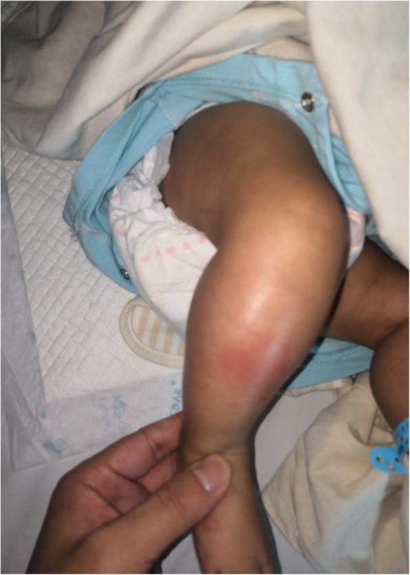
This clinical photograph displays the right lower leg of a 2-month-old infant in a hospital setting. The image demonstrates significant localized pathology characterized by marked edema (swelling) and overlying erythema (redness) on the anteromedial aspect of the proximal to mid-tibia region. The skin appears tense and shiny due to the underlying inflammatory process. A clinician's hand is visible stabilizing the ankle, providing scale and indicating a physical examination. The patient is situated on a medical underpad, wearing a diaper and hospital-issued clothing, which establishes the pediatric clinical context. This visual presentation is highly suggestive of acute inflammatory or infectious processes of the soft tissue or underlying bone, such as cellulitis or acute osteomyelitis. The educational focus is on the pediatric physical manifestation of musculoskeletal infection and the visual cues of inflammation—rubor (redness) and tumor (swelling)—in an infant.
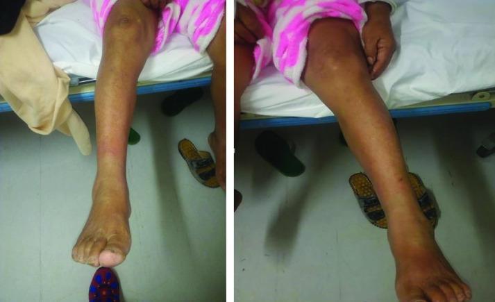
This clinical photograph displays a side-by-side comparison of a patient's lower extremities, illustrating unilateral pathology. The left leg (viewer's right) exhibits significant clinical abnormalities compared to the right leg. Key findings include diffuse erythema (redness) that is most prominent from the distal third of the leg extending down to the dorsal surface of the foot and ankle. There is also evident soft tissue swelling and edema, indicated by the increased girth and loss of definition in the ankle landmarks of the affected limb. The skin appears tense, though no discrete ulcerations or vesiculation are clearly visible in this view. This visual presentation is characteristic of inflammatory or infectious conditions such as cellulitis, deep vein thrombosis (DVT), or localized dependent edema. The image serves as an educational tool for medical students and clinicians to practice visual assessment of inflammatory signs and unilateral limb swelling in a clinical setting.
What Is Happening to This Leg
What You Can See in the Image
| Finding | Significance |
|---|---|
| Bright red, diffuse erythema from mid-calf to ankle | Active inflammation/infection |
| Taut, shiny, swollen skin | Significant edema from infection/inflammation |
| Ill-defined borders of redness | Classic for cellulitis (vs. sharp borders of erysipelas) |
| Brown/dusky discoloration around ankle and foot | Chronic venous stasis - longstanding poor circulation |
| Small vesicles/droplets near lower shin | Skin breakdown, possible early blister formation |
| Yellowed thickened heel skin | Chronic skin changes from poor perfusion |
Likely Diagnosis: Cellulitis
"Cellulitis is a suppurative inflammation involving the subcutaneous tissue... On the leg, tinea pedis is a common portal of entry. Mild local erythema and tenderness, malaise, fever and chills may be present... The erythema rapidly becomes intense and spreads. The area may be infiltrated and pit on pressure."
Why This Leg Was Vulnerable
Important Warning Signs to Watch For
- Red streaks running upward from the leg (lymphangitis - sign of spreading infection)
- Fever, chills, or feeling very unwell
- The redness continues to spread despite treatment
- Dark or black patches forming (possible necrotizing fasciitis - a surgical emergency)
- No improvement after 24-48 hours of antibiotics
Treatment (requires a doctor)
- Outpatient antibiotics (if no systemic toxicity): Dicloxacillin or cephalexin for 5 days is standard first-line treatment per Andrews' Diseases of the Skin
- If MRSA is suspected (e.g., previous MRSA, no response to first-line): trimethoprim-sulfamethoxazole or clindamycin
- Leg elevation to reduce swelling
- Treating underlying venous insufficiency (compression stockings once infection resolves)
- Hospital admission with IV antibiotics if fever is present, the patient is diabetic/immunocompromised, or the infection is rapidly spreading
This is a medical condition that needs prompt attention from a doctor. Please do not delay seeking care, as cellulitis can spread rapidly and become life-threatening if untreated.