Now compiling the comprehensive integrated response. This is a large but highly organized answer:
🧬 INTEGRATED METABOLIC CYCLES - Master Reference for NEET-PG / USMLE / INICET
PART 1: CARBOHYDRATE METABOLISM
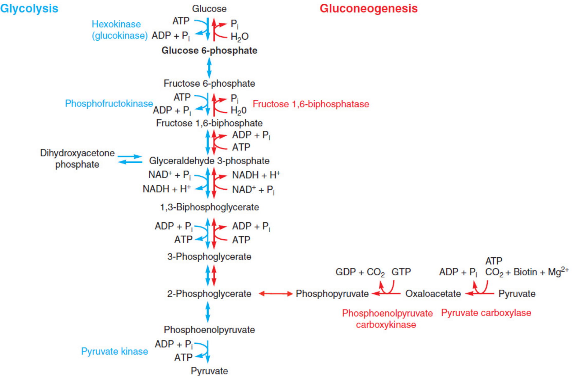
1.1 GLYCOLYSIS
Location: Cytoplasm | Oxygen requirement: Anaerobic (can proceed without O₂)
Overall equation:
Glucose + 2 NAD⁺ + 2 ADP + 2Pi → 2 Pyruvate + 2 NADH + 2H⁺ + 2 ATP
Glycolysis (blue, left) vs. Gluconeogenesis (red, right) - note the 3 irreversible bypass points
Steps and Key Enzymes
| Step | Substrate → Product | Enzyme | Cofactor | Notes |
|---|
| 1 | Glucose → G-6-P | Hexokinase (tissues) / Glucokinase (liver) | Mg²⁺, ATP | Hexokinase: low Km, inhibited by G-6-P; Glucokinase: high Km, induced by insulin |
| 3 | F-6-P → F-1,6-BP | Phosphofructokinase-1 (PFK-1) | Mg²⁺, ATP | Rate-limiting step of glycolysis |
| 5 | F-1,6-BP → DHAP + G-3-P | Aldolase | - | Splits 6C into two 3C |
| 6 | G-3-P ⇌ DHAP | Triose phosphate isomerase | - | - |
| 7 | G-3-P → 1,3-BPG | Glyceraldehyde-3-phosphate dehydrogenase | NAD⁺ | Inhibited by arsenate |
| 8 | 1,3-BPG → 3-PG | Phosphoglycerate kinase | ADP | Substrate-level phosphorylation |
| 10 | PEP → Pyruvate | Pyruvate Kinase | K⁺, Mg²⁺ | Irreversible; 3rd rate-limiting step |
Three Irreversible (Rate-Limiting) Steps - HIGH YIELD
- Hexokinase/Glucokinase - Glucose → G-6-P
- PFK-1 - F-6-P → F-1,6-BP (THE key regulatory step)
- Pyruvate Kinase - PEP → Pyruvate
Regulation of PFK-1 (Master Regulator)
| Activators | Inhibitors |
|---|
| AMP, ADP (low energy signal) | ATP (high energy) |
| Fructose 2,6-bisphosphate (insulin ↑) | Citrate (TCA running well) |
| Pi | Glucagon → ↓ F-2,6-BP → inhibits PFK-1 |
MNEMONIC - PFK-1 activators: "FAME" - F-2,6-BP, AMP, Mg²⁺, Energy low states
Net ATP Yield
- Aerobic glycolysis: 2 ATP net (+ 2 NADH fed to ETC → 5 ATP more)
- Anaerobic: Only 2 ATP net
Clinical Correlations
🔴 Pyruvate Kinase Deficiency (PKD)
- Most common hereditary non-spherocytic hemolytic anemia (after G6PD deficiency)
- RBCs cannot undergo aerobic metabolism → rely entirely on glycolysis
- PK deficiency → ↓ ATP in RBCs → rigid membrane → hemolysis
- Lab: ↑ 2,3-BPG (accumulates), Heinz bodies absent, Coombs negative
- INICET/NEET-PG: "2,3-BPG elevated - right shift of O₂-Hb dissociation curve"
🔴 Arsenic Poisoning
- Arsenate replaces phosphate in glyceraldehyde-3-phosphate dehydrogenase step
- 1,3-BPG → 1-arseno-3-PG (bypasses substrate-level phosphorylation at step 7)
- Net ATP = 0 from glycolysis; patient presents with Mees' lines, GI symptoms, polyneuropathy
🔴 Fructose Intolerance vs Fructosuria
- Fructokinase deficiency → Essential fructosuria (benign, fructose in urine)
- Aldolase B deficiency → Hereditary Fructose Intolerance (HFI): fructose-1-P accumulates → hypoglycemia, liver failure, renal tubular acidosis
- HFI patients develop aversion to sweet foods (protective)
🔴 Warburg Effect (Oncology)
- Cancer cells preferentially use aerobic glycolysis even in presence of O₂
- PET scan exploits ↑ glucose uptake by tumor cells (↑ ¹⁸F-FDG uptake)
- Basis of FDG-PET in tumor staging
🔴 Lactate Acidosis
- Type A: Tissue hypoxia (shock, cardiac arrest) - pyruvate → lactate, NAD⁺ regenerated
- Type B: Drugs (metformin, linezolid), liver failure
- Lactate level: proportional to tissue oxygen deficit - used to gauge shock severity
Metabolic Disorders Table - Glycolysis
| Disorder | Deficient Enzyme | Accumulated Metabolite | Key Features |
|---|
| Pyruvate Kinase Deficiency | Pyruvate Kinase | Pyruvate, 2,3-BPG | Hemolytic anemia, RBC, AR inheritance |
| Essential Fructosuria | Fructokinase | Fructose | Benign, incidental finding |
| Hereditary Fructose Intolerance | Aldolase B | Fructose-1-P | Hypoglycemia, liver failure, vomiting after sweets |
| Galactosemia (Classic) | Galactose-1-P uridyltransferase | Gal-1-P, Galactitol | Cataracts, liver failure, E. coli sepsis in neonates |
| Galactokinase Deficiency | Galactokinase | Galactitol | Cataracts only, milder |
1.2 GLUCONEOGENESIS
Location: Liver (primary, 90%) and renal cortex (10%) | Occurs when: Fasting, prolonged exercise, high protein diet
Substrates (glucogenic):
- Lactate (Cori cycle) - from muscle
- Alanine (Alanine cycle) - from muscle
- Glycerol - from fat hydrolysis
- Oxaloacetate - from TCA intermediates
- Glucogenic amino acids (all except Leu, Lys which are purely ketogenic)
- Propionyl-CoA (from odd-chain fatty acid oxidation)
Unique Bypass Enzymes of Gluconeogenesis
| Bypassed Glycolytic Step | Gluconeogenic Enzyme | Location | Cofactor |
|---|
| Pyruvate kinase | Pyruvate Carboxylase (Pyr → OAA) then PEPCK (OAA → PEP) | Mito / Cyto | Biotin (PC), GTP (PEPCK) |
| PFK-1 | Fructose-1,6-bisphosphatase (F-1,6-BP → F-6-P) | Cytoplasm | Mg²⁺ |
| Hexokinase | Glucose-6-phosphatase (G-6-P → Glucose) | ER lumen | - |
HIGH YIELD: Glucose-6-phosphatase is present only in liver, kidney, intestine - that is why only these organs can export free glucose. Muscle CANNOT release glucose into blood!
Regulation of Gluconeogenesis
| Stimulated by | Inhibited by |
|---|
| Glucagon (↑ cAMP) | Insulin |
| Cortisol | AMP (inhibits F-1,6-BPase) |
| Fasting | Fructose-2,6-bisphosphate |
| High NADH:NAD⁺ ratio (alcoholism) | - |
Clinical Correlations - Gluconeogenesis
🔴 Alcohol and Hypoglycemia
- Ethanol → Acetaldehyde → Acetate; both steps use NAD⁺ → massive ↑ NADH
- High NADH:NAD⁺ → OAA converted to malate (not available for gluconeogenesis)
- Pyruvate → Lactate (not pyruvate for gluconeogenesis)
- Result: Hypoglycemia + Lactic acidosis in alcoholics (especially after fasting)
🔴 Biotin Deficiency
- Pyruvate Carboxylase (needs biotin) impaired → gluconeogenesis fails
- Presents with: alopecia, dermatitis, neurological symptoms
- Also: Pyruvate cannot enter TCA → lactic acidosis
- Caused by: eating raw egg whites (avidin binds biotin) OR TPN without biotin
🔴 Von Gierke Disease (Type I GSD) - USMLE FAVORITE
- Glucose-6-phosphatase deficiency
- Glycogen and fat accumulate in liver and kidney
- Classic: Fasting hypoglycemia, lactic acidosis, hyperuricemia (gout), hyperlipidemia, hepatomegaly
- Doll-like face, protuberant abdomen
- NO response to glucagon (can't release glucose from G-6-P)
🔴 Cori Cycle
- Muscle: Glucose → Lactate (anaerobic glycolysis)
- Lactate travels to liver via blood
- Liver: Lactate → Pyruvate → Glucose (gluconeogenesis)
- Glucose returns to muscle
- Net: transfers energy deficit from muscle to liver
1.3 TCA CYCLE (Krebs / Citric Acid Cycle)
Location: Mitochondrial matrix | Occurs when: Aerobic conditions
Purpose: Oxidize Acetyl-CoA → CO₂ + NADH + FADH₂ + GTP
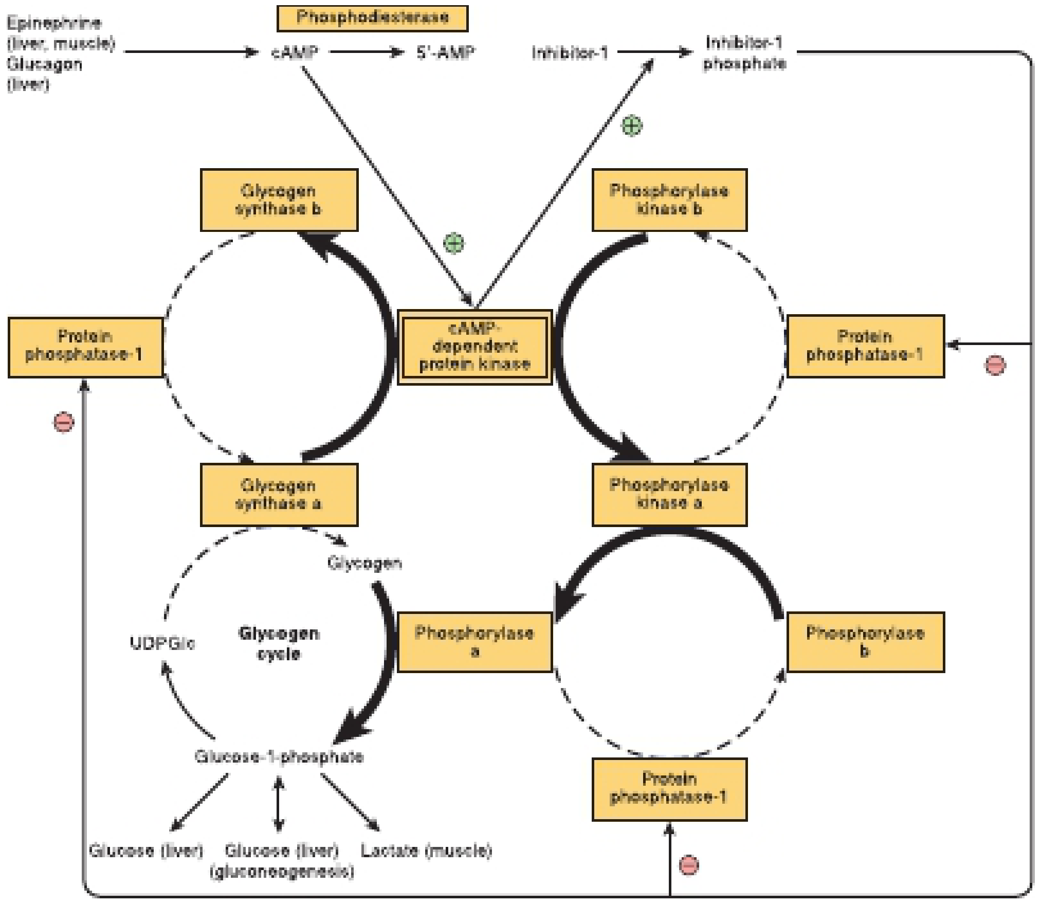
The cAMP-mediated coordinated control of glycogen synthesis and breakdown - note reciprocal regulation
TCA Cycle Steps
| Step | Reaction | Enzyme | Cofactor | Product |
|---|
| 1 | Acetyl-CoA + OAA → Citrate | Citrate Synthase | - | Citrate (6C) |
| 2 | Citrate → Isocitrate | Aconitase | Fe-S | Isocitrate |
| 3 | Isocitrate → α-KG | Isocitrate Dehydrogenase | NAD⁺, Mn²⁺ | NADH + CO₂ (Rate Limiting Step) |
| 4 | α-KG → Succinyl-CoA | α-Ketoglutarate Dehydrogenase | TPP, Lipoate, CoA, FAD, NAD⁺ | NADH + CO₂ |
| 5 | Succinyl-CoA → Succinate | Succinyl-CoA Synthetase | GDP/ADP | GTP (substrate-level phosphorylation) |
| 6 | Succinate → Fumarate | Succinate Dehydrogenase | FAD | FADH₂ (Complex II of ETC) |
| 7 | Fumarate → Malate | Fumarase | H₂O | Malate |
| 8 | Malate → OAA | Malate Dehydrogenase | NAD⁺ | NADH |
Per Turn of TCA (from 1 Acetyl-CoA)
- 3 NADH, 1 FADH₂, 1 GTP, 2 CO₂
- ATP equivalent: 3×2.5 + 1×1.5 + 1 = 10 ATP per acetyl-CoA
Rate-Limiting Step
Isocitrate Dehydrogenase - the committed step of the cycle
Regulation of TCA
| Activators | Inhibitors |
|---|
| ADP, Ca²⁺, NAD⁺ | ATP, NADH |
| Low energy state | High energy state |
| Substrate availability | Succinyl-CoA (product inhibition of α-KG DH) |
Key Cofactors (ALL 5 for PDH and α-KG DH)
- Thiamine (B1) - TPP
- Riboflavin (B2) - FAD
- Niacin (B3) - NAD⁺
- Pantothenic acid (B5) - CoA
- Lipoic acid - Lipoamide
- Mg²⁺ - for Isocitrate DH
MNEMONIC: "The Lovely Nastily Flavored Compound" = Thiamine, Lipoate, NAD (niacin), FAD (riboflavin), CoA (pantothenate)
PYRUVATE DEHYDROGENASE COMPLEX (PDH) - Bridge from Glycolysis to TCA
Reaction: Pyruvate + CoA + NAD⁺ → Acetyl-CoA + CO₂ + NADH
Same 5 cofactors as α-KG DH (TPP, Lipoate, CoA, FAD, NAD⁺)
Regulation:
- Activated by: Ca²⁺, ADP, NAD⁺, CoA, Pyruvate (substrate), Insulin (dephosphorylates PDH kinase)
- Inhibited by: Acetyl-CoA, NADH, ATP, PDH kinase (phosphorylates and inactivates PDH)
Clinical Correlations - TCA/PDH
🔴 Thiamine (B1) Deficiency - USMLE/NEET HIGH YIELD
- PDH and α-KG DH require TPP (thiamine pyrophosphate)
- Deficiency → pyruvate and α-KG cannot be oxidized → ACCUMULATE
- α-Ketoglutarate = same as α-ketoglutaric acid (used to measure B1 deficiency)
- Dry Beriberi: peripheral neuropathy (sensorimotor)
- Wet Beriberi: high-output cardiac failure, dilated cardiomyopathy
- Wernicke's Encephalopathy: confusion, ophthalmoplegia, ataxia (classic triad)
- Korsakoff Psychosis: confabulation, anterograde amnesia (mammillary body degeneration)
- Wernicke-Korsakoff seen in alcoholics - give IV thiamine BEFORE glucose!
🔴 PDH Deficiency
- Accumulation of pyruvate and lactate → lactic acidosis
- Neurological damage, developmental delay
- Blood: Elevated lactate, pyruvate, alanine
- Treatment: High-fat, low-carb diet; thiamine supplements
🔴 Isocitrate Dehydrogenase (IDH) Mutations - Oncology
- IDH1 (cytoplasm), IDH2 (mitochondria) mutations
- Gain-of-function mutation → produces 2-hydroxyglutarate (oncometabolite)
- Found in: Gliomas (grade II/III), AML, cholangiocarcinoma
- IDH-mutant gliomas have better prognosis
- Treatment: Ivosidenib (IDH1), Enasidenib (IDH2)
🔴 Aconitase Inhibition
- Fluorocitrate (from fluoroacetate, rat poison) → inhibits aconitase → TCA halted
- Citrate accumulates → inhibits PFK-1 → glycolysis also fails
- "Citrate trap" - double inhibition of energy metabolism
🔴 Succinate Dehydrogenase (SDH) Mutations
- SDH = Complex II of ETC (only TCA enzyme embedded in inner mito membrane)
- SDHB/SDHD mutations → Hereditary Paraganglioma/Pheochromocytoma syndrome
- SDH is a tumor suppressor; loss → Warburg-like shift
1.4 GLYCOGEN METABOLISM
Glycogenesis (Synthesis): Fed state, insulin-driven, liver + muscle
Glycogenolysis (Breakdown): Fasting/stress, glucagon/epinephrine-driven
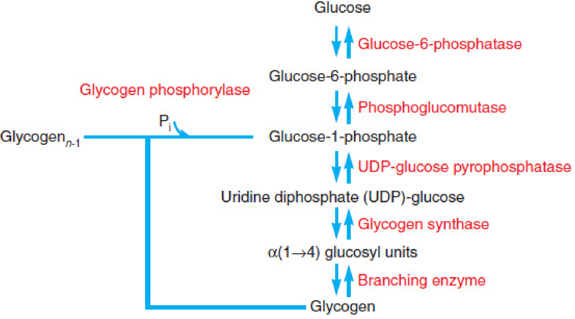
Central role of glucose-6-phosphate in glycogen metabolism
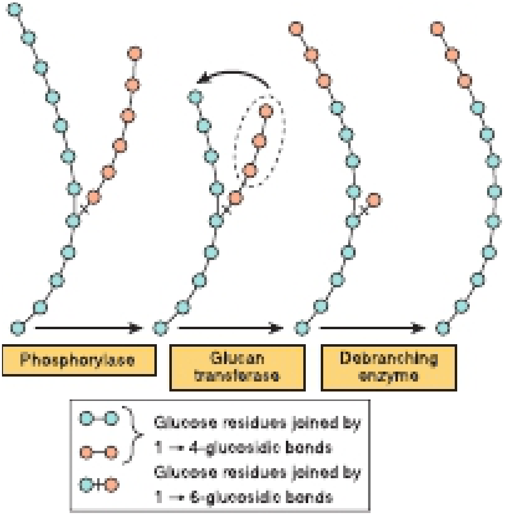
Glycogenolysis: Phosphorylase removes 1→4 links, debranching enzyme handles 1→6 branch points
Glycogenesis Steps
| Step | Enzyme | Notes |
|---|
| Glucose → G-6-P | Hexokinase/Glucokinase | |
| G-6-P → G-1-P | Phosphoglucomutase | |
| G-1-P + UTP → UDP-Glucose | UDP-glucose pyrophosphorylase | |
| UDP-Glucose → Glycogen chain | Glycogen Synthase | Rate-limiting step; adds α(1→4) links; requires a primer |
| Branch formation | Branching Enzyme | Transfers ≥6 residues to form α(1→6) |
Glycogenolysis Steps
| Step | Enzyme | Notes |
|---|
| Glycogen → G-1-P | Glycogen Phosphorylase | Rate-limiting; cleaves α(1→4); cofactor = Pyridoxal Phosphate (B6) |
| Debranching | Debranching enzyme (2 activities) | Glucan transferase + α-1,6-glucosidase |
| G-1-P → G-6-P | Phosphoglucomutase | |
| G-6-P → Glucose (liver only) | Glucose-6-phosphatase | NOT present in muscle; muscle uses G-6-P internally |
Regulation - Reciprocal Control by cAMP
| Signal | Effect on Glycogen Synthase | Effect on Phosphorylase | Net Result |
|---|
| Glucagon/Epinephrine (↑ cAMP) | Inactivated (phosphorylated) | Activated (phosphorylated) | Glycogenolysis |
| Insulin | Activated (dephosphorylated) | Inactivated | Glycogenesis |
| Ca²⁺ (muscle contraction) | - | Activated via Phosphorylase Kinase | Glycogenolysis |
| Glucose-6-phosphate | Activates synthase | Inhibits phosphorylase | Glycogenesis |
Glycogen Storage Diseases - COMPLETE TABLE (HIGH YIELD)
| Type | Eponym | Deficient Enzyme | Gene | Organ Affected | Key Clinical Features | Exam Tip |
|---|
| 0 | - | Glycogen Synthase | GYS2 | Liver | Fasting hypoglycemia, hyperketonemia | Opposite: too LITTLE glycogen |
| Ia | Von Gierke | Glucose-6-phosphatase | G6PC | Liver, Kidney | Fasting hypoglycemia, lactic acidosis, hyperuricemia (gout), hyperlipidemia, hepatomegaly, doll-like facies | No response to glucagon; hepatocellular adenoma risk |
| Ib | Von Gierke variant | G-6-P translocase | SLC37A4 | Liver, Kidney | Same as Ia + neutropenia + infections | Transportopathy |
| II | Pompe | Acid Maltase (α-1,4 glucosidase) | GAA | Lysosomes - all tissues | Infant: hypotonia, cardiomegaly, death by 2 yrs; Adult: myopathy | Only GSD with lysosomal enzyme deficiency; Treatment: Alglucosidase alfa (ERT) |
| III | Cori / Forbes | Debranching enzyme | AGL | Liver, Muscle | Milder Von Gierke; limit dextrin accumulates | Fasting hypoglycemia, hepatomegaly |
| IV | Andersen | Branching enzyme | GBE1 | Liver | Progressive cirrhosis, liver failure, abnormal long-chain glycogen | Also called Amylopectinosis |
| V | McArdle | Muscle Phosphorylase | PYGM | Muscle only | Exercise intolerance, cramps, myoglobinuria, NO rise in lactate after exercise (ischemic forearm exercise test) | Blood glucose NORMAL; serum CK elevated |
| VI | Hers | Liver Phosphorylase | PYGL | Liver | Mild hypoglycemia, hepatomegaly; benign course | |
| VII | Tarui | Phosphofructokinase (muscle) | PFKM | Muscle, RBC | Like McArdle + hemolytic anemia | PFK deficiency in RBCs too |
| IX | - | Phosphorylase kinase | PHKA | Liver, Muscle | Mild, X-linked | |
MNEMONIC for GSD types I-V: "Very Poor Carbohydrate Absorption Mechanism"
- I = Von gierke (V = Very)
- II = Pompe (P = Poor)
- III = Cori (C = Carbohydrate)
- IV = Andersen (A = Absorption)
- V = McArdle (M = Mechanism)
1.5 PENTOSE PHOSPHATE PATHWAY (HMP Shunt)
Location: Cytoplasm | Active in: Liver, RBCs, lactating mammary gland, adrenal cortex, testis
Two phases:
- Oxidative (irreversible): G-6-P → Ribulose-5-P + 2 NADPH + CO₂
- Non-oxidative (reversible): Interconversion of sugar phosphates
Key Enzymes
| Enzyme | Role | Clinical Relevance |
|---|
| Glucose-6-Phosphate Dehydrogenase (G6PD) | Rate-limiting; G-6-P → 6-Phosphogluconate | G6PD deficiency = most common enzyme deficiency worldwide |
| 6-Phosphogluconate Dehydrogenase | → Ribulose-5-P + CO₂ + NADPH | - |
| Transketolase | Non-oxidative phase; requires Thiamine (B1) | Activity measured in B1 deficiency (RBC transketolase activity) |
| Transaldolase | Non-oxidative phase | - |
Functions of PPP
- NADPH production - for: Glutathione reduction (GSH), Fatty acid synthesis, Cholesterol synthesis, Cytochrome P450, Respiratory burst (NADPH oxidase), Nitric oxide synthesis
- Ribose-5-phosphate - for nucleotide synthesis (DNA/RNA)
Regulation
- Controlled by NADPH levels (product inhibition of G6PD)
- When NADPH is oxidized → pathway activated
- Insulin ↑ G6PD expression
Clinical Correlations
🔴 G6PD Deficiency - USMLE/NEET CLASSIC
- X-linked recessive (most common enzyme deficiency in humans, affects ~400 million people)
- NADPH deficient → cannot regenerate GSH → oxidized hemoglobin → Heinz bodies
- RBC membrane damage → intravascular hemolysis → hemoglobinuria (dark urine)
- Triggers: Primaquine, dapsone, nitrofurantoin, sulfonamides, fava beans, infection (most common trigger!)
- Peripheral smear: Heinz bodies, bite cells (macrophages nibble Heinz bodies)
- Coombs test: NEGATIVE (not immune-mediated)
- G6PD variants: G6PD A- (African), G6PD Mediterranean (severe), G6PD Canton (Asian)
🔴 Chronic Granulomatous Disease (CGD)
- Defective NADPH oxidase in neutrophils (not G6PD, but uses same NADPH)
- Cannot produce superoxide (O₂⁻) → cannot kill catalase-positive organisms
- Recurrent infections with: S. aureus, Aspergillus, Nocardia, Pseudomonas, Serratia
- DHR (dihydrorhodamine) flow cytometry: diagnostic
- Treatment: Prophylactic TMP-SMX, itraconazole; IFN-γ; bone marrow transplant (cure)
🔴 Transketolase and Thiamine (B1)
- Transketolase in PPP requires TPP; activity used to diagnose subclinical thiamine deficiency
- RBC transketolase activity test: ↓ activity in thiamine deficiency
PART 2: PROTEIN/AMINO ACID METABOLISM
2.1 TRANSAMINATION
Location: Liver primarily (also kidney, heart)
Purpose: Transfer amino group from amino acid to α-keto acid (reversible)
Key Reactions
Reaction: Amino acid + α-Ketoglutarate ⇌ α-Keto acid + Glutamate
Enzymes:
- ALT (Alanine aminotransferase / SGPT): Alanine + α-KG ⇌ Pyruvate + Glutamate (liver-specific)
- AST (Aspartate aminotransferase / SGOT): Aspartate + α-KG ⇌ OAA + Glutamate (liver + heart + muscle)
- Cofactor: Pyridoxal phosphate (Vitamin B6) - for ALL aminotransferases
Alanine-Glucose Cycle (Cahill Cycle)
- Muscle: Pyruvate + glutamate → Alanine (via ALT) → travels to liver
- Liver: Alanine → Pyruvate (transamination) → Glucose (gluconeogenesis)
- Nitrogen delivered to liver as alanine for urea synthesis
- Glucose returned to muscle via blood
- Net: Transfers N from muscle to liver without releasing free NH₃ into blood
Clinical Correlations
🔴 ALT vs AST in Liver Disease
- Viral hepatitis: ALT > AST (AST:ALT ratio <1)
- Alcoholic hepatitis: AST:ALT ratio >2:1 (AST elevated due to B6 deficiency and mitochondrial damage; classic USMLE)
- Liver cirrhosis: AST > ALT (scar tissue)
- MI: AST elevated (cardiac muscle), ALT normal
🔴 Pyridoxine (B6) Deficiency
- Impairs ALL aminotransferases
- Dermatitis, glossitis, cheilosis, peripheral neuropathy, sideroblastic anemia (B6 needed for heme synthesis too - ALA synthase)
- Isoniazid (INH) causes B6 deficiency (INH is a B6 antagonist) → always supplement B6 with INH
2.2 UREA CYCLE (Ornithine Cycle / Krebs-Henseleit Cycle)
Location: Liver ONLY (hepatocytes) | Start in: Mitochondria, Finish in: Cytoplasm
Purpose: Convert toxic ammonia → non-toxic urea → excreted in urine
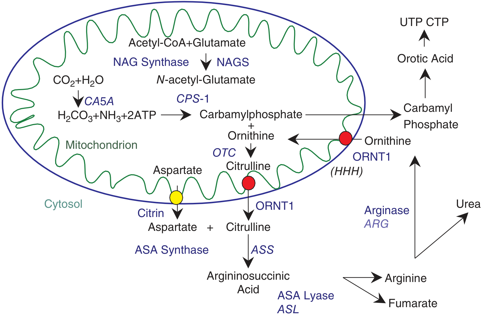
The urea cycle - note CPS-I and OTC in mitochondria; remainder in cytosol
Steps of the Urea Cycle
| Step | Enzyme | Location | Cofactor | Notes |
|---|
| 1 | NH₃ + CO₂ + 2ATP → Carbamoyl phosphate | CPS-I | N-Acetylglutamate (NAG), Mg²⁺ | Mitochondria; Rate-Limiting Step |
| 2 | Carbamoyl-P + Ornithine → Citrulline | OTC (Ornithine Transcarbamylase) | - | Mitochondria; most common defect |
| 3 | Citrulline + Aspartate → Argininosuccinate | Argininosuccinate Synthetase (ASS) | ATP | Cytoplasm; needs N from aspartate |
| 4 | Argininosuccinate → Arginine + Fumarate | Argininosuccinate Lyase (ASL) | - | Cytoplasm; fumarate → TCA |
| 5 | Arginine → Ornithine + Urea | Arginase | Mn²⁺ | Cytoplasm; urea released |
Key Points
- 2 N atoms in urea: 1 from NH₃ (CPS-I step), 1 from aspartate (step 3)
- Net energy cost: 3 ATP per cycle (2 for CPS-I + 1 for ASS)
- NAG activates CPS-I - formed from acetyl-CoA + glutamate by NAG synthase
- Arginine is essential amino acid in urea cycle defects (body cannot make enough)
- Fumarate links urea cycle to TCA cycle
Regulation of Urea Cycle
- CPS-I activated by: N-Acetylglutamate (NAG) - obligate activator
- NAG synthesis increased by: Arginine, high protein intake
- Long-term: Gene induction by glucocorticoids, high protein diet
Ammonia Detoxification in Brain (NOT liver)
- Brain: Glutamate + NH₃ → Glutamine (glutamine synthetase)
- Glutamine is the non-toxic transport form of NH₃ in blood
- High NH₃ depletes α-KG from TCA → ↓ energy production in neurons → encephalopathy
Urea Cycle Defects - COMPLETE TABLE
| Defect | Enzyme | Inheritance | Key Lab Finding | Distinguishing Feature |
|---|
| OTC Deficiency | Ornithine transcarbamylase | X-linked (only XLR urea cycle defect) | ↑ Orotic acid in urine, ↑ NH₃, ↓ citrulline | Most common; excess carbamoyl-P → pyrimidine synthesis → orotic acid |
| CPS-I Deficiency | Carbamoyl phosphate synthetase I | AR | ↑ NH₃, ↓ citrulline, Normal orotic acid | No orotic acid (problem is before carbamoyl-P is made) |
| NAG Synthase Deficiency | N-Acetylglutamate synthase | AR | ↑ NH₃, ↓ citrulline, Normal orotic acid | Treatable with N-carbamoyl-glutamate (NAG analogue) |
| Citrullinemia Type I | Argininosuccinate Synthetase | AR | ↑↑ Citrulline, ↑ NH₃, ↑ orotic acid | Citrulline accumulates massively |
| Argininosuccinicaciduria | Argininosuccinate Lyase | AR | ↑ Argininosuccinate, ↑ citrulline | Hair abnormality (trichorrhexis nodosa) |
| Argininemia | Arginase | AR | ↑↑ Arginine, spastic diplegia | Spastic tetraplegia, intellectual disability; late onset |
| HHH Syndrome | Ornithine transporter (ORNT1) | AR | ↑ Ornithine, ↑ NH₃, Homocitrullinuria | Ornithine cannot enter mitochondria |
Key Differentiating Points (EXAM)
- ↑ Orotic acid: OTC deficiency (ONLY X-linked), Citrullinemia
- Normal orotic acid: CPS-I, NAG synthase deficiency
- OTC vs CPS-I: Both have ↑ NH₃ and ↓ citrulline, but OTC has ↑ orotic acid
- All present with: lethargy, vomiting, encephalopathy shortly after birth; ↑ plasma glutamine
Clinical Correlations
🔴 Hyperammonemia - Management
- Dietary: Low-protein diet, essential amino acid supplementation
- Sodium benzoate / sodium phenylbutyrate: alternative nitrogen excretion pathways
- Arginine supplementation (essential in cycle defects)
- Dialysis for acute crisis
- Rifaximin for hepatic encephalopathy (reduces gut NH₃ production)
- Liver transplant (definitive for urea cycle defects)
🔴 Liver Failure and Hyperammonemia
- Portosystemic shunting bypasses hepatocytes → NH₃ reaches brain
- Hepatic encephalopathy: flapping tremor (asterixis), fetor hepaticus (musty odor), confusion → coma
- Precipitants: GI bleed, infection, constipation, sedatives, high-protein meal
🔴 Reye's Syndrome
- Mitochondrial dysfunction (often post-viral + aspirin use in children)
- Impairs urea cycle enzymes + fatty acid oxidation
- Presents: vomiting, encephalopathy, hepatic failure, hypoglycemia
- NO jaundice (hepatocellular necrosis minimal)
- Lab: ↑ NH₃, ↑ ALT/AST, ↑ PT, normal bilirubin
2.3 AMINO ACID CATABOLISM & SELECTED METABOLIC DISORDERS
Phenylalanine - Tyrosine Pathway
Phe → Tyr (Phenylalanine hydroxylase + BH4 cofactor)
Tyr → DOPA → Dopamine → NE → Epinephrine (catecholamine synthesis)
Tyr → Thyroxine (T3, T4)
Tyr → Melanin (tyrosinase)
Tyr → Homogentisate → Maleylacetoacetate → Fumarate + Acetoacetate (catabolism)
| Disorder | Deficient Enzyme | Accumulated | Clinical Features | Inheritance |
|---|
| PKU (Phenylketonuria) | Phenylalanine Hydroxylase (or BH4) | Phenylalanine, Phenylpyruvate | Intellectual disability, seizures, fair skin/hair/eyes, musty odor, eczema | AR |
| Malignant PKU | BH4 (Tetrahydrobiopterin) synthesis or recycling | Phe | Same + neurotransmitter deficiency (DOPA, serotonin) | AR |
| Alcaptonuria | Homogentisate oxidase | Homogentisic acid | Black urine (oxidized homogentisic acid), ochronosis (dark pigment in cartilage), arthritis | AR |
| Albinism | Tyrosinase | Melanin not made | Lack of pigment in skin, hair, eyes; ↑ skin cancer risk | AR |
| Tyrosinemia Type I | Fumarylacetoacetate hydrolase (FAH) | Succinylacetone | Liver failure, renal tubular acidosis, cabbage odor, ↑ AFP | AR |
Branch-Chain Amino Acids (BCAA) - Leucine, Isoleucine, Valine
Maple Syrup Urine Disease (MSUD)
- Defective α-Keto acid dehydrogenase (branched-chain)
- Same cofactors as PDH: TPP, lipoate, CoA, FAD, NAD⁺
- Accumulation of leucine, isoleucine, valine and their keto-acids
- Maple syrup smell in urine/cerumen
- Neonatal seizures, encephalopathy, feeding difficulty
- Thiamine is adjunct treatment (cofactor)
- Dietary: Restriction of Leu, Ile, Val
Homocysteine Metabolism
| Disorder | Deficient Enzyme | Accumulated | Key Features | Treatment |
|---|
| Homocystinuria (Classic) | Cystathionine β-synthase | Homocysteine | Marfanoid habitus, ectopia lentis (DOWNWARD), intellectual disability, thromboembolic events, early atherosclerosis | Pyridoxine (B6), folate, methionine restriction |
| Homocystinuria (MTHFR) | MTHFR (methylenetetrahydrofolate reductase) | Homocysteine | Mild-moderate; associated with neural tube defects (pregnancy), CVD risk | Folate, betaine |
| Elevated Hcy (B12/Folate deficiency) | Methionine synthase (needs B12, methylfolate) | Homocysteine, Methylmalonic acid (B12 only) | Megaloblastic anemia, subacute combined degeneration (B12) | B12, Folate |
Ectopia Lentis:
- Marfan syndrome: superotemporal displacement
- Homocystinuria: inferonasal (downward/medial) displacement
- Weill-Marchesani: inferonasal
- Sulphite oxidase deficiency: variable
Organic Acidemias
| Disorder | Defect | Accumulates | Features |
|---|
| Methylmalonic Acidemia | Methylmalonyl-CoA Mutase (needs B12-adenosylcobalamin) | Methylmalonic acid | Metabolic acidosis, hyperammonemia, neutropenia |
| Propionic Acidemia | Propionyl-CoA Carboxylase (needs Biotin) | Propionic acid | Severe metabolic acidosis, hyperammonemia |
| Isovaleric Acidemia | Isovaleryl-CoA Dehydrogenase | Isovaleric acid | "Sweaty feet" odor, metabolic acidosis |
PART 3: LIPID METABOLISM
3.1 FATTY ACID SYNTHESIS (Lipogenesis)
Location: Cytoplasm | Occurs in: Liver, adipose tissue, lactating mammary gland
Substrate: Acetyl-CoA (from glucose/amino acids via PDH or β-oxidation overflow)
Shuttle: Acetyl-CoA cannot cross inner mitochondrial membrane → citrate shuttle (citrate exits mito → cleaved to acetyl-CoA + OAA in cytoplasm by ATP citrate lyase)
Steps
| Step | Enzyme | Cofactor | Product | Notes |
|---|
| 1 | Acetyl-CoA + CO₂ + ATP → Malonyl-CoA | Acetyl-CoA Carboxylase (ACC) | Malonyl-CoA | Rate-limiting step; requires Biotin |
| 2-7 | Malonyl-CoA + NADPH → FA elongation | Fatty Acid Synthase (FAS) | Palmitate (16C) | 7 cycles; uses 7 NADPH per cycle |
Overall: Acetyl-CoA + 7 Malonyl-CoA + 14 NADPH → Palmitate + 7 CO₂ + 8 CoA
Regulation of Fatty Acid Synthesis
| Activators of ACC | Inhibitors of ACC |
|---|
| Citrate (allosteric) | Palmitoyl-CoA (product inhibition) |
| Insulin (activates via dephosphorylation) | Glucagon/Epinephrine (↑ cAMP → phosphorylates ACC → inactive) |
| High carbohydrate diet | Malonyl-CoA inhibits CPT-I (prevents FA oxidation simultaneously) |
KEY EXAM POINT: Malonyl-CoA INHIBITS Carnitine Palmitoyl Transferase-I (CPT-I), preventing entry of fatty acids into mitochondria for β-oxidation. Thus: when synthesis is ON, oxidation is OFF - mutual exclusivity
NADPH Sources for Fatty Acid Synthesis
- Pentose Phosphate Pathway (major source in liver)
- Malate enzyme (cytoplasmic malic enzyme)
- Isocitrate dehydrogenase (cytoplasmic)
3.2 FATTY ACID OXIDATION (β-Oxidation)
Location: Mitochondrial matrix (long-chain FA) | Peroxisomes: Very long chain FA (VLCFA)
Activation: Fatty acid + CoA + ATP → Acyl-CoA + AMP + PPi (by Acyl-CoA Synthetase, on outer mitochondrial membrane)
Carnitine Shuttle (Entry into Mitochondria)
- CPT-I (outer membrane): Acyl-CoA + Carnitine → Acylcarnitine (rate-limiting for FA oxidation)
- Translocase: Carries acylcarnitine across inner membrane
- CPT-II (inner membrane): Regenerates Acyl-CoA inside matrix
Malonyl-CoA inhibits CPT-I = the switch between fat synthesis and oxidation
β-Oxidation Cycle Steps
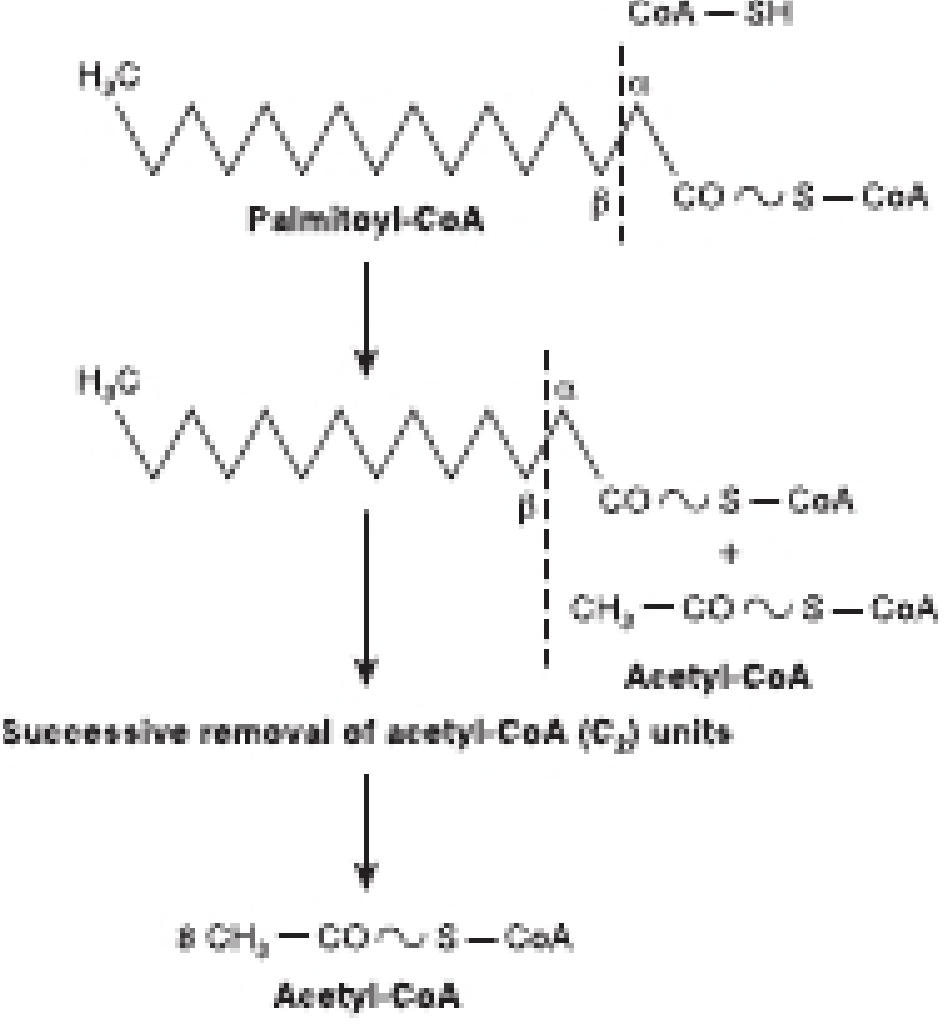
Beta-oxidation: successive removal of 2-carbon acetyl-CoA units from palmitoyl-CoA
| Step | Reaction | Enzyme | Cofactor | Product |
|---|
| 1 | Acyl-CoA → Enoyl-CoA | Acyl-CoA Dehydrogenase | FAD | FADH₂ |
| 2 | Enoyl-CoA + H₂O → 3-Hydroxyacyl-CoA | Enoyl-CoA Hydratase | H₂O | - |
| 3 | 3-Hydroxyacyl-CoA → 3-Ketoacyl-CoA | 3-Hydroxyacyl-CoA Dehydrogenase | NAD⁺ | NADH |
| 4 | 3-Ketoacyl-CoA + CoA → Acetyl-CoA + Acyl-CoA (n-2) | Thiolase | CoA | Acetyl-CoA |
Per cycle: 1 FADH₂ + 1 NADH + 1 Acetyl-CoA
For Palmitate (C16): 7 cycles → 7 FADH₂ + 7 NADH + 8 Acetyl-CoA → NET 106 ATP
Metabolic Disorders - Beta-Oxidation
| Disorder | Defect | Key Features | Lab | Notes |
|---|
| MCAD Deficiency (Most common FA oxidation disorder) | Medium-Chain Acyl-CoA Dehydrogenase | Hypoketotic hypoglycemia during fasting, vomiting, encephalopathy, sudden death | ↑ Octanoylcarnitine (C8) on newborn screen, ↑ Medium-chain FA in urine | Most common; diagnosed on newborn screening; AR |
| LCHAD | Long-chain 3-hydroxyacyl-CoA DH | Hypoglycemia, cardiomyopathy, peripheral neuropathy, AFLP in mother (carrier) | ↑ Long-chain acylcarnitines | Maternal AFLP associated |
| VLCAD | Very Long-chain Acyl-CoA DH | Cardiomyopathy, hypoglycemia | ↑ C14:1 acylcarnitine | |
| Primary Carnitine Deficiency | OCTN2 (plasma membrane carnitine transporter) | Hypoketotic hypoglycemia, cardiomyopathy, muscle weakness | ↓ Serum carnitine, ↑ acylcarnitines | Treatment: Carnitine supplementation |
| CPT-I Deficiency | CPT-I (liver) | Hypoketotic hypoglycemia, liver dysfunction | ↑ Free carnitine, ↓ acylcarnitines | Opposite pattern from most FA disorders |
| CPT-II Deficiency | CPT-II (muscle) | Myopathy, myoglobinuria with exercise | ↑ Long-chain acylcarnitines | |
| Jamaican Vomiting Sickness | - (external toxin) | Hypoglycin A (from unripe ackee fruit) → inhibits MCAD and SCHAD | Hypoketotic hypoglycemia, vomiting, encephalopathy | Not inherited |
3.3 KETONE BODY METABOLISM
Location: Synthesis: Liver mitochondria | Utilization: Brain, heart, muscle, kidney (NOT liver - lacks ketolytic enzyme)
Conditions promoting ketosis: Fasting >24h, uncontrolled DM Type 1, prolonged exercise, ketogenic diet, alcoholism
Ketogenesis Steps (in liver mitochondria)
| Step | Reaction | Enzyme | Notes |
|---|
| 1 | 2 Acetyl-CoA → Acetoacetyl-CoA | Thiolase | |
| 2 | Acetoacetyl-CoA + Acetyl-CoA → HMG-CoA | HMG-CoA Synthase | Rate-limiting step of ketogenesis; mitochondrial |
| 3 | HMG-CoA → Acetoacetate + Acetyl-CoA | HMG-CoA Lyase | |
| 4 | Acetoacetate → β-Hydroxybutyrate | β-Hydroxybutyrate DH | Reversible; uses NADH |
| - | Acetoacetate → Acetone + CO₂ | Non-enzymatic decarboxylation | Fruity breath in DKA |
3 ketone bodies: Acetoacetate, β-Hydroxybutyrate (predominant in DKA), Acetone
Ketone Body Utilization (Peripheral tissues)
- β-Hydroxybutyrate → Acetoacetate (β-HB DH)
- Acetoacetate + Succinyl-CoA → Acetoacetyl-CoA + Succinate (Succinyl-CoA:3-ketoacid CoA transferase = Thiophorase - absent in liver!)
- Acetoacetyl-CoA → 2 Acetyl-CoA → TCA
Clinical Correlations
🔴 Diabetic Ketoacidosis (DKA)
- Type 1 DM (insulin deficiency) → ↑ glucagon → ↑ lipolysis → ↑ FA to liver → ↑ ketogenesis
- Malonyl-CoA LOW → CPT-I active → FA enter mitochondria
- pH <7.3, anion gap metabolic acidosis
- Ketones: β-HB >> Acetoacetate in DKA (ratio 3:1 normally, up to 10:1 in DKA)
- Nitroprusside test detects acetoacetate ONLY (NOT β-HB) → may UNDERESTIMATE DKA severity
- Treatment: Insulin + fluids + potassium replacement
🔴 Starvation Ketosis
- Glucose depletes after 24h → insulin ↓, glucagon ↑
- Brain adapts to use ketone bodies after 3-4 days (reduces protein catabolism)
- After prolonged starvation: brain gets 70% energy from ketones
- Controlled; pH usually normal (buffering capacity adequate)
🔴 Alcoholic Ketoacidosis
- Ethanol → ↑ NADH:NAD⁺ → favors β-HB over acetoacetate
- Nitroprusside test may be NEGATIVE despite severe ketosis (measures only acetoacetate!)
- Anion gap metabolic acidosis + history of alcohol + vomiting
3.4 CHOLESTEROL SYNTHESIS (Mevalonate Pathway)
Location: Liver (primary), intestine | Occurs in: Cytoplasm + ER
Substrate: Acetyl-CoA → Mevalonate → Cholesterol
Key Steps
| Step | Enzyme | Notes |
|---|
| 2 Acetyl-CoA → Acetoacetyl-CoA | Thiolase | |
| Acetoacetyl-CoA + Acetyl-CoA → HMG-CoA | HMG-CoA Synthase | Cytoplasmic (≠ mitochondrial) |
| HMG-CoA → Mevalonate | HMG-CoA Reductase | Rate-limiting step - TARGET OF STATINS |
| Mevalonate → Isopentenyl pyrophosphate | Multiple steps | Requires ATP, NADPH |
| IPP → Squalene → Lanosterol → Cholesterol | Multiple steps | |
Regulation of HMG-CoA Reductase
| Activated by | Inhibited by |
|---|
| Insulin | Glucagon |
| Thyroxine | Cholesterol (product inhibition, via SREBP) |
| - | Statins (competitive inhibitors) |
| - | PCSK9 (degrades LDL receptors - PCSK9 inhibitors increase LDL clearance) |
Other products of Mevalonate Pathway
- Ubiquinone (CoQ10) - electron carrier ETC
- Dolichol - N-glycosylation of proteins
- Farnesyl/Geranylgeranyl pyrophosphate - prenylation of Ras (oncoproteins)
- Heme A (modified porphyrin)
STATIN SIDE EFFECT NOTE: Statins inhibit HMG-CoA Reductase → ↓ CoQ10 → myopathy (rhabdomyolysis risk). Also ↓ farnesyl-PP → cannot prenylate Ras (anti-tumor side benefit)
Lipoprotein Metabolism
| Lipoprotein | Origin | Main Lipid | Apoprotein | Function | Receptor |
|---|
| Chylomicrons | Intestinal enterocytes | Dietary TG | ApoB-48, ApoC-II, ApoE | Transport dietary fat (exogenous) | LPL (activated by ApoC-II) |
| VLDL | Liver | Endogenous TG | ApoB-100, ApoC-II, ApoE | Transport hepatic TG | LPL |
| IDL | VLDL remnant | TG + CE | ApoB-100, ApoE | Intermediate; cleared by liver | LDL receptor |
| LDL | IDL | Cholesterol esters | ApoB-100 | Deliver cholesterol to tissues | LDL receptor (deficient in FH) |
| HDL | Liver + intestine | Protein-rich | ApoA-I | Reverse cholesterol transport | SR-BI |
Key Enzymes:
- Lipoprotein Lipase (LPL): Hydrolyzes TG in chylomicrons and VLDL; activated by ApoC-II; inhibited by ApoC-III; deficiency → hypertriglyceridemia (Type I hyperlipoproteinemia)
- LCAT (Lecithin-Cholesterol Acyltransferase): Esterifies cholesterol in HDL; activator = ApoA-I
- CETP (Cholesterol Ester Transfer Protein): Transfers CE from HDL to VLDL/LDL in exchange for TG
- Hepatic Lipase: Converts IDL → LDL; removes TG from HDL
Clinical Correlations - Cholesterol/Lipoproteins
🔴 Familial Hypercholesterolemia (FH)
- LDL receptor mutation (most common: loss-of-function AR/AD)
- Heterozygous FH: LDL 200-400 mg/dL; Homozygous FH: LDL >600-1000 mg/dL
- Xanthomas (tendon, especially Achilles), xanthelasma, corneal arcus <45yrs
- Early MI (males by 40s, females by 50s in heterozygous)
- Treatment: Statins + Ezetimibe + PCSK9 inhibitors (evolocumab, alirocumab); LDL apheresis
🔴 Familial Hypertriglyceridemia
- LPL deficiency (Type I) or ApoC-II deficiency: very high TG (>1000), eruptive xanthomas, lipemia retinalis, pancreatitis
- Type V: combined VLDL + chylomicron elevation
🔴 Abetalipoproteinemia
- MTP (microsomal triglyceride transfer protein) deficiency → cannot assemble chylomicrons or VLDL
- Fat malabsorption, acanthocytes, progressive ataxia/neuropathy (Vitamin E deficiency)
- Absence of ApoB-containing lipoproteins
🔴 Tangier Disease
- ABCA1 transporter deficiency → cholesterol cannot be effluxed to HDL
- Very low/absent HDL, orange tonsils, hepatosplenomegaly, peripheral neuropathy
Lipid Storage Disorders (Sphingolipidoses and Lipidoses)
| Disorder | Deficient Enzyme | Accumulated Lipid | Key Clinical Features | Inheritance |
|---|
| Gaucher Disease (Type I - most common lysosomal storage disorder) | β-Glucocerebrosidase | Glucocerebroside | Gaucher cells (crinkled tissue paper cytoplasm), hepatosplenomegaly, bone pain, pancytopenia; NO CNS in Type I | AR; ERT: Imiglucerase; SRT: Miglustat |
| Niemann-Pick | Sphingomyelinase | Sphingomyelin | Hepatosplenomegaly, cherry-red spot (50%), foam cells; Types A (severe, early death) and B | AR |
| Tay-Sachs | Hexosaminidase A | GM2 ganglioside | Cherry-red spot, progressive neurodegeneration, NO organomegaly, Ashkenazi Jewish | AR |
| Sandhoff Disease | Hexosaminidase A + B | GM2 + globoside | Same as Tay-Sachs + organomegaly | AR |
| Krabbe Disease | Galactocerebrosidase | Galactocerebroside + psychosine | Peripheral neuropathy, globoid cells; infantile onset | AR |
| Metachromatic Leukodystrophy (MLD) | Arylsulfatase A | Sulfatide | Demyelination, peripheral neuropathy, developmental regression; metachromatic deposits | AR |
| Fabry Disease | α-Galactosidase A | Globotriaosylceramide (Gb3) | Angiokeratomas, neuropathic pain, corneal opacity (cornea verticillata), cardiomyopathy, renal failure | X-linked |
| Farber Disease | Ceramidase | Ceramide | Hoarse cry, joint swelling, subcutaneous nodules | AR |
| Wolman Disease | Lysosomal acid lipase | Cholesterol esters + TG | Adrenal calcification, liver failure, early death | AR |
| GM1 Gangliosidosis | β-Galactosidase | GM1 + keratan sulfate | Dysostosis multiplex, cherry-red spot, coarse facies | AR |
| Mucopolysaccharidoses (MPS) | Various glycosaminoglycan enzymes | GAGs | Coarse features, organomegaly, skeletal dysplasia, corneal clouding; see separate table | AR (except Hunter: XLR) |
Mucopolysaccharidoses (MPS) - Quick Reference
| Type | Eponym | Enzyme | GAG Accumulated | Key Features |
|---|
| I H | Hurler | α-L-Iduronidase | HS + DS | Coarse facies, corneal clouding, hepatosplenomegaly, intellectual disability, cardiac |
| I S | Scheie | α-L-Iduronidase | HS + DS | Mild, corneal clouding, normal intelligence |
| II | Hunter | Iduronate sulfatase | HS + DS | X-linked (only XLR MPS); NO corneal clouding; "hunts at night" (pebbly skin, deafness) |
| III | Sanfilippo | Multiple (4 subtypes) | HS | Severe intellectual disability, mild somatic features |
| IV | Morquio | N-acetylgalactosamine-6-sulfatase (A) or β-Galactosidase (B) | KS + CS | Short stature, atlantoaxial instability (dens hypoplasia → risk of cord compression), normal intelligence, NO corneal clouding except B |
| VI | Maroteaux-Lamy | Arylsulfatase B | DS | Coarse features, NO intellectual disability |
| VII | Sly | β-Glucuronidase | HS + DS + CS | Hydrops fetalis |
MNEMONIC for Hunter Syndrome (Type II): "Hunter hunts with X (crossbow)" = X-linked; no corneal clouding (hunter needs to SEE = NO corneal haze)
PART 4: INTEGRATION OF ALL CYCLES
The Metabolic Integration Map
CARBOHYDRATES          PROTEINS            LIPIDS
    |                     |                   |
Glucose             Amino Acids         Triglycerides
    |                     |                   |
Glycolysis         Transamination       Lipolysis (HSL)
    |                     |                   |
Pyruvate -------> α-KG/OAA -------> Glycerol → Gluconeogenesis
    |                              Fatty Acids
    ↓ PDH                              |
Acetyl-CoA <---------------------- β-Oxidation
         \                              |
          \                             |
           TCA Cycle <-------- OAA ← Propionyl-CoA
          /    \                  (odd-chain FA)
    NADH/FADH₂  GTP
          |
        ETC → ATP
          |
     Ketone Bodies (liver excess acetyl-CoA)
Key Integration Points
| Junction | Molecules | Connects |
|---|
| Acetyl-CoA | Central metabolite | Carb + Lipid + Protein all converge here |
| Pyruvate | Metabolic crossroads | Glycolysis → TCA (PDH), Gluconeogenesis (PC), Lactate (LDH), Alanine (ALT) |
| Oxaloacetate (OAA) | TCA/Gluconeogenesis | Must be replenished (anaplerosis) from amino acids; "OAA is the spark plug of TCA" |
| α-Ketoglutarate | TCA/Amino acid | Accepts/donates NH₂ in transamination; connects N metabolism to TCA |
| Succinyl-CoA | TCA/Heme/BCAA | Porphyrin synthesis, methylmalonyl-CoA, odd-chain FA |
| Fumarate | TCA/Urea cycle | Urea cycle feeds fumarate to TCA (Bicyclic connection) |
| G-6-P | Glycolysis/PPP/Glycogen | Central hub of carbohydrate metabolism |
| Glucose-6-phosphatase | Gluconeogenesis/Glycogenolysis | Absent in muscle; explains why muscle can't export glucose |
Anaplerotic Reactions (Replenishing TCA intermediates)
| Source | Reaction | Enzyme | Clinical |
|---|
| Pyruvate → OAA | Pyruvate + CO₂ → OAA | Pyruvate Carboxylase (Biotin) | Critical for GNG; impaired in biotin deficiency |
| Glutamate → α-KG | Glutamate → α-KG + NH₃ | Glutamate DH | Links amino acid and TCA |
| Aspartate → OAA | Transamination | AST | AST reaction connects urea cycle to TCA |
| Propionyl-CoA → Succinyl-CoA | 3 steps | Propionyl-CoA carboxylase + Methylmalonyl-CoA mutase | Requires biotin (PCC) and B12 (MCM); deficiency → organic acidemia |
| Odd-chain FA → Succinyl-CoA | β-oxidation + 3 steps | - | Only FA carbon that enters gluconeogenesis |
MASTER COFACTOR TABLE
| Cofactor | Vitamin | Key Enzymes | Deficiency Disease |
|---|
| TPP (Thiamine pyrophosphate) | B1 | PDH, α-KG DH, BCKA DH, Transketolase | Beriberi, Wernicke-Korsakoff |
| FAD/FMN | B2 (Riboflavin) | Multiple DH enzymes in ETC and TCA | Glossitis, corneal vascularization, normocytic anemia |
| NAD⁺/NADP⁺ | B3 (Niacin) | Almost all dehydrogenases | Pellagra (4Ds: Diarrhea, Dermatitis, Dementia, Death) |
| CoA (Coenzyme A) | B5 (Pantothenic acid) | PDH, α-KG DH, Fatty acid metabolism | Rare; burning feet syndrome |
| PLP (Pyridoxal Phosphate) | B6 (Pyridoxine) | ALL aminotransferases, ALA synthase, Glycogen phosphorylase, Serine DH | Sideroblastic anemia, peripheral neuropathy, convulsions; INH-induced |
| Biotin | B7 | Pyruvate Carboxylase, ACC, Propionyl-CoA Carboxylase, MCC | Alopecia, dermatitis, neurological symptoms; raw egg whites |
| Folate (THF) | B9 | Thymidylate synthase, purine synthesis, methionine synthesis (with B12) | Megaloblastic anemia, NTD (fetal) |
| Cobalamin (B12) | B12 | Methionine synthase (homocysteine→methionine), Methylmalonyl-CoA mutase | Megaloblastic anemia, subacute combined degeneration, elevated MMA |
| Lipoic acid | - | PDH, α-KG DH, Glycine cleavage system | - |
| Mg²⁺ | - | Hexokinase, PFK-1, Pyruvate Kinase, all ATPases | - |
| Mn²⁺ | - | Isocitrate DH, Arginase | - |
MASTER RATE-LIMITING STEPS TABLE
| Pathway | Rate-Limiting Enzyme | Key Activators | Key Inhibitors |
|---|
| Glycolysis | PFK-1 | AMP, F-2,6-BP, insulin | ATP, citrate, glucagon |
| Gluconeogenesis | Fructose-1,6-bisphosphatase | ATP, glucagon | AMP, F-2,6-BP, insulin |
| Glycogenesis | Glycogen Synthase | G-6-P, insulin, dephosphorylation | Glucagon/Epi (phosphorylation) |
| Glycogenolysis | Glycogen Phosphorylase | AMP, Ca²⁺, epinephrine, glucagon | G-6-P, glucose, insulin, ATP |
| TCA Cycle | Isocitrate Dehydrogenase | ADP, Ca²⁺, NAD⁺ | ATP, NADH |
| Pyruvate → Acetyl-CoA | Pyruvate Dehydrogenase Complex | Ca²⁺, ADP, NAD⁺, CoA, insulin | Acetyl-CoA, NADH, ATP, PDH kinase |
| Pentose Phosphate Pathway | G6PD | NADP⁺ | NADPH |
| Fatty Acid Synthesis | Acetyl-CoA Carboxylase | Citrate, insulin | Palmitoyl-CoA, glucagon, epinephrine, malonyl-CoA (feedback) |
| Beta-Oxidation | CPT-I (entry) | Glucagon, fasting | Malonyl-CoA |
| Ketogenesis | HMG-CoA Synthase (mito) | Fasting, low insulin | Fed state, insulin |
| Cholesterol Synthesis | HMG-CoA Reductase | Insulin, thyroxine | Glucagon, statins, cholesterol (SREBP) |
| Urea Cycle | CPS-I | N-Acetylglutamate | - |
| Fatty Acid Elongation (Mito) | Thiolase (in reverse) | - | - |
INTER-CYCLE CONNECTIONS (HIGHLY TESTED)
| Connection | Mechanism | Clinical Relevance |
|---|
| Urea Cycle + TCA | Fumarate produced in urea cycle enters TCA; OAA → aspartate for urea cycle | Hyperammonemia depletes TCA intermediates; liver failure affects both |
| Glycolysis + PPP | G-6-P is the branch point; competition for G-6-P | G6PD deficiency: G-6-P shunted to glycolysis only |
| FA Synthesis + Cholesterol | Both use Acetyl-CoA + NADPH + HMG-CoA | Statins inhibit only cholesterol synthesis step (HMG-CoA reductase) |
| β-Oxidation + Ketogenesis | FA oxidation generates acetyl-CoA → ketone bodies when oxaloacetate is limiting | Starvation/DM1: OAA used for gluconeogenesis → acetyl-CoA cannot enter TCA → ketogenesis |
| Glycolysis + FA Synthesis | Acetyl-CoA for FA synthesis comes from glucose via PDH | High carb diet → ↑ lipogenesis; role of citrate shuttle |
| Cori Cycle | Lactate (muscle) → glucose (liver) | Shuttles energy; requires gluconeogenesis in liver |
| Methionine Cycle + Folate Cycle | Methionine synthase (B12) uses MTHF to regenerate methionine from Hcy | B12 deficiency = folate trap; both → megaloblastic anemia |
| Propionyl-CoA + TCA | Odd-chain FA/BCAA → Propionyl-CoA → Methylmalonyl-CoA → Succinyl-CoA | B12 deficiency blocks this step → methylmalonic acidemia |
| Glucose-Alanine Cycle | Muscle sends alanine → liver → glucose returned | Reduces NH₃ toxicity; maintains glucose during fasting |
CLINICAL CROSS-CORRELATION TABLE (MULTI-SUBJECT - NEET PG / USMLE)
| Metabolic Defect | Biochemistry | Medicine/Pediatrics | Surgery/Obs | Pharmacology |
|---|
| PDH Deficiency / B1 deficiency | Pyruvate → acetyl-CoA blocked | Wernicke-Korsakoff, beriberi | Bariatric surgery → B1 deficiency | Give IV thiamine before glucose in Wernicke's |
| G6PD Deficiency | PPP impaired, ↓ NADPH | Hemolytic anemia triggers: infections, drugs | - | Avoid primaquine, dapsone, sulfonamides, nitrofurantoin |
| HMG-CoA Reductase (statins) | ↓ cholesterol synthesis | Familial hypercholesterolemia, statin myopathy | Pre-op lipid management | Statin myopathy: ↑ CK, rhabdomyolysis; monitor LFTs |
| Glycogen storage (Von Gierke) | Glucose-6-phosphatase | Fasting hypoglycemia, hepatomegaly | - | Cornstarch therapy (slow glucose release) |
| Urea Cycle (OTC) | X-linked; NH₃ buildup | Encephalopathy in neonates | - | Sodium benzoate, sodium phenylbutyrate, rifaximin |
| Fatty acid oxidation (MCAD) | No ketones during fasting | Reye-like presentation, sudden death | Identified on newborn screen | Avoid fasting; carnitine supplementation debated |
| DKA | Insulin deficiency → ketosis | Type 1 DM management, osmotic diuresis | DKA in surgery/trauma patient | Insulin + K⁺ replacement; nitroprusside test only detects acetoacetate |
| Phenylketonuria | Phe hydroxylase | Intellectual disability if untreated | Maternal PKU → fetal malformations | Low-Phe diet; tetrahydrobiopterin (BH4) for responsive cases; pegvaliase |
| Homocystinuria | CBS deficiency | Thromboembolic events, lens dislocation | DVT risk in surgery | Pyridoxine, betaine, folate |
| Gaucher Disease | β-Glucocerebrosidase | Bone crises, pancytopenia, Gaucher cells | Splenomegaly requiring splenectomy | ERT (imiglucerase); SRT (miglustat) |
| Acute intermittent porphyria | ALA synthase dysregulation | Episodic neurovisceral attacks | Avoid surgery triggers (barbiturates, fasting) | Attacks triggered by: cytochrome P450 inducers, fasting, estrogens |
STORAGE DISORDER SUMMARY TABLE - CARBOHYDRATES
| Disease | Type | Enzyme | Storage Material | Organs | Treatment |
|---|
| Von Gierke (GSD I) | GSD | Glucose-6-phosphatase | Glycogen + Fat (liver/kidney) | Liver, Kidney | Cornstarch, allopurinol, ACE-I for nephropathy |
| Pompe (GSD II) | GSD (lysosomal) | Acid Maltase (α-1,4-glucosidase) | Glycogen in lysosomes | All tissues (heart, muscle) | Alglucosidase alfa (ERT) |
| Cori/Forbes (GSD III) | GSD | Debranching enzyme | Limit dextrin | Liver, Muscle | High-protein diet |
| Andersen (GSD IV) | GSD | Branching enzyme | Amylopectin-like glycogen | Liver | Liver transplant |
| McArdle (GSD V) | GSD | Muscle phosphorylase | Glycogen in muscle | Muscle | Sucrose before exercise, high protein diet |
| Galactosemia | Sugar metabolism | Gal-1-P uridyltransferase | Gal-1-P, Galactitol | Liver, Brain, Lens | Galactose-free diet (no milk) |
| HFI | Sugar metabolism | Aldolase B | Fructose-1-P | Liver, Kidney | Fructose/sucrose/sorbitol-free diet |
STORAGE DISORDER SUMMARY TABLE - LIPIDS (Sphingolipidoses)
| Disease | Enzyme | Stored Lipid | Key Feature | Inheritance | Therapy |
|---|
| Gaucher I | β-Glucocerebrosidase | Glucocerebroside | Crinkled-paper cells, no CNS | AR | ERT (imiglucerase), SRT (miglustat) |
| Niemann-Pick A/B | Sphingomyelinase | Sphingomyelin | Cherry-red spot (A), foam cells | AR | No proven ERT for A; miglustat for C |
| Tay-Sachs | Hexosaminidase A | GM2 ganglioside | Cherry-red spot, NO organomegaly | AR | Supportive |
| Sandhoff | Hex A + B | GM2 + globoside | Like Tay-Sachs + organomegaly | AR | Supportive |
| Krabbe | Galactocerebrosidase | Galactocerebroside | Globoid cells, peripheral neuropathy | AR | HSCT (early) |
| MLD | Arylsulfatase A | Sulfatide | Metachromatic deposits, demyelination | AR | HSCT, gene therapy |
| Fabry | α-Galactosidase A | Globotriaosylceramide | Angiokeratomas, renal failure | X-linked | ERT (agalsidase), migalastat |
| Farber | Ceramidase | Ceramide | Hoarse cry, joint nodules | AR | Supportive |
| Wolman | Lysosomal acid lipase | Cholesterol esters + TG | Adrenal calcification | AR | Sebelipase alfa |
STORAGE DISORDER SUMMARY TABLE - AMINO ACID / PROTEIN
| Disease | Defect | Stored/Accumulated | Key Feature | Inheritance |
|---|
| PKU | Phenylalanine hydroxylase | Phenylalanine, phenylpyruvate | Intellectual disability, musty odor, fair features | AR |
| Alcaptonuria | Homogentisate oxidase | Homogentisic acid | Black urine, ochronosis, arthritis | AR |
| Homocystinuria | Cystathionine β-synthase | Homocysteine | Marfanoid, lens dislocation (DOWN), thrombosis | AR |
| MSUD | BCKA dehydrogenase | Leu, Ile, Val + keto-acids | Maple syrup urine, neurological crisis | AR |
| Tyrosinemia I | FAH | Succinylacetone | Liver failure, renal tubular acidosis, ↑ AFP | AR |
| Methylmalonic Acidemia | Methylmalonyl-CoA mutase | Methylmalonic acid | Metabolic acidosis, hyperammonemia | AR |
| Propionic Acidemia | Propionyl-CoA carboxylase | Propionic acid | Severe acidosis, B12 unresponsive | AR |
EXTRA HIGH-YIELD POINTS FOR NEET-PG / INICET
Unique Enzyme Locations:
- PDH complex: inner mitochondrial membrane (matrix side)
- Fatty acid synthase: cytoplasm
- HMG-CoA synthase: cytoplasm (cholesterol) vs mitochondria (ketones) - TWO separate enzymes!
- Glucose-6-phosphatase: ER lumen (only in liver, kidney, intestine)
- Succinate dehydrogenase: only TCA enzyme on inner mitochondrial membrane (also Complex II of ETC)
"Two HMG-CoA Synthases" - KEY DISTINCTION:
- Mitochondrial HMG-CoA Synthase → Ketogenesis (in liver mito)
- Cytoplasmic HMG-CoA Synthase → Cholesterol synthesis (in liver cytoplasm/ER)
Glucagon vs Insulin Effects:
| Effect | Glucagon | Insulin |
|---|
| Blood glucose | ↑ | ↓ |
| Glycolysis | ↓ (↓ PFK-1 via ↓ F-2,6-BP) | ↑ |
| Gluconeogenesis | ↑ | ↓ |
| Glycogenesis | ↓ | ↑ |
| Glycogenolysis | ↑ | ↓ |
| Fatty acid synthesis | ↓ | ↑ |
| Beta-oxidation | ↑ | ↓ |
| Ketogenesis | ↑ | ↓ |
| Lipolysis (HSL) | ↑ | ↓ |
| Protein catabolism | ↑ | ↓ |
The "Well-Fed" State: Insulin dominates - glucose oxidized, glycogen formed, fatty acids synthesized, proteins synthesized
The "Fasted" State: Glucagon dominates - glycogenolysis, gluconeogenesis, fatty acid oxidation, ketogenesis, protein catabolism for gluconeogenic substrates
Sources: Harper's Illustrated Biochemistry 32nd Ed, Lippincott's Biochemistry 8th Ed, Harrison's Principles of Internal Medicine 22nd Ed, Guyton & Hall Medical Physiology, Adams & Victor's Neurology 12th Ed, Basic Medical Biochemistry - Lieberman & Ricer 6th Ed, Mulholland & Greenfield's Surgery 7th EdYour credits are exhausted. Your daily credits reset tomorrow, or you can purchase a top-up for more.