
Cardiopulmonary bypass for md anaesthesia exam
Reading File
Finding Sources
Finding Sources
The best sources are Miller's Anesthesia and Morgan & Mikhail. Let me read the key CPB sections in parallel.
Reading File
Reading File
Reading File
Good. Now let me read deeper sections covering the key CPB exam topics in parallel.
Reading File
Reading File
Excellent - getting great content. Let me continue reading the critical sections on anticoagulation, management phases, complications, and conduct of CPB from Morgan & Mikhail:
Reading File
Reading File
Excellent content. Let me now fetch the CPB circuit image and read the anticoagulation/complications sections:
Loading Image
Reading File
I now have comprehensive content. Let me compile a thorough, exam-focused response.
Cardiopulmonary Bypass (CPB) — MD Anaesthesia Exam Notes
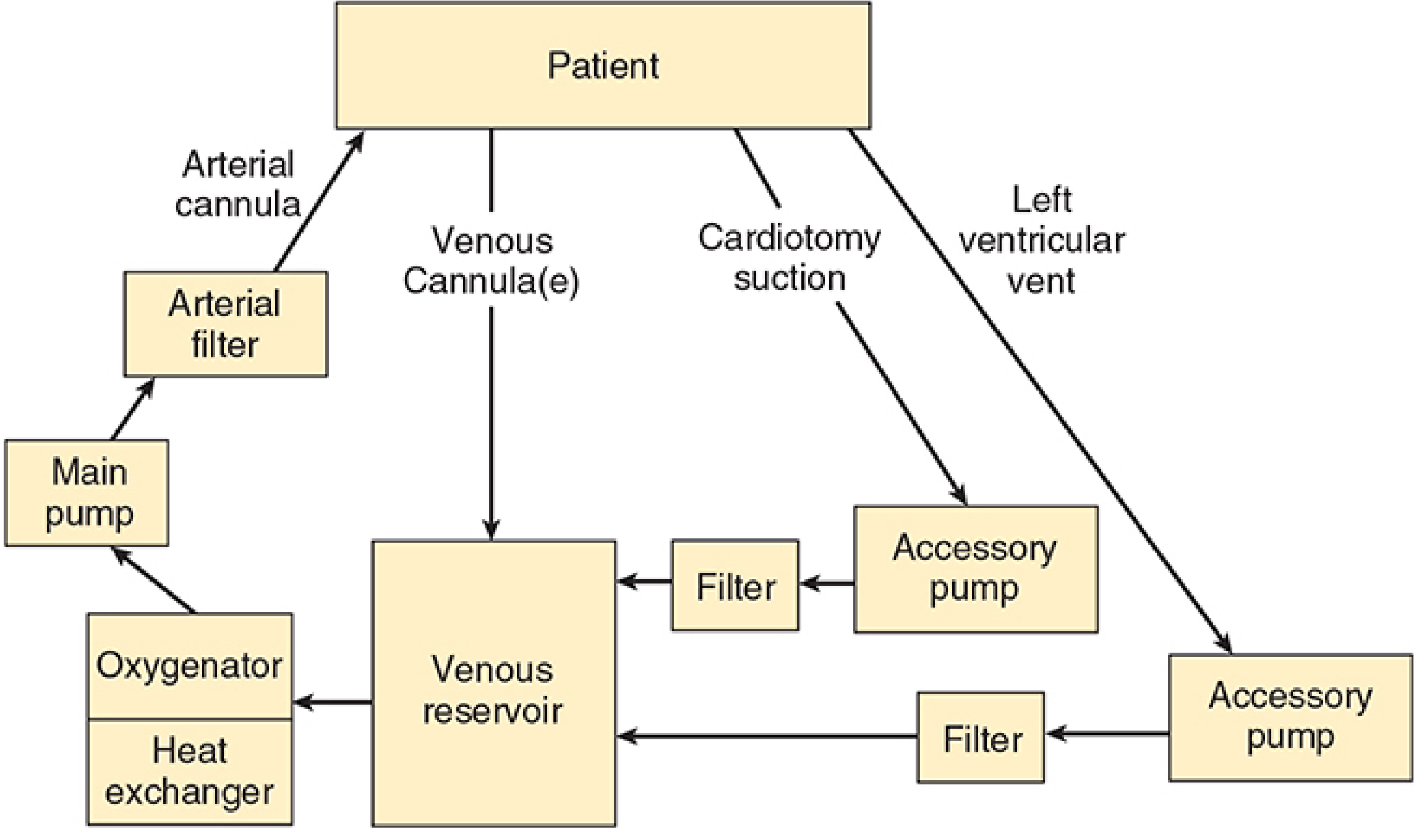
1. Definition & Purpose
CPB diverts venous blood away from the heart (via cannulae in the right atrium/vena cavae), oxygenates it, removes CO₂, and returns it via an arterial cannula (usually ascending aorta or femoral artery). It provides both artificial circulation and ventilation, allowing the surgeon a bloodless, motionless field.
Key non-physiological features:
- Mean arterial pressure lower than normal
- Flow is usually non-pulsatile
- Varying degrees of systemic hypothermia
- Hemodilution from circuit priming
2. Components of the CPB Circuit
| Component | Function |
|---|---|
| Venous reservoir | Receives deoxygenated blood by gravity drainage from RA/IVC/SVC; acts as buffer |
| Oxygenator | Gas exchange via thin silicone gas-permeable membrane; volatile agent can be added |
| Heat exchanger | Cools/rewarems blood by water conduction; filter traps bubbles during rewarming |
| Main pump | Propels blood (roller or centrifugal) |
| Arterial line filter | Traps particulate/gaseous emboli before blood returns to patient |
| Accessory pumps | Cardiotomy suction, LV vent, cardioplegia delivery |
Pump Types — Key Exam Distinction
| Feature | Roller pump | Centrifugal pump |
|---|---|---|
| Mechanism | Positive displacement; compresses tubing | Spinning cones — centrifugal force |
| Flow | Fixed per RPM (non-pulsatile) | Pressure-sensitive; needs flowmeter |
| Air embolism risk | Can pump air — dangerous if reservoir empty | Cannot pump air — safer |
| Haemolysis | More trauma to RBCs | Less traumatic |
| Position in circuit | After oxygenator | Between reservoir and oxygenator |
3. Circuit Priming
- Volume: 1200–1800 mL for adults (crystalloid — usually lactated Ringer's)
- Common additives: colloid (albumin/starch), mannitol (diuresis), heparin (500–5000 units), bicarbonate
- Causes hemodilution → haematocrit falls to 22–27% at onset of bypass
- Blood added to prime for: neonates, infants, severely anaemic adults
4. Anticoagulation for CPB
Heparin
- Required to prevent clotting in the extracorporeal circuit
- Dose: 300–400 units/kg IV (target ACT > 400–480 seconds before bypass)
- Mechanism: potentiates antithrombin III → inhibits factors IIa (thrombin), Xa, IXa, XIa, XIIa
- Monitoring: Activated Clotting Time (ACT)
- Normal ACT: < 130 s
- Target on CPB: > 400–480 s (most centres use > 480 s)
Protamine Reversal
- Dose: ~1 mg per 100 units of heparin given (typically 3–4 mg/kg)
- Adverse reactions:
- Hypotension (most common) — from histamine release or complement activation
- Anaphylaxis (especially in patients with fish allergy or prior protamine exposure, e.g. NPH insulin users)
- Pulmonary hypertension (catastrophic — complement-mediated thromboxane release)
- If heparin not adequately reversed → coagulopathy post-bypass
5. Myocardial Protection
Goal: provide bloodless, motionless field while minimising ischaemic injury.
Cardioplegia
- Chemical solution that arrests myocardial electrical activity
- Delivered antegrade (into aortic root/coronary ostia) or retrograde (via coronary sinus)
- Composition:
- High potassium (20–40 mEq/L) → depolarises and arrests the heart in diastole
- Cold temperature (4°C) reduces metabolic demand
- Other additives: blood, bicarbonate, magnesium, glutamate, aspartate
| Cardioplegia Type | Contents | Notes |
|---|---|---|
| Cold crystalloid | Saline + K⁺ | Standard; arrest + hypothermia |
| Cold blood | 4:1 blood:crystalloid | Better oxygen delivery |
| Warm blood (Buckberg) | Normothermic blood + K⁺ | Continuous delivery; induction/terminal warm shot |
| Del Nido | Crystalloid-based | Popular in paeds/adults; single dose lasts ~60 min |
Topical hypothermia
- Ice slush around the heart provides additional protection against rewarming
6. Temperature Management
| Phase | Core Temp | Purpose |
|---|---|---|
| Mild hypothermia | 32–35°C | Reduces metabolic demand |
| Moderate hypothermia | 25–32°C | Most CABG/valve surgery |
| Deep hypothermia (DHCA) | ≤18°C | Allows circulatory arrest (complex congenital, aortic arch) |
- DHCA at 15–18°C allows up to 60 minutes of safe circulatory arrest
- Brain protection during DHCA: ice packing around head, methylprednisolone 30 mg/kg, mannitol 0.5 g/kg
pH Management During Hypothermia
| Strategy | Principle | Use |
|---|---|---|
| Alpha-stat | Maintain pH 7.40 at 37°C uncorrected; allows cerebral autoregulation | Adults; most CABG |
| pH-stat | Add CO₂ to maintain pH 7.40 corrected for actual temperature; causes cerebral vasodilation | Better neurological outcomes in paediatric DHCA |
7. Conduct of CPB — Phases
Pre-bypass
- Heparinisation (confirm ACT > 400–480 s before cannulation)
- Arterial cannula inserted first (ascending aorta), then venous (RA or bicaval)
- TEE inserted; lines zeroed; baseline ACT, ABG, K⁺, glucose
Initiation of Bypass
- Surgeon: "Going on bypass"
- Anesthesiologist: stops ventilation (lungs collapse), turns off volatile agent (will be delivered via oxygenator)
- Perfusionist: gradually increases pump flow to 2.2–2.4 L/min/m² (full flow)
- Monitor: arterial line pressure, venous reservoir level, SvO₂, urine output, temperature
On Bypass
- Maintain MAP 50–80 mmHg — use phenylephrine (vasoconstriction) or sodium nitroprusside (vasodilation)
- Acceptable haematocrit: ≥21–25% (lower targets may be acceptable with hypothermia)
- Glucose: maintain < 180 mg/dL (hyperglycaemia worsens neurological outcome)
- K⁺ must be monitored frequently (cardioplegia loads potassium → hyperkalaemia risk)
- Cross-clamp applied to ascending aorta before cardioplegia delivery
- Pulmonary artery catheter must be withdrawn 2–3 cm (migrates distally during CPB → risk of PA rupture)
- Volatile agent delivery via oxygenator vaporiser (prevent awareness on CPB)
- Cooling → surgery → rewarming
Weaning from Bypass
Sequence:
- Rewarm to ≥37°C nasopharyngeal (bladder/rectal ≥35°C)
- Resume ventilation (confirm bilateral air entry, ETCO₂, good lung compliance)
- Defibrillate if needed (most hearts restart spontaneously or with defibrillation at 20 J)
- Ensure: sinus rhythm (or pace at 80–90 bpm), adequate filling, acceptable ABG/electrolytes
- Gradually reduce pump flow while heart takes over
- Decannulate → protamine administration (slow IV over 10–15 min; surgeon should be warned as it can cause pulmonary hypertension)
Failure to wean — causes: inadequate surgical repair, poor myocardial protection, metabolic derangement, tamponade. Management: inotropes, IABP, LVAD.
8. Monitoring During CPB
| Monitor | Purpose |
|---|---|
| Arterial line (radial/femoral) | Continuous MAP (radial may underread post-bypass) |
| CVP | Trend; also venous cannula function |
| PAC (if used) | LV filling, CO, SvO₂ |
| TEE | Ventricular function, de-airing, surgical result, valves |
| ACT | Adequacy of heparinisation |
| ABG, electrolytes | Acid-base, K⁺, Ca²⁺, glucose |
| Urinary output | Renal perfusion (target > 0.5 mL/kg/h) |
| Temperature | ≥2 sites: core (nasopharyngeal/bladder) + peripheral |
| Cerebral oximetry (NIRS) | Brain O₂ delivery; especially for aortic arch/carotid disease |
| Transcranial Doppler | Emboli detection; associated with neurocognitive decline |
9. Physiological Consequences of CPB
| System | Effect | Mechanism |
|---|---|---|
| Inflammatory | SIRS — activation of complement, cytokines, neutrophils | Blood–artificial surface contact |
| Haematological | Dilutional coagulopathy; platelet dysfunction; ↓ clotting factors | Hemodilution + activation/consumption |
| Renal | AKI — oliguria, ATN | Low flow, emboli, inflammatory mediators |
| Neurological | Stroke, cognitive dysfunction ("pumphead") | Macroembolism, microemboli, hypoperfusion |
| Pulmonary | Post-pump lung — ↑ interstitial fluid, atelectasis, ↑ A-a gradient | Inflammatory mediators, ischaemia-reperfusion |
| Endocrine | Hyperglycaemia, ↑ cortisol, ↑ catecholamines | Surgical stress response |
| GI | Gut ischaemia, pancreatitis | Non-pulsatile flow, emboli |
10. Complications of CPB
| Complication | Key Points |
|---|---|
| Air embolism | Roller pump + empty reservoir; deairing manoeuvres (Trendelenburg, needle aspiration) |
| Massive gas embolism | Oxygenator failure; immediate circulatory arrest, retrograde perfusion |
| Neurological injury | Stroke (~1–3% CABG), cognitive dysfunction (20–40%) |
| Coagulopathy | Dilution + platelet dysfunction + residual heparin; treat with FFP, platelets, cryoprecipitate, protamine |
| Protamine reaction | Hypotension, anaphylaxis, pulmonary HTN |
| Haemolysis | Red urine — excessive suction, prolonged bypass, roller pump |
| Vasoplegic syndrome | Severe vasoplegia post-CPB — treat with noradrenaline ± vasopressin ± methylene blue |
| Aortic dissection | Aortic cannulation complication |
| Low cardiac output | Poor myocardial protection, MI, inadequate repair |
11. Anesthetic Agents During CPB
- Induction: Etomidate (haemodynamic stability), ketamine (in compromised patients), midazolam, fentanyl/sufentanil, propofol
- Maintenance on CPB: Volatile agent via oxygenator (isoflurane, sevoflurane) + fentanyl infusion
- Awareness risk on CPB is real — BIS monitoring and volatile agent monitoring in gas outflow line are useful
- Volatile agent preconditioning: isoflurane/sevoflurane protect against ischaemia-reperfusion injury (reduce infarct size)
- Muscle relaxants: non-depolarising (pancuronium, rocuronium); avoid succinylcholine after CPB (hyperkalaemia risk with rhabdomyolysis)
12. Special Populations
Paediatric CPB
- Circuit volume is ~3× infant's blood volume → blood priming required
- Higher flow rates: up to 200 mL/kg/min
- MAP lower: 20–50 mmHg acceptable
- DHCA used for complex congenital repairs
- pH-stat preferred for neurological protection during DHCA
- Modified ultrafiltration (MUF) post-bypass: removes inflammatory cytokines, reduces oedema
- Intense inflammatory response → corticosteroids given prophylactically
- Ca²⁺ replacement more important (impaired calcium homeostasis in young children)
HIT (Heparin-Induced Thrombocytopaenia)
- Absolute contraindication to heparin
- Alternatives: bivalirudin (direct thrombin inhibitor; preferred for CPB), argatroban
- Monitor: ACT or ecarin clotting time
13. High-Yield Exam Facts
| Fact | Value |
|---|---|
| ACT target before CPB | > 400–480 s |
| Normal ACT | < 130 s |
| Heparin dose | 300–400 units/kg |
| Protamine dose | ~1 mg/100 units heparin |
| Circuit prime volume (adult) | 1200–1800 mL |
| Hct after prime | 22–27% |
| Target pump flow | 2.2–2.4 L/min/m² |
| MAP target on CPB | 50–80 mmHg |
| Safe DHCA time at 15°C | up to 60 minutes |
| PA catheter: withdraw by | 2–3 cm during CPB |
| Cardioplegia K⁺ | 20–40 mEq/L |
Sources:
- Morgan and Mikhail's Clinical Anesthesiology, 7e, Chapter 22 (pp. 811–860)
- Barash, Cullen & Stoelting's Clinical Anesthesia, 9e, Chapter 39 (pp. 3281–3360)
- Miller's Anesthesia, 10e, relevant CPB sections
This is a shared conversation. Sign in to Orris to start your own chat.