Complete ulna bone
Reading File
Finding Sources
Searching Images
ulna bone anatomy complete labeled diagram
Reading File
Finding Sources
Loading Image
Reading File
Searching Images
human ulna bone labeled anterior posterior surfaces borders shaft styloid process
Reading File
Reading File
Loading Image
The ulna is one of the two long bones of the forearm (medial side in anatomical position, little-finger side).
Complete ulna anatomy (quick summary)
-
Type: Long bone
-
Location: Medial forearm, parallel to radius
-
Main parts:
- Proximal end
- Olecranon process (forms tip of elbow, triceps insertion)
- Coronoid process
- Trochlear (semilunar) notch for articulation with humeral trochlea
- Radial notch for proximal radioulnar joint
- Shaft
- Triangular in most of proximal/mid part
- 3 borders: anterior, posterior, interosseous
- 3 surfaces: anterior, posterior, medial
- Interosseous border gives attachment to interosseous membrane
- Distal end
- Head of ulna
- Styloid process (ulnar collateral ligament attachment)
- Articulates with radius and TFCC region; ulna is separated from carpal bones by articular disc
- Proximal end
-
Function
- Major stabilizing/load-sharing bone of forearm
- Key in pronation/supination with radius via proximal and distal radioulnar joints
Source: Imaging anatomy text and atlas Volume 3, pp. 96-97.

If you want, I can give you this as:
- exam-style labeled points,
- mnemonics, or
- an OSPE viva answer format.
Complete ulnar bone ulnar bone image
Complete ulnar bone (ulna)
The ulna is the medial long bone of the forearm (little-finger side in anatomical position).
It has upper end, shaft, and lower end.
It has upper end, shaft, and lower end.
1) Upper (proximal) end
- Olecranon process
- Forms the bony tip of elbow
- Insertion of triceps brachii
- Coronoid process
- Projects anteriorly
- Gives attachment to brachialis and flexor origins
- Trochlear (semilunar) notch
- Between olecranon and coronoid
- Articulates with trochlea of humerus
- Radial notch
- On lateral side of coronoid
- Articulates with head of radius (proximal radioulnar joint)
- Ulnar tuberosity
- Distal to coronoid; brachialis insertion area
2) Shaft
- Shape: mostly triangular in cross-section proximally
- Borders (3):
- Anterior
- Posterior
- Interosseous (lateral) border for interosseous membrane attachment
- Surfaces (3):
- Anterior
- Posterior
- Medial
- Provides origins/attachments for multiple forearm flexor-extensor muscles
3) Lower (distal) end
- Head of ulna
- Articulates with ulnar notch of radius (distal radioulnar joint)
- Styloid process
- Medial-posterior projection
- Attachment for ulnar collateral ligament
- Separated from carpal bones by articular disc (TFCC)
Functions
- Stabilizes forearm during pronation/supination
- With radius, transmits forces between hand and elbow
- Forms major part of elbow hinge with humerus
Ulnar bone image (labeled)
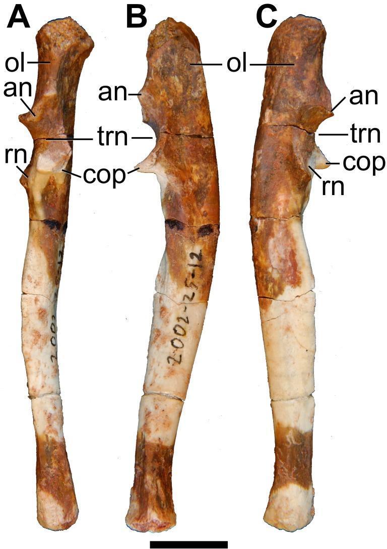
Labels in image:
- ol = olecranon
- an = anconeal region/process
- trn = trochlear notch
- cop = coronoid process
- rn = radial notch
If you want, I can also provide a clean exam diagram labeling list (exact order to label in practical exams).
This is a shared conversation. Sign in to Orris to start your own chat.