
Leukoaraiosis
Reading File
Leukoaraiosis refers to white matter changes seen on brain imaging - typically periventricular and subcortical white matter hypodensities on CT or hyperintensities on MRI (T2/FLAIR). It reflects chronic small vessel ischemic disease and is associated with cognitive decline, gait disturbance, and increased stroke risk.
Searching sources...
Finding Sources
Finding Sources
Searching PubMed
leukoaraiosis white matter hyperintensities
Good - I have relevant file paths. Let me read the most detailed neurology and radiology sections.
Reading File
Reading File
Good content. Let me get more from both files - especially the continuation of the radiology text and the clinical consequences/grading sections.
Reading File
Reading File
Now let me get the clinical consequences and grading from Bradley's neurology text, and fetch the key MRI image.
Reading File
Loading Image
Let me fetch the key MRI image from Bradley's neurology and also look for clinical consequences (cognitive decline, grading scales).
Loading Image
Finding Sources
Reading File
I now have comprehensive content. Let me also fetch the MRI image showing leukoaraiosis from the radiology textbook.
Loading Image
I have enough comprehensive information to provide a thorough answer. Here is the full evidence-based summary:
Leukoaraiosis
Definition and Terminology
Leukoaraiosis (from Greek: leuko = white, araiosis = rarefaction) is a radiological term coined by Hachinski et al. (1987) referring to diffuse or patchy signal abnormalities in the cerebral white matter. The terms white matter hyperintensities (WMH), microvascular ischemic changes, and chronic small vessel disease are used interchangeably. These are among the most common abnormal findings on MRI in the adult and elderly population.
Imaging Characteristics
CT
- Ill-defined hypodensities in the periventricular and subcortical white matter
- Sensitivity is low; MRI (especially FLAIR) is far superior
MRI - the preferred modality
- T2/FLAIR: hyperintense signal in the deep hemispheric and subcortical white matter (the primary diagnostic sequence)
- T1: may or may not show hypointensity; lesions visible on T1 may be more clinically significant
- DWI: identifies superimposed acute lacunar infarcts; new silent infarcts develop every few months in active small vessel disease
- GRE / SWI / T2*: detects associated microhaemorrhages (cerebral microbleeds) as small foci of susceptibility artefact - not visible on CT
Distribution
- Periventricular (PV) zone - particularly at frontal and occipital horns
- Deep white matter, often symmetrical in parietal and frontal lobes
- Subcortical white matter with sparing of U-fibers (arcuate fibers)
- Basal ganglia, thalami, ventral pons (when lacunar infarcts coexist)
- External capsules (also characteristically involved)
Morphology
- Punctate to small focal lesions with indistinct "cotton-wool" borders, no edema, no mass effect
- May become confluent, especially periventricularly
- A "beads-on-string" band-like pattern parallel to the lateral ventricles is characteristic
- Chronic, stable appearance - changes only very gradually on serial scans over years
Normal vs. Pathological threshold
- A uniformly thin linear periventricular rim (ependymitis granularis) at the frontal horn tips = normal age-related change
- Generally, one WMH per decade of life is considered within normal range; beyond this threshold, lesions are pathological
Pathophysiology
The dominant mechanism is gradual narrowing or occlusion of small penetrating vessels (diameter < 200 µm) - long perforating arterioles of the deep white matter. Depending on vessel calibre:
| Vessel size | Outcome |
|---|---|
| Larger perforating artery occluded | Lacunar infarct (< 1.5 cm, cavity-like when mature) |
| Smaller arterioles occluded | Ischaemic demyelination + gliosis = leukoaraiosis |
Pathologically, lesions show focal demyelination and gliosis. Vessel walls exhibit arteriosclerotic changes and commonly amyloid deposits. Lumen is narrowed or occluded. Associated central atrophy worsens with higher lesion load.
Causes
| Frequency | Cause |
|---|---|
| Common | Ischaemic microangiopathy (hypertension, diabetes, hyperlipidaemia, smoking, age) |
| Uncommon | Cerebral amyloid angiopathy (CAA), CADASIL, vasculitis |
| Rare | Fabry disease, post-radiation leukoencephalopathy, venous causes |
CADASIL (cerebral autosomal dominant arteriopathy with subcortical infarcts and leukoencephalopathy) is the key hereditary mimic - characterised by confluent WMH that characteristically involve the anterior temporal lobe white matter including subcortical U-fibers, helping distinguish it from sporadic ischaemic disease.
More than 95% of those over 65 years have some degree of white matter lesions on MRI, though usually limited in extent.
Risk Factors
- Age (incidence rises from age 40 onward)
- Hypertension (the strongest modifiable risk factor - confirmed in Framingham and Cardiac Health studies)
- Diabetes mellitus
- Hypercholesterolaemia
- Smoking
Clinical Consequences
Leukoaraiosis was initially thought to be of indeterminate significance but is now well recognised as clinically detrimental, with a dose-response relationship between lesion burden and outcomes:
| Domain | Effect |
|---|---|
| Cognition | Executive dysfunction, episodic memory impairment, decreased processing speed; associated with incident MCI |
| Gait | Progressive gait deterioration, increased fall risk |
| Urinary | Urgency incontinence (disruption of frontal-subcortical bladder control pathways) |
| Mood | Incident depression |
| Disability | Progression to functional dependence |
| Stroke risk | Marker of vascular fragility; risk factor for post-thrombolysis symptomatic intracerebral haemorrhage |
| Dementia | Risk factor for dementia after stroke; associated with vascular dementia and Binswanger disease |
When extensive and progressive with clinical dementia, the condition is termed Binswanger disease (subcortical arteriosclerotic encephalopathy).
Associated Findings
- Cerebral microbleeds: small foci of haemosiderin deposition, best seen on SWI/GRE; present in ~6% of asymptomatic older people; marker of vascular fragility
- Lacunar infarcts: mature lacunae are < 1.5 cm, sharply delineated, cavity-like; the gliotic hyperintense rim on FLAIR distinguishes them from perivascular spaces (Virchow-Robin spaces)
- Brain atrophy: progressive, correlating with WMH load
Key Imaging - Wallerian Degeneration (associated finding)
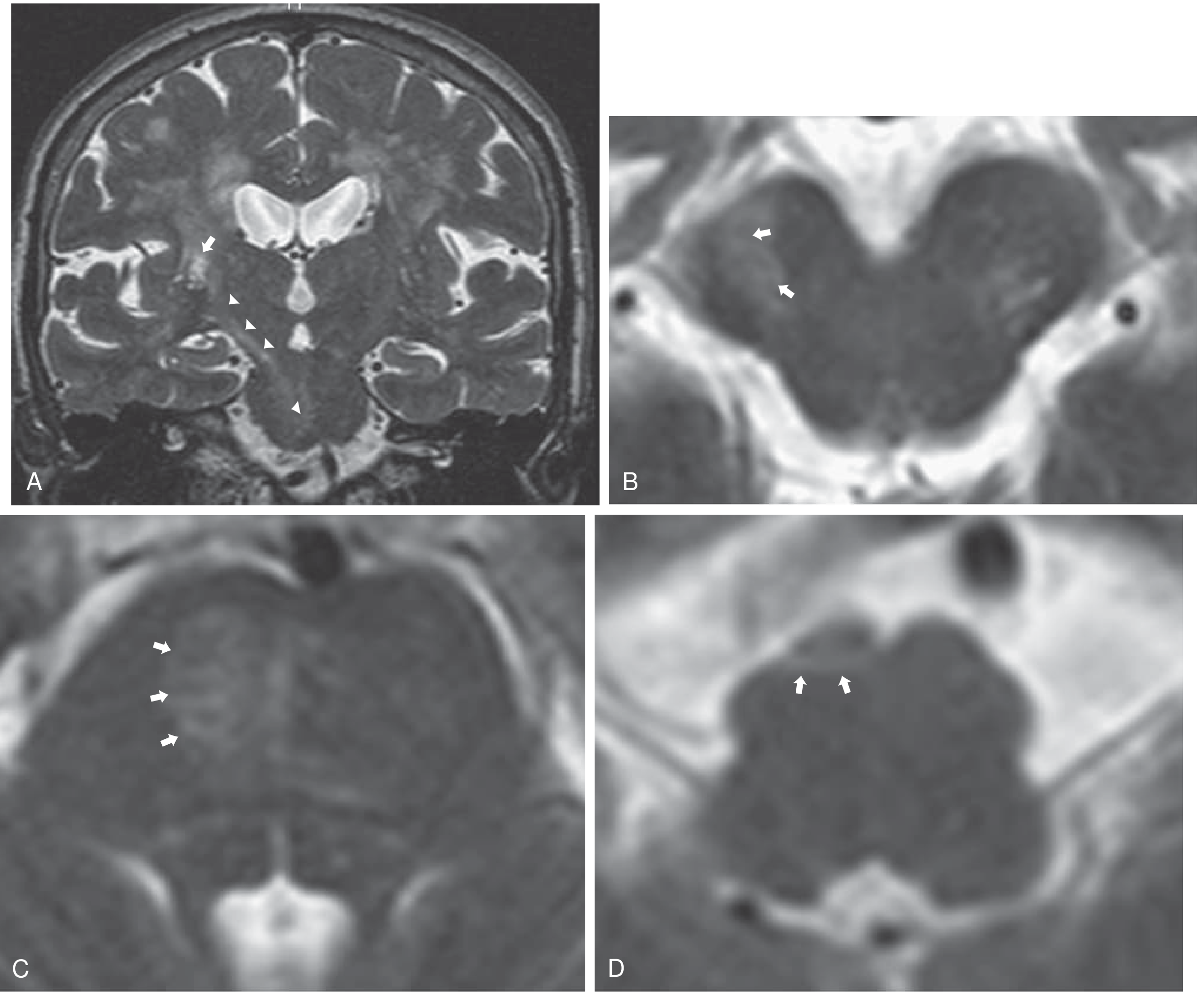
Wallerian degeneration following a right internal capsule lacunar infarct - a common coexisting finding in small vessel disease (Bradley and Daroff's Neurology, Fig. 40.39)
Management
There are no treatments that reverse leukoaraiosis once established. Management is directed at risk factor control to slow progression:
- Aggressive blood pressure control (most important)
- Glycaemic control in diabetes
- Statin therapy for hyperlipidaemia
- Smoking cessation
- Antiplatelet therapy (in ischaemic cerebrovascular disease context) - though microbleed burden may influence decisions
- Anticoagulation decisions must weigh microbleed burden and WMH severity (risk of haemorrhagic transformation)
Recent Evidence
A 2024 meta-analysis (PMID 39303876) confirmed an association between elevated pulse wave velocity (a marker of arterial stiffness) and white matter hyperintensity volume in older adults across both cross-sectional and longitudinal studies, supporting vascular stiffness as a modifiable pathway in WMH progression.
Sources: Bradley and Daroff's Neurology in Clinical Practice; Grainger & Allison's Diagnostic Radiology; Kaplan & Sadock's Comprehensive Textbook of Psychiatry; Rosen's Emergency Medicine
This is a shared conversation. Sign in to Orris to start your own chat.