
Parkinson
What aspect of Parkinson's disease are you interested in? · Who is this for?
"Parkinson disease"[MeSH Terms] AND treatment
Parkinson disease substantia nigra Lewy bodies pathology
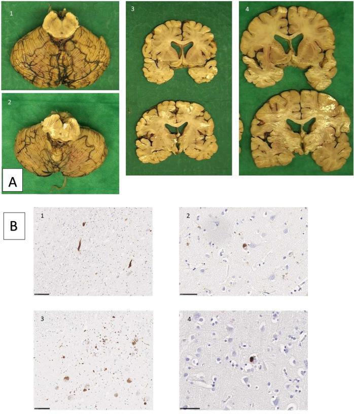
This composite image presents neuropathological findings from two siblings with Parkinson’s disease. Panel A contains macroscopic brain specimens: images 1 and 2 show the substantia nigra and cerebellum with evidence of depigmentation; images 3 and 4 display coronal sections through the basal ganglia and cortical regions, demonstrating mild ventricular enlargement and cortical atrophy. Panel B presents microscopic immunohistochemistry for alpha-synuclein pathology. Images B1 and B3 illustrate the substantia nigra, highlighting dark-stained Lewy neurites (thread-like processes) and classical Lewy bodies (rounded cytoplasmic inclusions). Images B2 and B4 show cortical sections containing cortical Lewy bodies, which appear as less defined, round inclusions within neuronal soma. The pathology demonstrates the characteristic widespread distribution of alpha-synuclein aggregates in genetic or familial Parkinson’s disease, involving both brainstem and cortical regions. This visual resource is intended for educational use in neurology and pathology to distinguish between macroscopic neurodegeneration and microscopic proteinaceous inclusions.
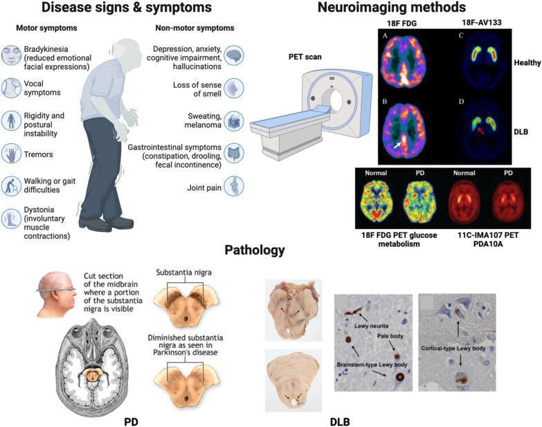
A comprehensive educational infographic summarizing the clinical features, neuroimaging, and pathology of Lewy Body (LB) diseases, specifically Parkinson's disease (PD) and Dementia with Lewy Bodies (DLB). The 'Disease signs & symptoms' section illustrates an elderly patient with classic PD posture, listing motor symptoms like bradykinesia, tremors, and rigidity, alongside non-motor symptoms such as cognitive impairment and hyposmia. The 'Neuroimaging methods' section compares PET scan modalities: 18F-FDG scans showing posterior cortical hypometabolism in DLB compared to healthy controls, and 18F-AV133 and 11C-IMA107 scans demonstrating dopaminergic and phosphodiesterase (PDA10A) alterations in PD and DLB. The 'Pathology' section features midbrain cross-sections highlighting the depigmentation of the substantia nigra characteristic of PD. It also includes histological images identifying hallmark protein aggregates: brainstem-type Lewy bodies, cortical-type Lewy bodies, Lewy neurites, and pale bodies. This diagram serves as a diagnostic and pathobiological overview of synucleinopathies for medical education.
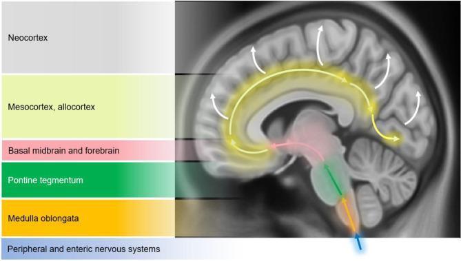
This medical schematic diagram illustrates the Braak staging model of alpha-synuclein pathology progression in Parkinson's disease. The image features a sagittal view of the human brain overlaid with color-coded regions and directional arrows to depict the caudo-rostral spread of Lewy bodies and Lewy neurites. The progression is categorized into six stages starting from a blue arrow representing the peripheral and enteric nervous systems. The pathological ascent is shown sequentially through the medulla oblongata (orange), the pontine tegmentum (green), and into the basal midbrain and forebrain (pink), which includes the substantia nigra. From these subcortical structures, yellow arrows indicate the spread to the temporal mesocortex and allocortex (light yellow), eventually reaching the neocortex (grey) as shown by white arrows. This visual model highlights how neurodegeneration moves from the lower brainstem to higher cortical regions, correlating with the clinical transition from motor symptoms to cognitive dysfunction and dementia.
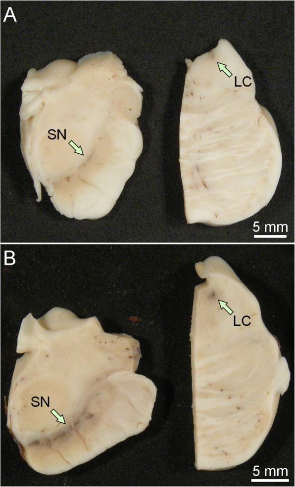
This clinical photograph shows a macroscopic comparison of post-mortem human brain tissue specimens from the midbrain and pons. The image is divided into two panels: (A) representing a brain with Lewy body disease and (B) representing a healthy control. Green arrows indicate two critical anatomical landmarks: the substantia nigra (SN) in the midbrain sections and the locus coeruleus (LC) in the pontine sections. In the control sample (B), both the SN and LC exhibit distinct, dark pigmentation due to the presence of neuromelanin. In the Lewy body disease sample (A), there is a significant and visible loss of this neuromelanin pigment, resulting in a pale appearance of these nuclei. This depigmentation is a hallmark pathological finding in synucleinopathies such as Parkinson's disease and Dementia with Lewy Bodies, reflecting the progressive loss of dopaminergic neurons in the substantia nigra and noradrenergic neurons in the locus coeruleus. A 5 mm scale bar is provided for anatomical reference.
Parkinson's Disease - Medical Student Overview
Epidemiology
Pathophysiology
Clinical Features
Motor (the classic triad - remember "TRB")
| Feature | Description |
|---|---|
| Tremor | Resting ("pill-rolling"), 4-6 Hz, asymmetric onset; presenting symptom in ~70%. Asymmetric rest tremor is virtually pathognomonic of PD. |
| Rigidity | Lead-pipe or cogwheel (ratchety resistance on passive movement). |
| Bradykinesia | Slowness of movement - often described by patient as "weakness" despite normal strength. |
Additional motor features
- Micrographia (small handwriting)
- Masked facies (reduced facial expression)
- Shuffling, narrow-based gait with reduced arm swing
- Postural instability - typically a late finding; if early, consider Parkinson-plus
Non-motor features (often overlooked but common)
- Dysphagia - subjective in ~35%, objective (videofluoroscopy) in up to 82%. Silent aspiration occurs in 15-33%
- Autonomic dysfunction: orthostatic hypotension, constipation, bladder dysfunction
- Sleep disorders: REM sleep behavior disorder (may precede motor symptoms by years)
- Anosmia (hyposmia)
- Depression and anxiety
- Dementia - occurs in advanced disease; if dementia is early, consider DLB
Diagnosis
- Bradykinesia PLUS at least one of: rigidity, 4-6 Hz rest tremor, or postural instability
- Exclusion of other causes
- Supporting features: asymmetric onset, rest tremor, excellent levodopa response
Differential Diagnosis
| Condition | Key distinguishing features |
|---|---|
| Essential tremor | Action/intention tremor (not resting), usually symmetric, no rigidity or bradykinesia |
| Progressive supranuclear palsy (PSP) | Vertical gaze palsy (esp. downward), early falls, axial rigidity, poor levodopa response |
| Dementia with Lewy bodies (DLB) | Early dementia + visual hallucinations; levodopa may help somewhat |
| Multiple system atrophy (MSA) | Autonomic failure + cerebellar/parkinsonian features; poor levodopa response |
| Vascular parkinsonism | Step-wise progression, lower-body predominance, MRI shows infarcts; poor levodopa response |
| Drug-induced parkinsonism | Key: usually reversible. Caused by dopamine-blocking drugs: neuroleptics, metoclopramide, prochlorperazine, haloperidol, amiodarone, valproic acid |
Up to 20% of patients initially diagnosed with PD ultimately have an alternative diagnosis. - Textbook of Family Medicine 9e
Neuropathology (visual)
Treatment
Principle
Drug Classes
1. Levodopa + Carbidopa (Sinemet) - Gold Standard
- Levodopa is the dopamine precursor; it crosses the blood-brain barrier (dopamine itself does not)
- Carbidopa is a peripheral DOPA decarboxylase inhibitor - it prevents levodopa from being converted to dopamine in the periphery, reducing side effects (nausea, vomiting, orthostatic hypotension) and increasing CNS availability
- In the US: levodopa/carbidopa = Sinemet; in other countries: levodopa/benserazide = Madopar
- Also available with a COMT inhibitor as Stalevo (levodopa + carbidopa + entacapone)
- Remains the most effective treatment and the benchmark for all new therapies
- Wearing-off: duration of benefit progressively shortens with time; benefits begin to mirror the drug's short half-life (60-90 min)
- On-off phenomenon: unpredictable sudden switches from mobile "on" to parkinsonian "off" state
- Peak-dose dyskinesias: involuntary choreiform movements at peak plasma concentration; can be disabling in advanced disease
- Harrison's Principles of Internal Medicine 22E
2. Dopamine Agonists
- Act directly on striatal dopamine receptors; used as monotherapy in early disease or as adjuncts to levodopa
- Examples: pramipexole, ropinirole (non-ergot, preferred); bromocriptine (ergot)
- Longer half-life than levodopa - less likely to cause wearing-off and dyskinesias when used early
- Side effects: nausea, somnolence, hallucinations, impulse control disorders (gambling, hypersexuality)
3. MAO-B Inhibitors
- Block monoamine oxidase type B, which metabolizes dopamine in the brain - extend dopamine's effect
- Selegiline, rasagiline, safinamide
- Used in early disease as monotherapy or as adjuncts; modest symptomatic benefit
- Possible (unproven) neuroprotective effect
4. COMT Inhibitors
- Block catechol-O-methyltransferase, which metabolizes levodopa and dopamine
- Entacapone (peripheral), tolcapone (central + peripheral; hepatotoxic - requires monitoring)
- Used to smooth out wearing-off fluctuations in patients on levodopa
5. Anticholinergics
- Benztropine, trihexyphenidyl
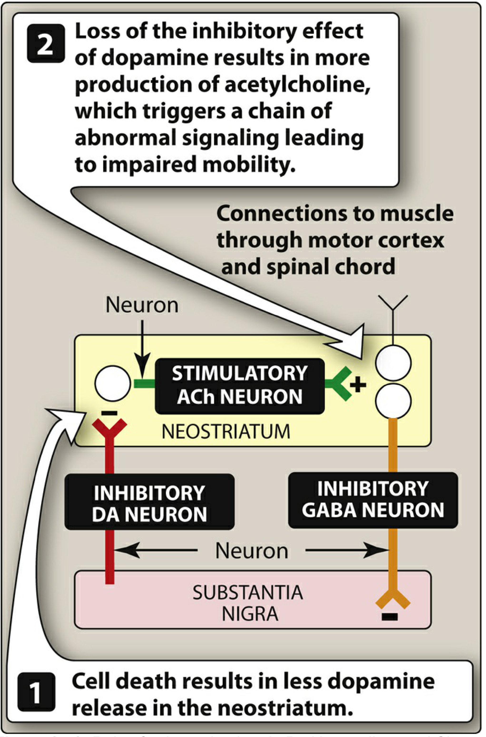
- Reduce cholinergic overactivity that results from dopamine deficiency
- Most useful for tremor in young patients; avoid in elderly due to cognitive side effects (confusion, urinary retention, constipation)
6. Amantadine
- NMDA receptor antagonist; also has weak dopaminergic and anticholinergic activity
- Useful for mild early symptoms AND for treating levodopa-induced dyskinesias in advanced disease
- Side effects: livedo reticularis, ankle edema, hallucinations
7. Advanced / Surgical
- Deep Brain Stimulation (DBS) of the subthalamic nucleus (STN) or globus pallidus interna (GPi) - most effective surgical option for motor fluctuations and dyskinesias refractory to medication
- Levodopa-carbidopa intestinal gel (LCIG/Duopa) - continuous intraduodenal infusion for advanced PD with severe fluctuations
- rTMS - repetitive transcranial magnetic stimulation; under investigation, currently modest evidence
Drug-induced Parkinsonism - Important!
Non-dopaminergic Features in Advanced PD
- Falls and freezing of gait
- Autonomic failure
- Dementia
- Sleep disorders
Recent Research (2024-2026)
- GBA1 gene variants are the most common genetic risk factor for PD; a 2025 MDSGene systematic review (PMID 39927608) describes genotype-phenotype relationships - GBA1-PD tends to have earlier onset, more cognitive decline, and faster progression
- Gut microbiome is emerging as a potential contributor to PD pathogenesis; a 2025 meta-analysis in Nature Communications (PMID 40335465) using machine learning identified consistent gut microbial alterations associated with PD, reinforcing the gut-brain axis hypothesis consistent with Braak's staging model
Key Mnemonics & High-yield Points
| Point | Detail |
|---|---|
| Classic triad | Tremor (resting) + Rigidity + Bradykinesia (TRB) |
| Rest tremor | 4-6 Hz "pill-rolling"; asymmetric onset virtually pathognomonic |
| Pathology | Lewy bodies = alpha-synuclein; substantia nigra pars compacta loss |
| First-line treatment | Levodopa/carbidopa (Sinemet) |
| Why carbidopa? | Prevents peripheral conversion of levodopa to dopamine; reduces nausea/hypotension |
| Motor complications | Wearing-off, on-off phenomenon, peak-dose dyskinesias |
| Dyskinesia treatment | Amantadine; consider DBS |
| Drug-induced parkinsonism | Metoclopramide, haloperidol, prochlorperazine - reversible |
| Parkinson-plus red flag | Early dementia, early falls, vertical gaze palsy, no levodopa response |