
Double bubble sign in xray
double bubble sign X-ray duodenal atresia
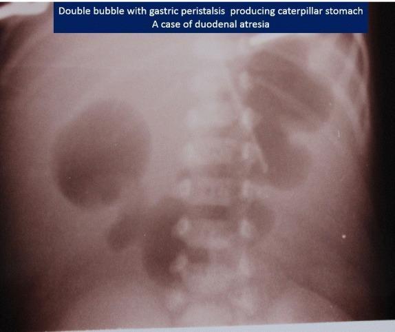
This abdominal X-ray of a neonate demonstrates the classic 'double bubble' sign, highly indicative of duodenal atresia. Two large, air-filled radiolucencies are visible in the upper abdomen: a larger gastric bubble on the patient's left and a smaller duodenal bubble on the right of the midline. A notable feature in this specific image is the 'caterpillar sign,' where the dilated gastric wall shows multiple waves of peristalsis (undulating borders), suggesting an attempt by the stomach to overcome the distal obstruction. There is a complete absence of bowel gas distal to the duodenum throughout the rest of the abdomen, signifying high intestinal obstruction. This diagnostic image is a key educational resource for pediatric radiology and neonatal surgery, illustrating the pathophysiology of congenital intestinal malformations and the radiographic markers used to differentiate them from other causes of neonatal emesis.
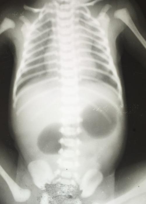
This abdominal X-ray of a neonate demonstrates the classic 'double bubble' sign, a hallmark radiographic finding for duodenal obstruction. The image reveals two prominent, rounded, radiolucent (air-filled) areas in the upper abdomen: one representing the distended stomach and the other representing the dilated proximal duodenum. These two gas bubbles are separated by the pylorus. Notably, there is a complete absence of distal bowel gas (a 'gasless' distal abdomen), which strongly suggests complete atresia rather than a partial stenosis. The skeletal structures shown, including the ribs and spine, are consistent with a newborn. In clinical practice, this presentation is most commonly associated with congenital duodenal atresia, frequently seen in infants with Trisomy 21 (Down syndrome), or other obstructive pathologies such as annular pancreas or malrotation with Ladd bands. This diagnostic image is essential for pediatric surgical evaluation and confirms a high gastrointestinal obstruction requiring immediate clinical management.
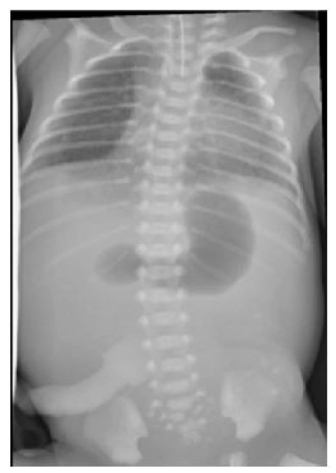
This diagnostic abdominal and chest X-ray of a neonate demonstrates the classic 'double bubble' sign, highly characteristic of duodenal atresia. The image shows two prominent, gas-distended radiolucent structures in the upper abdomen: a larger lucency on the left representing the dilated stomach and a smaller lucency to the right representing the dilated proximal duodenum. Crucially, there is a complete absence of distal bowel gas throughout the lower abdomen and pelvis, indicating a high-grade intestinal obstruction. The thoracic portion of the radiograph shows the ribcage and spine, with an orogastric tube visible, though its distal tip is not clearly identified within the stomach bubble. This imaging is a hallmark for neonatal surgical emergencies and is frequently associated with congenital conditions such as Down syndrome or VACTERL association, particularly when seen alongside tracheo-oesophageal fistula or anorectal malformations.
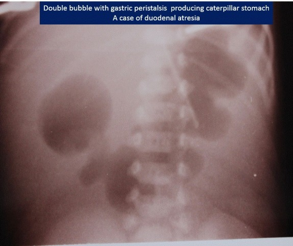
**Imaging Modality:** Plain abdominal radiograph (X-ray), supine view. **Anatomical Region:** Upper abdomen. **Observed Pathology:** Duodenal atresia. **Characteristic Visual Features:** The image demonstrates the classic "double bubble" sign, characterized by two prominent, air-filled radiolucencies in the upper abdomen. One bubble represents the dilated stomach (left), and the second represents the dilated proximal duodenum (right). A hallmark "caterpillar sign" is visible, where the gastric contour exhibits multiple undulations or hyperperistaltic waves against the obstruction, resembling the body of a caterpillar. There is a complete absence of distal bowel gas, indicating a high-grade or total intestinal obstruction. **Key Diagnostic Features:** - **Double Bubble Sign:** Diagnostic of proximal duodenal obstruction. - **Caterpillar Sign:** Indicates active gastric peristalsis attempting to overcome the distal duodenal atresia. - **Gasless Distal Abdomen:** Confirms the complete nature of the atresia, distinguishing it from partial stenosis or malrotation where some distal gas may be present. **Clinical Context:** This presentation is pathognomonic for congenital duodenal atresia in a neonatal patient.
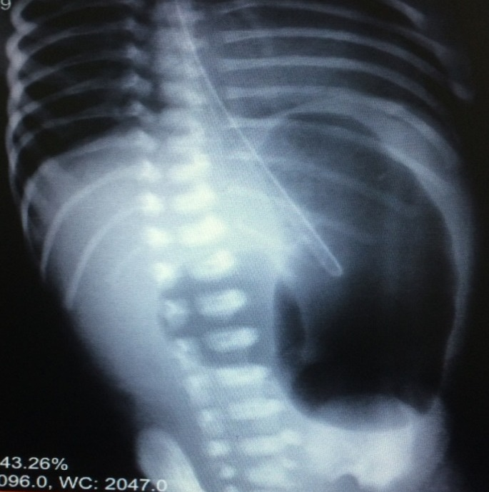
**Imaging Modality:** Plain abdominal radiograph (X-ray), anteroposterior (AP) view. **Anatomical Region:** Neonatal/pediatric abdomen and lower thorax. **Observed Pathology:** The image demonstrates the classic "double bubble" sign, highly suggestive of proximal intestinal obstruction, most commonly associated with duodenal atresia. **Characteristic Visual Features:** - **Double Bubble Sign:** Two prominent, air-filled, dilated structures are visible in the upper abdomen. The larger lucency on the left represents the dilated stomach, while the smaller lucency to the right of the midline represents the dilated proximal duodenum. - **Gas Distribution:** There is a complete absence of distal bowel gas (gasless abdomen) beyond the second "bubble," indicating a high-grade or complete obstruction. - **Supportive Structures:** An enteric (nasogastric/orogastric) tube is visible, with its distal tip positioned within the gastric lucency. - **Skeletal Landmarks:** The visible ribs and vertebral bodies show immature ossification centers consistent with a neonate. **Key Diagnostic Features:** The simultaneous dilation of the stomach and proximal duodenum with an absence of distal intestinal air is the pathognomonic radiographic hallmark of duodenal atresia, stenosis, or annular pancreas in a newborn.
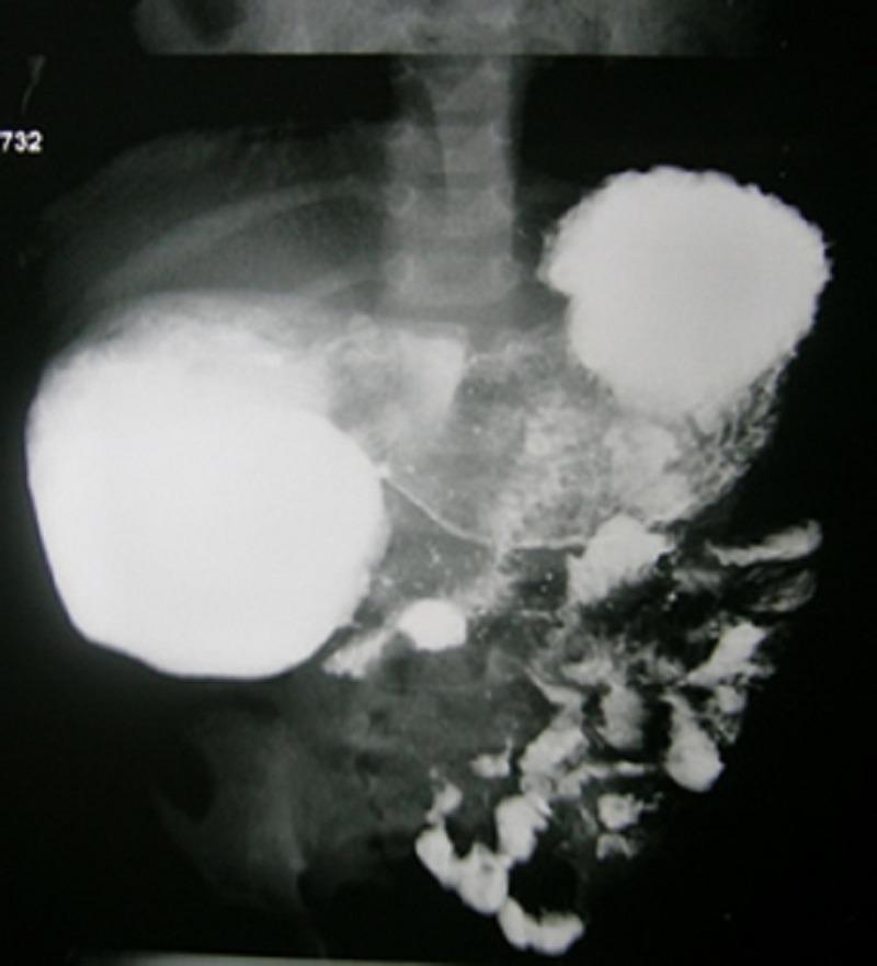
This diagnostic image is an abdominal X-ray from an upper gastrointestinal (UGI) contrast study, likely in a pediatric patient given the skeletal development. The radiograph demonstrates a 'double bubble' sign characterized by two large, well-defined, radiopaque collections of contrast material in the upper abdomen. The proximal bubble corresponds to a dilated stomach, and the second, more right-sided bubble represents a dilated proximal duodenum. Distal to these structures, there is a sparse, mottled, and fragmented distribution of contrast material within the lower intestinal loops. This pattern suggests a partial or high-grade intestinal obstruction, consistent with a fenestrated duodenal membrane or web in the second part of the duodenum. The presence of some contrast distal to the primary dilatation indicates the obstruction is not complete, distinguishing it from total duodenal atresia. Educational value lies in the radiological identification of neonatal/pediatric intestinal obstruction and the interpretation of contrast transit in congenital duodenal anomalies.
Double Bubble Sign on X-Ray
What Is It?
- Bubble 1 (larger, left): Dilated stomach
- Bubble 2 (smaller, right of midline): Dilated proximal duodenum (1st part / duodenal bulb)
- Separated by: The pylorus
- Key feature: Complete absence of distal bowel gas below the second bubble
Classic Radiographic Appearance
Primary Diagnosis: Duodenal Atresia (DA)
Embryological Basis
Location
- In 80% of cases, the obstruction is just distal to the ampulla of Vater - resulting in bilious vomiting
- Pre-ampullary obstructions (~15-20%) cause non-bilious vomiting
Differential Diagnoses (Causes of the Double Bubble)
| Cause | Notes |
|---|---|
| Duodenal atresia | Most common; complete obstruction; no distal gas |
| Duodenal stenosis / web | Partial obstruction; some distal gas may be present |
| Annular pancreas | Extrinsic ring of pancreatic tissue encircling the duodenum; clinically identical appearance |
| Malrotation with Ladd bands | Extrinsic compression; often with some distal gas |
| Preduodenal portal vein | Rare extrinsic compression |
Key distinction: Complete absence of distal gas = complete atresia. Presence of some distal gas = partial stenosis, web, or malrotation with volvulus. - Grainger & Allison's Diagnostic Radiology
Associated Conditions
| Association | Frequency |
|---|---|
| Down syndrome (Trisomy 21) | ~30% of DA patients |
| Congenital heart disease | ~20% |
| Malrotation | 20-30% |
| VACTERL association | Present in a subset |
| Oesophageal atresia (OA) | Coexists in up to 10% |
| Polyhydramnios | Common prenatal finding (proximal GI obstruction prevents fluid absorption) |
Clinical Presentation
- Neonate with repeated vomiting shortly after birth (bilious or non-bilious depending on level)
- Diagnosed prenatally on the 20-week gestation ultrasound in many cases (fluid-filled double bubble)
- Upper abdominal fullness / distension before orogastric decompression
Radiological Workup
- Plain abdominal X-ray - classic double bubble + gasless distal abdomen is sufficient to proceed to surgery without further imaging in a stable neonate - Fischer's Mastery of Surgery, p. 8125
- If distal gas is present - raises concern for: duodenal web, stenosis, or midgut volvulus (emergency!) - upper GI contrast study warranted
- If midgut volvulus suspected - take urgently to OR; do not delay for imaging
- Prenatal ultrasound - fluid-filled double bubble + polyhydramnios is the antenatal equivalent
- Tip: If orogastric decompression has already occurred, instill air via the OG tube before the X-ray to re-demonstrate the sign
Management
- Surgical correction is the definitive treatment: duodenoduodenostomy (diamond-shaped anastomosis)
- Pre-op: orogastric decompression, IV fluids, cardiac/chromosomal workup (given Down syndrome association)
- Outcomes are generally excellent with modern neonatal surgery