
Intra uterine growth retardation
Reading File
Now let me search the medical library for IUGR content in parallel.
Finding Sources
Finding Sources
Searching PubMed
"fetal growth restriction" OR "intrauterine growth retardation"
Reading File
Finding Sources
Excellent - there's a dedicated chapter in Creasy & Resnik's Maternal-Fetal Medicine. Let me pull that content now.
Finding Sources
Reading File
Finding Sources
Reading File
Finding Sources
Reading File
Reading File
Reading File
Reading File
I now have comprehensive content. Let me also read the etiology/causes section that I partially saw earlier.
Reading File
I now have comprehensive content from Creasy & Resnik's Maternal-Fetal Medicine. Let me compile the full answer.
Intrauterine Growth Retardation (IUGR) / Fetal Growth Restriction (FGR)
Source: Creasy & Resnik's Maternal-Fetal Medicine: Principles and Practice; Pfenninger and Fowler's Procedures for Primary Care
Definition
The best definition of fetal growth restriction (FGR) is a fetus that does not achieve its growth potential. The term IUGR has been largely replaced by FGR in modern literature, though they remain interchangeable clinically.
Historically, the classification used birth weight percentiles from population-based reference curves:
- Small for gestational age (SGA): birth weight < 10th percentile - used as a surrogate for FGR
- Appropriate for gestational age (AGA): 10th to 90th percentile
- Large for gestational age (LGA): > 90th percentile
However, SGA and FGR are not synonymous - many infants below the 10th percentile are simply constitutionally small, while some infants between the 10th and 90th percentiles may still have FGR if they haven't achieved their individual growth potential. This has led to interest in customized fetal growth curves that account for maternal race, height, weight, and parity.
FGR is not a homogeneous entity - different phenotypes behave differently. For instance, FGR in the context of preeclampsia has a distinct clinical course from idiopathic FGR.
Incidence
- WHO (1987): approximately 1/4 to 1/3 of all infants weighing < 2500 g at birth have sustained IUGR
- 4-8% of all infants born in developed countries
- 6-30% of infants in developing countries
Classification
Symmetric FGR (20% of FGR cases)
- All fetal measurements (head circumference, abdominal circumference, femur length) fall proportionately
- Suggests an early insult during the phase of cellular hyperplasia (early pregnancy)
- Caused by chromosomal abnormalities, congenital infections, teratogenic drugs
- Head circumference (HC) and femur length (FL) are small along with AC
- Not independently associated with increased perinatal morbidity (compared to AGA infants, when not associated with chromosomal or structural defects)
Asymmetric FGR (minority, but higher-risk group)
- Head and brain are relatively spared ("brain-sparing effect"); abdominal circumference is most affected
- Reflects later-onset placental insufficiency causing redistribution of blood to the brain
- Femur length parallels gestational age from LMP; FL may not be helpful in diagnosis
- Associated with higher risk for intrapartum and neonatal complications
Mixed FGR
- Features of both symmetric and asymmetric patterns
Staging System (Mari Staging)
The staging system classifies FGR using Doppler parameters and assists with management decisions:
| Stage | Criteria |
|---|---|
| Stage 0 | EFW < 10th percentile (or AC < 10th percentile) with normal Doppler |
| Stage 1 | UA PI or MCA PI (or both) abnormal |
| Stage 2 | Absent end-diastolic flow (AEDF) of umbilical artery |
| Stage 3 | Reversed end-diastolic flow (REDF) of umbilical artery |
| Stage 4 | Abnormal ductus venosus a-wave (absent or reversed) |
Each stage can be further labeled N (normal AFI) or A (abnormal AFI/oligohydramnios), along with gestational age and any maternal pathology.
Etiology and Risk Factors
Maternal Factors
- Pregestational diabetes with vasculopathy
- Chronic hypertensive disease, preeclampsia, gestational hypertension
- Antiphospholipid syndrome
- Chronic renal insufficiency
- Autoimmune disease (e.g., systemic lupus erythematosus)
- Severe nutritional deficiencies
- Pregnancy at high altitude
- Specific medications (e.g., beta blockers)
- Smoking, alcohol use, illicit drug use
Fetal Factors
- Multiple gestation (progressive decrease in fetal weight as number of offspring increases)
- Viral infections (rubella, CMV, varicella, toxoplasmosis, HIV, malaria, Zika virus)
- Aneuploidy: trisomy 13, 18, 21 (in descending order of severity of growth restriction)
- Structural abnormalities - 22.3% frequency of FGR in malformed infants; congenital heart defects and gastroschisis particularly associated
- Chromosomal microarray analysis (used in addition to karyotype) increases detection yield in FGR with structural anomalies by ~4-9%
Placental Factors
- Placental insufficiency / dysfunction (most common cause of asymmetric FGR)
- Placental abnormalities (e.g., circumvallate placenta, bilobed placenta, placenta previa)
Perinatal Mortality and Morbidity
- FGR is associated with increased fetal and neonatal mortality and morbidity at every gestational age
- Risk increases with severity of restriction
- Asymmetric FGR cases have higher risk for intrapartum and neonatal complications
- Combination of oligohydramnios + IUGR portends less favorable outcome
- Long-term consequences include: cerebral palsy (especially in severe or preterm FGR), neurodevelopmental impairment, and increased risk of adult diseases (cardiovascular disease, hypertension, diabetes - Barker/DOHaD hypothesis)
Diagnosis
Clinical Signs
- Poor increase in fundal height (> 4 cm difference from expected fundal height)
- Inadequate maternal weight gain (< 100-200 g/week in the third trimester)
- Clinical diagnosis alone is possible in only ~33% of pregnancies
Ultrasonography (preferred modality)
Ultrasound is far more accurate, especially if gestational age dating is precise (CRL at 9-11 weeks is most accurate - error ±5-7 days).
Key biometric parameters:
| Parameter | Role in IUGR |
|---|---|
| Abdominal Circumference (AC) | Most sensitive single measurement - affected early in asymmetric FGR as liver glycogen depleted |
| Head Circumference (HC) | More shape-independent than BPD; spared in asymmetric FGR; used in HC/AC ratio |
| Biparietal Diameter (BPD) | Not helpful alone; may underestimate GA in symmetric IUGR |
| Femur Length (FL) | Misleading in asymmetric FGR (parallels GA from LMP); low in symmetric FGR |
| Estimated Fetal Weight (EFW) | Hadlock formula most used in USA; predictive accuracy ±14.8-20.2% (±2 SD) |
HC/AC ratio is particularly useful for distinguishing symmetric from asymmetric FGR.
Oligohydramnios
- Sensitivity for IUGR diagnosis: ~16% in general population
- In high-risk populations: sensitivity can exceed 85%
- If oligohydramnios is present with no PROM or congenital anomalies, IUGR is the likely cause
- Oligohydramnios + IUGR = less favorable outcome; delivery should be considered at ≥ 36 weeks
Doppler Velocimetry
The sequence of Doppler changes in worsening FGR follows a recognizable progression:
- Umbilical artery (UA) PI increases - placental resistance rising (Stage 1)
- Middle cerebral artery (MCA) PI decreases - cerebral vasodilation / brain-sparing (Stage 1)
- Cerebroplacental ratio (CPR) becomes abnormal - MCA PI / UA PI
- Absent end-diastolic flow (AEDF) in UA (Stage 2)
- Reversed end-diastolic flow (REDF) in UA (Stage 3) - ominous
- Ductus venosus (DV) a-wave abnormality - absent or reversed a-wave indicates impending acidemia / fetal death (Stage 4)
- Umbilical vein pulsations - late ominous sign
MCA Doppler is useful particularly after 28 weeks. DV Doppler is most useful when UA has AEDF/REDF, giving information on severity; its role in timing delivery remains controversial (TRUFFLE trial showed no immediate benefit to waiting for DV abnormalities).
Antenatal Fetal Testing
- Non-stress test (NST): Decreased short-term variability is a late finding in FGR; not a good screening test
- Biophysical profile (BPP): Includes NST + 4 ultrasound parameters (fetal movement, tone, breathing movements, AFV); score 8-10 = reassuring
- Modified BPP (NST + AFV): Acceptable screening tool
- Serial growth scans every 2-3 weeks once FGR is suspected
Management
General Principles
- Identify and treat underlying maternal conditions
- Optimize maternal nutrition
- Smoking cessation
- Low-dose aspirin (started before 16 weeks) in patients with prior FGR or placental insufficiency - can reduce recurrence risk
- Serial Doppler and growth ultrasounds every 2-3 weeks
- Antenatal corticosteroids if preterm delivery anticipated (< 34 weeks)
Delivery Timing Guidelines (> 30 weeks)
| Stage | Gestational Age for Delivery |
|---|---|
| Stage 3 (reversed DF UA) | 30-32 weeks |
| Stage 2 (absent DF UA) | 33-34 weeks |
| Stage 1 (decreased diastolic flow) | 37 weeks |
| Stage 0, EFW ≥ 3rd percentile | 38-39 weeks |
| Stage 0, EFW < 3rd percentile | > 37 weeks (controversial) |
- Delivery should not be delayed beyond 39 weeks in any FGR - risk of fetal demise rises significantly at term
- At ≥ 36 weeks with oligohydramnios + IUGR, risk of intrauterine loss may mandate delivery
Route of Delivery
- No proven advantage of elective cesarean over vaginal delivery in FGR (even preterm)
- An unfavorable cervix is not a reason to avoid induction
- For cervical ripening: mechanical methods (balloon catheter) preferred over prostaglandins in FGR; if Bishop score > 6, oxytocin is used
- In premature FGR with reversed UA flow and unfavorable cervix: option of scheduled cesarean is reasonable (many of these fetuses will not tolerate labor)
- Magnesium sulfate intrapartum if delivery < 29 weeks (reduces neurodevelopmental impairment)
- Continuous intrapartum fetal monitoring is essential
Long-Term Outcomes
- Increased risk of cerebral palsy (especially in those with associated preterm birth or severe FGR)
- Neurodevelopmental and cognitive impairment (reduced verbal IQ scores)
- Barker hypothesis / Developmental Origins of Health and Disease (DOHaD): IUGR infants are at higher lifetime risk for hypertension, cardiovascular disease, and type 2 diabetes
Recent evidence note: A 2024 systematic review on metabolomic signatures in FGR (PMID 39528475) highlights new biomarker research in FGR diagnosis that may supplement Doppler in future practice. A 2025 systematic review on antiphospholipid syndrome in pregnancy (PMID 40128683) reinforces APS as a significant modifiable etiology.
- Creasy & Resnik's Maternal-Fetal Medicine: Principles and Practice, Chapter 44
- Pfenninger and Fowler's Procedures for Primary Care, 3rd Ed.
Forcep delivery
Finding Sources
Searching PubMed
"forceps delivery" obstetric
Reading File
Reading File
Reading File
Reading File
Reading File
Finding Sources
Finding Sources
Reading File
Reading File
I now have comprehensive content from three textbooks. Let me fetch the forceps delivery figure for visual clarity.
Loading Image
Loading Image
I now have all the content needed. Here is the comprehensive answer:
Forceps Delivery
Sources: Creasy & Resnik's Maternal-Fetal Medicine; Pfenninger & Fowler's Procedures for Primary Care; Textbook of Family Medicine 9e
Introduction and History
Obstetric forceps were first used by the Chamberlen family in the 17th century but were not widely accepted until 100 years later. William Smellie was the first to systematically teach the principles of forceps delivery and was also the first to recognize their potential dangers. Today, forceps use has declined significantly as cesarean section rates have risen, leading some authorities to describe forceps operations as "a species on the brink of extinction" due to a reduction in training and experience among junior clinicians.
The Simpson forceps (and Simpson-DeLee variant) is the most commonly used for low and outlet deliveries. Elliot forceps are also used for these categories.
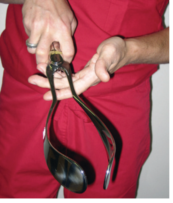
Anatomy of the Forceps
Each forceps has two mirror-image sides (left and right), each consisting of:
- Handle
- Shank
- Blade (with a cephalic curve to fit the fetal head and a pelvic curve to follow the birth canal)
The two blades articulate (lock) at the shank. The left blade is always inserted first, placed on the left side of the maternal pelvis.
Classification (ACOG 1988/1991 Criteria)
| Type | Criteria |
|---|---|
| Outlet forceps | Scalp visible at introitus without separating labia; fetal skull has reached pelvic floor; sagittal suture in AP diameter or R/L OA/OP; rotation ≤ 45° |
| Low forceps | Leading point of skull at station +2 cm or lower, not on pelvic floor; rotation ≤ 45° or > 45° (subcategories) |
| Mid-forceps | Station above +2 cm but head is engaged |
| High forceps | Head is unengaged - no longer performed in modern obstetrics |
Key clinical point: Mid-forceps deliveries (versus outlet/low forceps) are associated with lower cord pH values, higher incidence of fetal injury, and greater short-term maternal and perinatal morbidity. This distinction is critical in outcome data.
Prerequisites (ABCDEFGHIJ Mnemonic - ALSO Curriculum)
Before applying forceps, all of the following must be confirmed:
| Letter | Prerequisite |
|---|---|
| A | Anesthesia adequate (local, pudendal block, or regional) |
| B | Bladder empty (straight catheterize if needed) |
| C | Cervix completely dilated |
| D | Determine fetal head position (posterior fontanelle = Y-shape; anterior fontanelle = cross shape; feel which way the ear bends) |
| E | Equipment ready (suction bulb, cord clamp, instrument table) |
| F | Forceps ready and articulated properly |
| G | Gentle traction planned (Pajot's maneuver) |
| H | Handle elevated to follow the J-shaped pelvic curve |
| I | Incision (episiotomy) - assess need based on perineal distention |
| J | Jaw reachable = remove forceps |
Additional prerequisites from ACOG:
- Fetal head must be engaged
- Head position must be known
- Membranes must be ruptured
- No cephalopelvic disproportion
- Adequate maternal pelvis
Indications
Forceps delivery is indicated when a safe vaginal delivery is achievable but the second stage needs assistance:
Maternal indications:
- Arrest of labor in the second stage
- Maternal exhaustion
- Inability to push effectively
- Medical conditions requiring shortened second stage (cardiac disease, cerebrovascular disease, severe hypertension)
Fetal indications:
- Non-reassuring fetal heart rate pattern (fetal distress) in the second stage
- Prolapsed cord with imminent delivery
Contraindications
- Unengaged fetal head (absolute)
- Cervix not fully dilated
- Unknown fetal head position
- Fetal demineralization disorders (osteogenesis imperfecta)
- Fetal coagulopathy (e.g., known hemophilia)
- Malpresentations (face presentation with mentum posterior; brow presentation)
- Cephalopelvic disproportion
Technique (Occiput Anterior - OA Delivery)
Step 1 - Application:
- Articulate the forceps to ensure proper fit, then disarticulate.
- Take the left handle in the left hand, holding it like a pencil with the concave cephalic curve toward the vulva, shank directed upward perpendicular to the floor.
- Ease the left blade along the left side of the fetal head, using the right hand inside to protect the maternal sidewall and guide the blade into position.
- Insert the right blade symmetrically on the right side, using the left hand to guide.
- If correctly applied, the handles should fit together and lock easily.
Step 2 - Check application:
- Posterior fontanelle should be midway between the shanks (about 1 fingerbreadth above the interlocked shanks)
- Sagittal suture should be midline and midway between the shanks (perpendicular to the shanks)
- Fenestration should barely admit a fingertip between blade and fetal head
Step 3 - Traction (Pajot's Maneuver):
- The pelvic curve is a J-shaped curve from inlet to outlet
- One hand pulls the handles in the direction they extend (horizontal vector, outward)
- The other hand on the shaft pushes downward
- These two vectors sum to an outward-and-downward force initially
- As the crown moves under the symphysis, traction direction shifts upward
- Traction is applied with contractions and maternal pushing; released between contractions
Step 4 - Delivery and removal:
- Rotate head if needed to OA (done during uterine relaxation, just before a contraction; mild flexion aids rotation)
- When the jaw is reachable, remove the forceps - in the reverse order and direction of placement
- Assess the need for episiotomy based on perineal distention; removing forceps before head delivery may actually avoid the need for episiotomy
- Inspect vagina and cervix carefully for lacerations after delivery
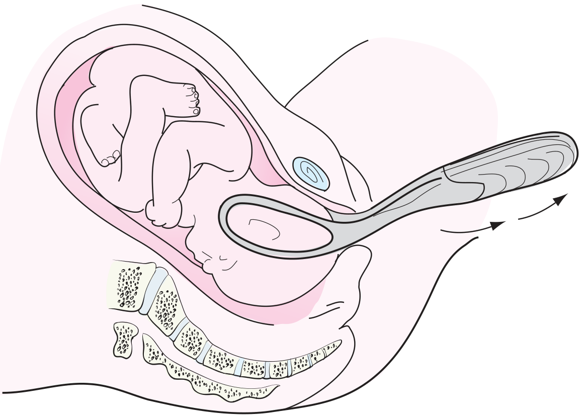
Occiput Posterior (OP) delivery modifications:
- Blades should be equidistant from the midline of the face and brow (not posterior fontanelle)
- Horizontal traction continues until the base of the infant's nose passes under the symphysis
- Greater perineal distention expected; a larger episiotomy may be needed
Complications
Maternal Complications
- Perineal, vaginal, and cervical lacerations (3rd and 4th degree tears)
- Bladder and urethral injury
- Urinary retention (identified as a risk factor in a 2024 meta-analysis, PMID 38970657)
- Postpartum hemorrhage
- Pelvic floor injury / long-term pelvic organ prolapse and incontinence
- Maternal injury is generally more frequent and more severe with forceps than with vacuum extraction
Neonatal Complications
- Facial nerve palsy (pressure on facial nerve from blades)
- Cephalohematoma (subperiosteal blood collection)
- Skull fracture (rare)
- Intracranial hemorrhage - a large California study (n=583,340) found no statistically significant difference in intracranial hemorrhage between vacuum, forceps, and cesarean delivery when fetal distress required operative delivery; however, combined use of both vacuum and forceps resulted in a rate of intracranial hemorrhage 7.4 times greater than spontaneous delivery and 3.4 times greater than vacuum alone
- Eye injuries (corneal abrasions, retinal hemorrhage - less common than with vacuum)
- Brachial plexus injury (rare)
- Sequential instrument use (vacuum then forceps) significantly increases both maternal and neonatal injury and should be avoided
Long-Term Neonatal Outcomes
- Multiple large studies found no significant difference in IQ or long-term neurodevelopmental outcomes between forceps delivery and spontaneous or cesarean delivery when modern criteria (post-1991 ACOG) are applied
- The earlier studies suggesting lower IQ scores with mid-forceps used older (1965) classification criteria - when reclassified by modern criteria, mid-forceps cases in those studies had significantly worse immediate outcomes
Forceps vs. Vacuum Extractor
| Feature | Forceps | Vacuum |
|---|---|---|
| Failure rate | Lower | Higher |
| Maternal trauma | More frequent/severe | Less frequent/less severe |
| Cervical/vaginal lacerations | More common | Less common |
| Intracranial hemorrhage | Similar | Similar (but less with vacuum alone) |
| Cephalohematoma | Less common | More common |
| Retinal hemorrhage | Less common | More common |
| Subgaleal hemorrhage | Less common | ~4% |
| Anesthesia requirement | More (regional preferred) | Less |
| Learning curve | Steeper | Easier to learn |
| Rotational ability | Yes (Kjelland's forceps) | Limited |
Current evidence favors the vacuum extractor as the instrument of first choice for operative vaginal delivery in most situations; however, forceps remain the instrument of choice in specific scenarios (e.g., face presentation with mentum anterior, preterm delivery, when rapid delivery is needed and rotation is required).
Mid-Forceps Delivery - Special Considerations
- Should be undertaken with caution
- Requires willingness to abandon the procedure and proceed to cesarean delivery if:
- Difficulty with proper application
- Head does not easily descend or rotate
- Use and teaching of mid-forceps under appropriate circumstances by adequately trained individuals is in accord with current ACOG recommendations
Creasy & Resnik's Maternal-Fetal Medicine: Principles and Practice, Chapter on Operative Vaginal Delivery
Pfenninger & Fowler's Procedures for Primary Care, 3rd Ed., Chapter 169
Textbook of Family Medicine, 9th Ed., Chapter 20
Advantage
Based on the context of our previous discussion on forceps delivery, here are the advantages of forceps delivery - drawn directly from the textbook content already retrieved:
Advantages of Forceps Delivery
Advantages Over Vacuum Extraction
| Advantage | Detail |
|---|---|
| Lower failure rate | Forceps deliver the infant more reliably; vacuum extraction fails more often and may require switching instruments |
| Rotational capability | Forceps (especially Kjelland's) can actively rotate a malpositioned head (e.g., occiput posterior or transverse); vacuum has limited rotational ability |
| Useful in specific presentations | Forceps are the instrument of choice for face presentation (mentum anterior), preterm delivery (protects the soft preterm skull), and aftercoming head in breech delivery |
| Less cephalohematoma | Vacuum is associated with more cephalohematomas and retinal hemorrhages than forceps |
| Less subgaleal hemorrhage | Subgaleal hemorrhage (~4%) is more associated with vacuum than forceps |
| Rapid delivery when urgent | Forceps can achieve delivery faster when fetal distress is acute and immediate extraction is needed |
| No cup dislodgement | Vacuum cups can pop off ("pop-off"), especially soft cups; forceps do not disengage |
| Applicable when scalp condition is a concern | Scalp lacerations and chignon from vacuum are avoided |
Advantages Over Cesarean Section
| Advantage | Detail |
|---|---|
| Less maternal morbidity in subsequent pregnancies | Forceps delivery results in less morbidity in subsequent pregnancies compared to cesarean (no uterine scar, no risk of placenta accreta spectrum, uterine rupture, or repeat cesarean) |
| Avoids surgical risks | No risk of surgical site infection, bowel/bladder injury, or anesthetic complications of general/spinal anesthesia for surgery |
| Faster recovery | Shorter hospital stay; quicker return to normal activity |
| Avoids neonatal respiratory morbidity | Infants delivered vaginally have lower rates of transient tachypnea of the newborn (TTN) and respiratory distress compared to elective cesarean |
| No abdominal incision | Preserves abdominal wall integrity |
| Lower risk when used appropriately | A large California study (n = 583,340) found that serious neonatal morbidity was significantly greater when cesarean was performed after a failed attempt at operative vaginal delivery - meaning a successful forceps delivery avoids this worst-case scenario |
General Advantages of Operative Vaginal Delivery (Forceps/Vacuum) as a Category
- Shortens the second stage of labor - beneficial when maternal pushing is ineffective (exhaustion, regional anesthesia, cardiac/cerebrovascular disease)
- Provides a controlled delivery with protection of the perineum compared to unassisted precipitous delivery
- Can be performed under regional anesthesia already in place, avoiding need for general anesthesia
Summary: When Forceps Are Specifically Preferred Over Vacuum
- Face presentation (mentum anterior) - vacuum cannot be applied to the face
- Preterm fetus (< 34 weeks) - forceps protect the soft skull better than vacuum suction
- Aftercoming head in breech delivery
- Need for active rotation > 45° (Kjelland's forceps)
- When the operator is more skilled with forceps than vacuum
- When rapid delivery is required and vacuum failure is not an option
Creasy & Resnik's Maternal-Fetal Medicine; Pfenninger & Fowler's Procedures for Primary Care, 3rd Ed.
Modes of termination of ectopic pregnancy
Finding Sources
Searching PubMed
"ectopic pregnancy" management treatment
Finding Sources
Reading File
Reading File
Finding Sources
Reading File
Finding Sources
Finding Sources
Reading File
Reading File
Reading File
Finding Sources
I now have comprehensive content from multiple sources. Here is the detailed answer:
Modes of Termination of Ectopic Pregnancy
Sources: Rosen's Emergency Medicine; Berek & Novak's Gynecology; Tintinalli's Emergency Medicine; Bailey & Love's Surgery
Overview
An ectopic pregnancy (most commonly tubal) cannot develop to term. Once implanted outside the uterus, it terminates by one of several natural (spontaneous) modes or is actively terminated by medical/surgical intervention. The natural modes are the classic "modes of termination."
Natural (Spontaneous) Modes of Termination
1. Tubal Rupture
The most dramatic and dangerous mode.
- As the trophoblast grows, it erodes through the tubal wall, causing rupture
- Leads to massive intraperitoneal hemorrhage - a surgical emergency
- Can occur with or without prior symptoms
- Cornual (interstitial) pregnancy is especially dangerous because the growing embryo can use the myometrial blood supply to grow larger (10-14 weeks) before rupture, causing catastrophic hemorrhage
- Important: Tubal rupture can occur even when β-hCG levels are falling
- Clinically: sudden severe abdominal pain, peritoneal signs, shoulder tip pain (diaphragmatic irritation from blood), haemodynamic collapse
- Site of rupture determines timing:
- Isthmic ectopic: ruptures early (~6 weeks) - narrow lumen
- Ampullary ectopic: ruptures later (~8 weeks) - wider, more distensible
- Interstitial/cornual ectopic: ruptures latest (~12-14 weeks) - surrounded by myometrium
2. Tubal Abortion
- The conceptus is extruded through the fimbriated end of the fallopian tube into the peritoneal cavity
- The trophoblast separates from the tubal wall; the embryo is expelled
- Blood leaks intermittently through the tubal wall or out of the fimbrial ends, with spillage into the peritoneal cavity
- Bleeding and symptoms are typically intermittent, unlike the sudden onset of rupture
- The extruded products may:
- Be reabsorbed spontaneously (common with small products)
- Re-implant in the peritoneal cavity as a secondary abdominal pregnancy (rare)
- Clinically: intermittent crampy pain, light-to-moderate vaginal bleeding, less acute than rupture
3. Spontaneous Resolution / Resorption (Involution)
- Some ectopic pregnancies simply die and are resorbed without rupture or abortion
- The trophoblast stops growing, β-hCG levels fall, and the pregnancy resolves spontaneously
- Most likely when:
- Initial β-hCG is very low (< 200 mIU/mL - reported 88% success rate of spontaneous remission)
- β-hCG ratio at 48 hours vs. initial level is < 0.87 (predicts a failing pregnancy of unknown location)
- Caution: Tubal rupture can still occur even as β-hCG falls - these patients must be followed with serial β-hCG until levels are undetectable
Summary Table: Natural Modes
| Mode | Mechanism | Clinical Features | Outcome |
|---|---|---|---|
| Tubal rupture | Trophoblast erodes tubal wall | Sudden severe pain, haemoperitoneum, shock | Surgical emergency |
| Tubal abortion | Expulsion through fimbrial end | Intermittent pain, peritoneal blood, adnexal mass | May resolve or re-implant |
| Spontaneous resorption | Trophoblast dies, reabsorbed | Falling β-hCG, minimal symptoms | Resolves without intervention |
Rare/Historical Natural Outcomes
| Outcome | Description |
|---|---|
| Lithopedion ("stone baby") | Dead abdominal pregnancy calcifies and is carried for years without symptoms |
| Tubal mole | Carneous mole formed in the tube after fetal death and blood clot organisation |
| Secondary abdominal pregnancy | Tubal abortion → re-implantation on peritoneum/omentum/bowel; can rarely grow to term |
Active (Treatment) Modes of Termination
1. Expectant Management
- For hemodynamically stable patients with very low and falling β-hCG (< 200 mIU/mL)
- Serial β-hCG monitoring until undetectable
- Risk of rupture persists even with falling levels - requires close follow-up
2. Medical Management - Methotrexate (MTX)
- Mechanism: Folate antagonist - kills rapidly dividing trophoblastic cells
- Single-dose protocol: MTX 50 mg/m² IM; β-hCG measured on days 4 and 7; if < 15% fall, repeat dose
- Multi-dose protocol: MTX on days 1, 3, 5, 7 alternating with folinic acid rescue
- Criteria for MTX (must meet ALL):
- Hemodynamically stable
- No signs of rupture
- β-hCG < 5000 mIU/mL (some guidelines allow up to 10,000)
- No fetal cardiac activity on ultrasound
- No contraindications (renal/hepatic disease, immunodeficiency, blood dyscrasias)
- "Separation pain" at days 3-7 is common (tubal distension from therapeutic tubal abortion/haematoma) - must distinguish from rupture pain
- A 2024 meta-analysis found letrozole alone or combined with MTX (PMID 39384737) may offer an alternative medical approach
3. Surgical Management
a) Salpingostomy (conservative)
- Linear incision over the ectopic; products removed; tube left open to heal by secondary intention
- Preserves the fallopian tube - preferred when contralateral tube is absent or damaged
- Risk of persistent trophoblast (4-15%) - requires post-op β-hCG monitoring; treated with MTX if needed
- Higher persistent ectopic rate with laparoscopic vs. open salpingostomy
b) Salpingectomy (radical/definitive)
- Removal of the entire affected fallopian tube
- Preferred when: tube is severely damaged, rupture has occurred, haemostasis is difficult, or patient has completed childbearing
- Lower risk of persistent trophoblast; lower recurrence risk in that tube
c) Approach:
- Laparoscopic (preferred when patient is stable): less blood loss, faster recovery, shorter hospital stay
- Laparotomy: for haemodynamically unstable/ruptured cases or failed laparoscopy
4. Special Cases
- Cervical ectopic: Methotrexate (systemic or local injection) is first-line; surgical intervention risks catastrophic haemorrhage
- Cornual/interstitial ectopic: Medical (MTX) preferred if diagnosed early; wedge resection or laparoscopic cornuectomy if surgery needed
- Abdominal (secondary) pregnancy: Laparotomy; placenta may be left in situ if removal risks haemorrhage
- Persistent trophoblastic tissue after salpingostomy: MTX is treatment of choice (tissue may not be confined to tube and may not be identifiable at re-operation)
Rosen's Emergency Medicine: Concepts and Clinical Practice; Berek & Novak's Gynecology; Tintinalli's Emergency Medicine: A Comprehensive Study Guide; Bailey & Love's Short Practice of Surgery, 28th Ed.
This is a shared conversation. Sign in to Orris to start your own chat.