
Minimally invasive retrosigmoid approach for petrous apex (MIRA) according to standard ENT textbook in simplified manner
retrosigmoid approach petrous apex anatomy surgical
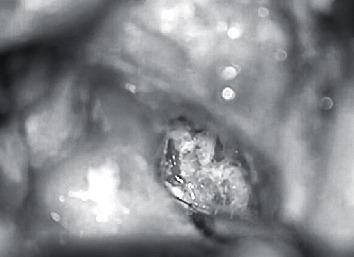
This intraoperative clinical photograph captures a surgical procedure in the otolaryngology domain, specifically an approach to the petrous apex. The image demonstrates a modified superior semicircular canal that has been surgically plugged, sealed, and partially amputated to facilitate access. The central focus is the petrous apex, where a cholesteatoma matrix is visible prior to its excision. Key anatomical landmarks include the exposed mastoid cavity and the deep surgical field of the temporal bone. The image illustrates advanced skull base surgery techniques, specifically the intentional occlusion of the semicircular canal as a pathway to reach deep-seated lesions of the petrous apex while attempting to preserve surrounding neural and inner ear structures. The photograph serves as an educational resource for understanding the surgical management of complex cholesteatomas and the necessary modifications to vestibular anatomy required for petrous apex exposure.
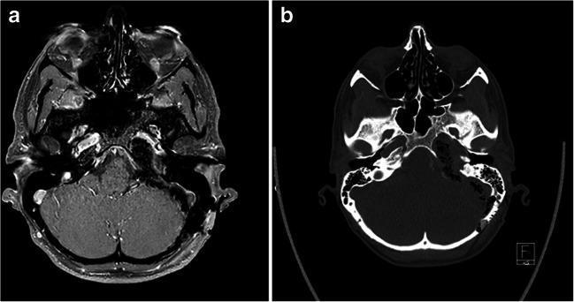
This composite figure presents postoperative neuroimaging in two modalities: (a) an axial T1-weighted MRI with Gadolinium contrast (Gd+) subtraction series and (b) a non-contrast axial CT scan. The images illustrate the result of a retrosigmoid intradural inframeatal petrosectomy (RESIP) for a grade II chondrosarcoma of the left petrous apex. In image (a), the MRI subtraction technique highlights the absence of significant pathological enhancement, confirming near-total resection of the tumor with high soft-tissue resolution of the brain parenchyma and cranial nerves. In image (b), the axial CT scan provides a detailed view of the bony anatomy of the skull base, showing the surgical defect in the left petrous bone and the extent of bone removal required to reach the petroclival space. Comparison between the two highlights the CT's superior visualization of osseous changes and the MRI's effectiveness in assessing residual soft-tissue tumor volume and inflammatory changes. The imaging confirms successful decompression and surgical management of a complex skull base lesion.
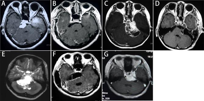
This composite of axial gadolinium-enhanced T1-weighted MRI scans illustrates various imaging classifications and postoperative outcomes for Petroclival Meningiomas (PCMs). The series compares preoperative tumor presentation (A, C, E, G) with postoperative results (B, D, F). Row 1 showcases the 'Petrous apex type' (A) with a left-sided mass near the temporal lobe, and its 'Simpson grade I' resection (B) showing complete removal via a subtemporal approach. The 'Tentorium type' (C) depicts a heterogeneous mass adjacent to the sigmoid sinus, with a 'Simpson grade II' resection (D) after a retrosigmoid approach. Row 2 highlights the 'Upper clivus type' (E), presenting a large midline mass involving the clivus, and the corresponding postoperative image (F) showing 'Simpson grade II' resection via an anterior sigmoid approach. Finally, image (G) demonstrates a 'Cavernous type' tumor involving the cavernous sinus and internal carotid artery region. The collection serves as an educational resource for neurosurgical planning, demonstrating how tumor location influences the choice of surgical approach (e.g., Kawase, retrosigmoid, subtemporal) and the achievable extent of resection.
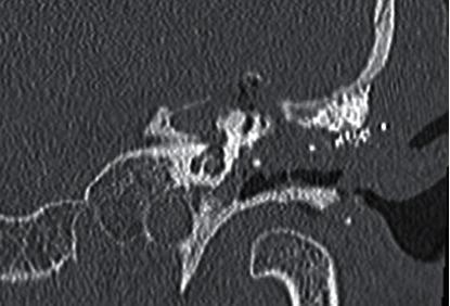
This diagnostic image is a coronal Computed Tomography (CT) scan focusing on the left temporal bone and petrous apex. The scan demonstrates postoperative changes following a surgical intervention for petrous apex pathology, such as a cholesteatoma. Key anatomical findings include an amputated superior semicircular canal, which appears truncated compared to normal morphology. There is a prominent postoperative bone defect in the petrous apex region, characterized by a loss of continuous bony structure and the absence of significant new bone formation within the cavity. Hyperdense material is visible along the superior aspect, corresponding to a titanium mesh previously implanted to stabilize the middle and posterior cranial fossae. The image illustrates the complex surgical anatomy encountered during an extended mastoidectomy and petrous apex approach. This material is highly relevant for neuroradiologists and otolaryngologists studying skull base surgery, surgical complications, and the management of deep temporal bone lesions.
MIRA - Minimally Invasive Retrosigmoid Approach for Petrous Apex
What Is MIRA?
As Cummings describes, MIRA is one of a series of neurotologic approaches that include the translabyrinthine, retrosigmoid, middle fossa, extended middle fossa, petrosal, and petrous apex drainage approaches - each chosen based on tumor size, location, and residual hearing.
- Cummings Otolaryngology Head & Neck Surgery
Anatomy You Must Know First
| Structure | Relevance to MIRA |
|---|---|
| Sigmoid sinus | Posterior boundary - retracted anteriorly |
| Posterior semicircular canal | Lateral limit of drilling - must NOT be violated |
| Internal auditory canal (IAC) | Target - posterior lip drilled to expose |
| Facial nerve (CN VII) | Monitored continuously; runs through IAC |
| Cochlear nerve (CN VIII) | Preserved for hearing |
| Cerebellum | Retracted gently after CSF drainage |
| CPA cistern | Drained of CSF to relax cerebellum |
Step-by-Step Surgical Technique (Simplified)
1. Patient Positioning
- Patient supine, head turned to the opposite side
- No head pins needed
- EMG electrodes placed in orbicularis oris and orbicularis oculi for facial nerve monitoring
- IV antibiotics given before incision
2. Skin Incision
- Curved postauricular incision, apex ~3 cm posterior to the postauricular crease
- Soft tissue and periosteum elevated from mastoid and adjacent occipital bone
- Self-retaining retractors inserted
3. Mastoidectomy (Key Distinction from Standard Retrosigmoid)
- A complete mastoidectomy is performed first - this is what makes MIRA different from a pure suboccipital approach
- The sigmoid sinus is skeletonized (bony shell removed)
- The middle fossa plate is identified and thinned superiorly
- Why do mastoidectomy? - It allows:
- Anterior retraction of the sigmoid sinus → better anterior CPA exposure
- Precise identification of the posterior semicircular canal → guides safe drilling of the posterior IAC lip
- Obliteration of mastoid air cells at closure (prevents CSF leak)
4. Bone Flap (Keyhole Craniotomy)
- A small bone flap is created posterior to the sigmoid sinus using an otologic drill
- The flap is preserved for replacement at closure
- Alternatively, where venous anatomy is favorable, a craniectomy with sinus skeletonization can be done without entering presigmoid air cells
5. Dural Opening
- Dura incised posterior to the sigmoid sinus
- Care taken to avoid injury to vessels adherent to the dura
- Critical step: Adequate CSF drainage from the CPA cistern before retracting the cerebellum
- Until sufficient CSF is drained, the cerebellum tends to herniate through the incision → risk of necrosis and hemorrhage
- Once drained, cerebellum is gently supported with retractors
6. Accessing the IAC and Petrous Apex
- Tumor in CPA visualized; if too large for facial nerve identification → debulk first
- Tumor dissected from facial nerve toward the porus acusticus
- For petrous apex extension: the posterior lip of the IAC is drilled using diamond burs
- A dural flap is created lateral to the porus and reflected medially
- Drilling continues until the lateral extent of the lesion is visible
- Hard limit: The posterior semicircular canal - drilling must stop here to preserve hearing
7. Endoscope-Assisted Component (the "Minimally Invasive" Part)
- After microscopic debulking, a 1.7 mm or 4 mm Hopkins rod (0° or 30°) is introduced through the small retrosigmoid craniotomy
- Endoscope allows visualization of:
- Intrameatal portion of the facial and cochlear nerves
- Residual tumor in the IAC fundus (not directly visible by microscope)
- Open mastoid air cells that need wax sealing
- This approach is described in Scott-Brown's as: "the aim is to obtain good control of the intrameatal portion of the nerve endoscopically, removing the residual acoustic neuroma within the inner canal"
8. Closure
- Dural edges reapproximated
- Mastoid and IAC obliterated with abdominal fat (prevents CSF leak)
- Bone flap replaced and secured with microplates or cranioplasty (hydroxyapatite cement or titanium)
- Why replace the bone flap? - Replacing it significantly reduces post-operative headaches, which are a known complication of the open retrosigmoid approach
- Wound closed in layers; compressive dressing applied
Hearing Preservation - The Key Advantage
- Success rates: 30-65% depending on patient selection
- Best results in: tumors <2 cm with good pre-op hearing and limited IAC involvement
- Principal limitation: the fundus of the IAC is not directly visualized by microscope alone (endoscope helps here)
- At the fundus, unlike the translabyrinthine approach (where the "Bill bar" definitively identifies CN VII), the facial nerve monitor is the primary guide
- Cummings: "Extension of tumor to the fundus is a relative contraindication to use of the retrosigmoid approach for hearing preservation"
Indications for MIRA at the Petrous Apex
| Lesion | Notes |
|---|---|
| Cholesterol granuloma | Most common petrous apex lesion; drainage preferred |
| Petrous apex cholesteatoma | Expansile; 94% present with hearing loss |
| Small-medium vestibular schwannoma (<2 cm) with serviceable hearing | Ideal case |
| Petroclival meningioma | Good exposure of petroclival region |
| Chondrosarcoma of petrous apex | RESIP variant used |
| CPA epidermoids | Non-enhancing; seen on DWI |
Comparison with Other Approaches
| Feature | Translabyrinthine | MIRA / Retrosigmoid | Middle Fossa |
|---|---|---|---|
| Hearing preservation | No (destroys labyrinth) | Yes (30-65%) | Yes (best for small intracanalicular) |
| Fundus visualization | Direct (via Bill bar) | Indirect (endoscope helps) | Direct |
| Tumor size | Any size | <2 cm ideal | Small intracanalicular |
| IAC drilling | Extradural | Intradural (key difference) | Extradural |
| Facial nerve risk | Lower | Moderate | Moderate |
| Headache risk | Lower | Higher (reduced by bone flap replacement) | Lower |
| Petrous apex access | Limited | Good | Limited |
Key Complications to Know
- Post-operative headache - Higher with retrosigmoid than translabyrinthine; reduced by replacing the bone flap
- CSF leak - Prevented by fat obliteration of air cells; endoscope helps find open air cells
- Cerebellar injury - From premature retraction before adequate CSF drainage
- Hearing loss - If posterior semicircular canal is violated, or blood supply to IAC disrupted
- Facial nerve injury - Monitored with EMG throughout; fundus drilling is highest-risk moment
Memory Aid (MIRA Steps)
- Positioning (supine, head turned)
- Mastoidectomy (skeletonize sigmoid, identify post. SCC)
- Dural incision (post. to sigmoid)
- CSF drainage (before cerebellar retraction)
- Drilling posterior IAC lip (to posterior SCC limit)
- Endoscope for fundus/IAC residual
- Closure with fat + bone flap replacement
- Cummings Otolaryngology Head & Neck Surgery, Chapter 179 (Arriaga & Brackmann) - Retrosigmoid Approach & Petrous Apex Lesions
- Scott-Brown's Otorhinolaryngology Head & Neck Surgery Vol 2 - Endoscopic Neurotologic Surgery chapter