
What is the use of acetabulum anteversion angle
acetabulum anteversion angle clinical significance orthopedics
acetabulum anteversion angle measurement hip joint
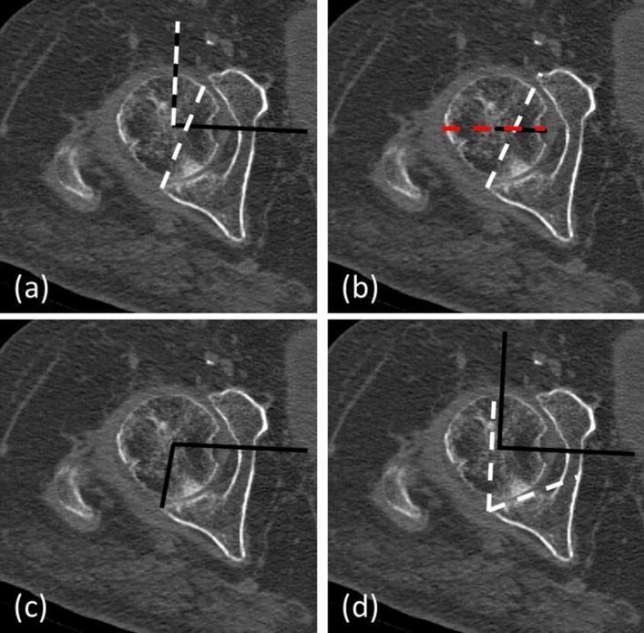
A set of four axial CT scan slices of the hip joint illustrating different methods for measuring acetabular orientation and femoral head coverage. The images show a spherical femoral head articulating with the bony acetabulum. Each panel demonstrates a specific radiological measurement: (a) Acetabular Anteversion Angle (AAA), defined by the angle between the intercapital center line (ICL90) and the anteversion line (AVL) connecting the acetabular lips; (b) Femoral head coverage/roofing, determined by the ratio of the femoral head contained within the AVL to the total femoral diameter; (c) Posterior Acetabular Sector Angle (PASA), measured from the femoral head center to the posterior acetabular wall edge; and (d) Posterior Wall Angle (PWA), determined by the angle between the ICL90 and the tangent to the posterior articular surface. These measurements are used in orthopedic surgery to assess hip stability and the risk of dislocation following Bipolar Hemiarthroplasty (BHA). The imaging modality is clinical CT, focused on musculoskeletal diagnostic assessment.
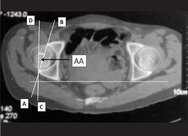
This diagnostic image is an axial computed tomography (CT) scan of the human pelvis at the level of the acetabulum, demonstrating the radiographic measurement of acetabular anteversion (AA). The scan shows the bilateral hip joints, including the femoral heads and the surrounding acetabular walls. Overlaying the right acetabulum (anatomically the patient's right, left on the image), several reference lines are drawn: Line AB connects the anterior and posterior acetabular margins, representing the orientation of the acetabular opening. Lines C and D are vertical reference lines aligned with the sagittal plane of the pelvis. The acute angle formed between the acetabular line (AB) and the sagittal reference line (CD) is labeled 'AA', indicating the degree of acetabular anteversion. This measurement is clinically significant in orthopedic surgery and radiology for evaluating hip joint morphology, diagnosing impingement or dysplasia, and planning total hip arthroplasty to ensure stable component positioning.
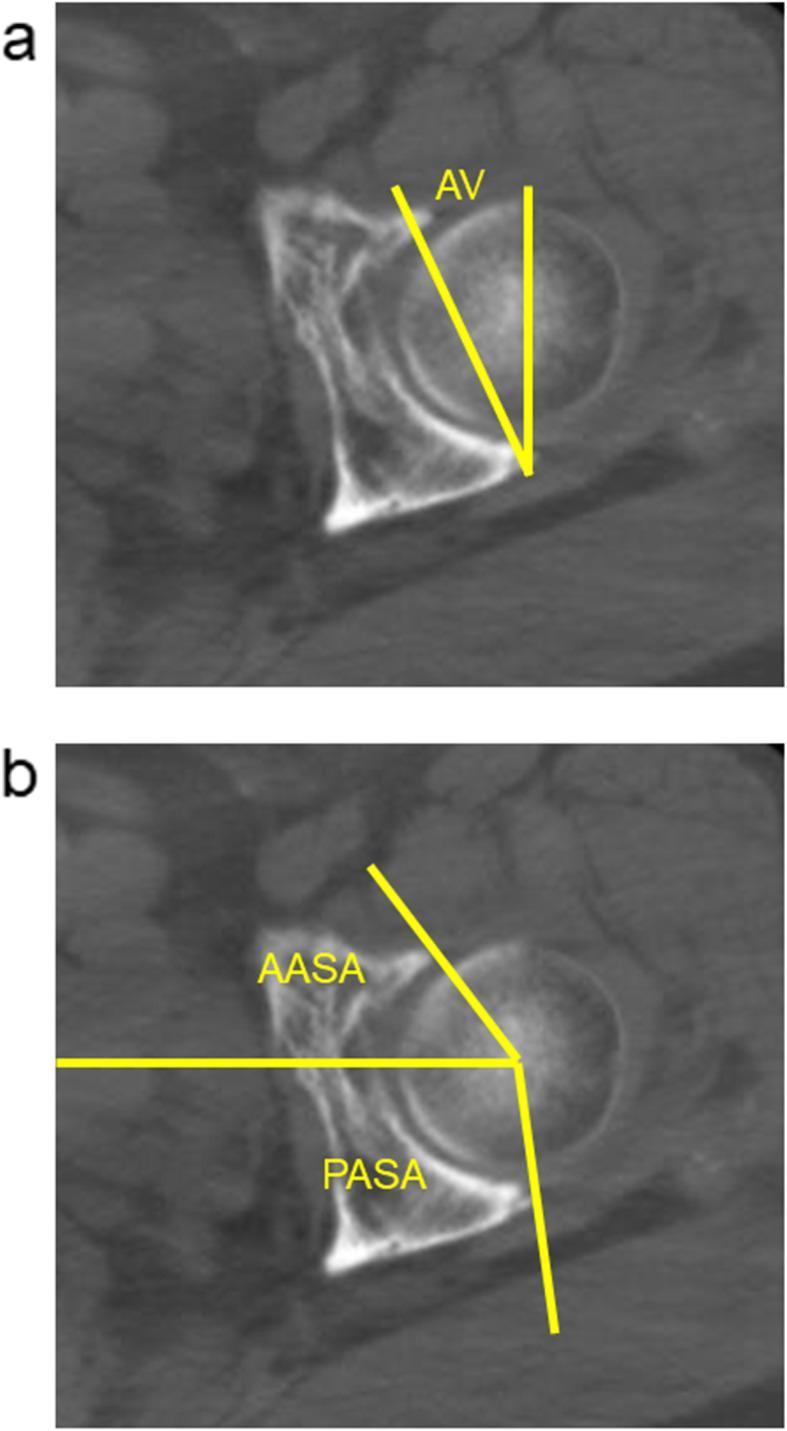
This diagnostic image consists of two axial CT slices (labeled a and b) of the human hip joint at the level of the femoral head center, demonstrating standardized methods for assessing 3D acetabular morphology. Image 'a' illustrates the measurement of acetabular anteversion (AV) using two intersecting yellow lines at the acetabular margins. Image 'b' demonstrates the measurement of the anterior acetabular sector angle (AASA) and the posterior acetabular sector angle (PASA). These angles are defined by a horizontal reference line passing through the center of the femoral head and lines extending to the anterior and posterior acetabular rims, respectively. These measurements are clinically significant in orthopedics for evaluating bony coverage of the femoral head, diagnosing acetabular dysplasia, and planning or evaluating surgical interventions such as rotational acetabular osteotomy (RAO). The images provide a high-contrast view of cortical bone against surrounding soft tissue, essential for precise morphometric analysis of the acetabulum.
Acetabular Anteversion Angle - Definition and Clinical Uses
What It Is
- Native acetabulum: approximately 15-25 degrees of anteversion
- Anterior wall covers ~19% of the femoral head; posterior wall covers ~43%
Clinical Uses
1. Assessment of Hip Stability and Femoral Head Coverage
- Femoral head coverage ratio (percent of head within the acetabular cup)
- Posterior Acetabular Sector Angle (PASA) and Anterior Acetabular Sector Angle (AASA) - derived measurements to quantify hip stability
2. Diagnosis of Acetabular Dysplasia
- Anterior undercoverage of the femoral head
- Posterior overcoverage - the posterior wall crosses laterally to the center of rotation
- Increased risk of anterior hip instability and dislocation
- Acetabular dysplasia with anterior wall deficiency (anterior wall coverage <15%)
3. Total Hip Arthroplasty (THA) - Component Positioning ("Safe Zone")
- Lewinnek's safe zone (the traditional standard): 15° ± 10° anteversion (i.e., 5-25°) combined with 40° ± 10° abduction (inclination)
- When within the safe zone: dislocation rate is ~1.5%; outside it: dislocation risk increases to 6.1% (some reports up to 61% in high-risk cases)
- Miller's Review recommends: cup anteversion 20-30°; Campbell's recommends: 10-20° of anteversion with 35-45° of abduction
| Malposition | Risk |
|---|---|
| Retroversion (too little anteversion) | Posterior dislocation, anterior impingement |
| Excess anteversion (>25-30°) | Anterior dislocation |
| High theta angle (vertical cup) | Posterior-superior dislocation, edge loading, accelerated wear |
- Miller's Review of Orthopaedics, 9th Ed, p. 4840-4858
- Campbell's Operative Orthopaedics, 15th Ed, p. 5727-5750
4. Combined Anteversion (Cup + Stem)
- Combined anteversion target: 25-50° (cup anteversion + stem anteversion)
- Stem anteversion target: 10-15°
- This concept is particularly important in hip dysplasia (DDH) reconstruction, where both components are often abnormal
5. Hip-Spine Relationship
- For every 1° of posterior pelvic tilt, acetabular anteversion increases by 0.7-0.8°
- From standing to sitting: anteversion changes by ~15.6° total
- Lumbar spinal fusion or degenerative stiffness removes this accommodation, placing THA patients at high risk for impingement or dislocation - surgeons must adjust the target cup anteversion accordingly
6. Planning Corrective Osteotomies
- Periacetabular osteotomy (PAO) - reorienting the acetabulum to normalize anteversion and improve femoral head coverage
- Rotational acetabular osteotomy (RAO)
- Adequate correction of anteversion improves joint mechanics and delays or prevents osteoarthritis
7. Postoperative Radiological Evaluation
- Plain radiographs: Superimposed anterior/posterior margins = ~0° anteversion; an elliptical appearance indicates anteversion present
- CT scan: The gold standard for precise measurement, identifying retroversion or excessive anteversion that may explain instability or impingement symptoms
Summary Table
| Clinical Use | Why AAA Matters |
|---|---|
| Hip stability assessment | Determines anterior vs. posterior femoral head coverage |
| Acetabular dysplasia diagnosis | Excessive anteversion = anterior undercoverage, instability |
| FAI diagnosis | Retroversion = pincer-type impingement |
| THA cup placement | Safe zone (Lewinnek) prevents dislocation |
| Combined anteversion planning | Cup + stem must balance each other |
| Hip-spine patients | Spinal stiffness alters functional anteversion |
| Corrective osteotomy planning | PAO targets normalize anteversion in dysplasia |
| THA postoperative evaluation | Confirms or detects cup malposition |