Y /MY RIBS IS PAININ/G IN THE SIDE ABOVE THE WAIST

Reading File
Finding Sources
Searching the Web

pain in side of ribs above waist causes diagnosis

Finding Sources
Finding Sources
Reading File
Finding Sources
Reading File
Searching Images

flank pain side rib anatomy diagram causes
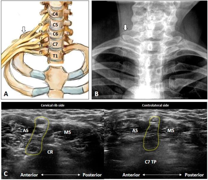
This composite image illustrates the anatomy and diagnosis of a cervical rib through three modalities. Panel A is an anatomical diagram showing the cervical vertebrae (C4-T1), the first and second ribs, and the brachial plexus (C5-T1). A white arrow identifies a cervical rib emerging from the C7 vertebra. Panel B is a posterior-anterior (PA) neck X-ray demonstrating a radiopaque bony projection (cervical rib) at the C7 level, indicated by a white arrow. Panel C provides a comparative ultrasound study in the transverse plane. The left image (Cervical rib side) shows a hyperechoic structure (CR) with posterior acoustic shadowing located between the anterior scalene (AS) and middle scalene (MS) muscles, encroaching upon the brachial plexus (yellow dashed circle). The right image (Contralateral side) shows the normal anatomy of the C7 transverse process (C7 TP) and the interscalene groove containing the brachial plexus. These images demonstrate how a cervical rib can narrow the interscalene space, contributing to neurogenic thoracic outlet syndrome (TOS).
This composite image illustrates the anatomy and diagnosis of a cervical rib through three modalities. Panel A is an anatomical diagram showing the cervical vertebrae (C4-T1), the first and second ribs, and the brachial plexus (C5-T1). A white arrow identifies a cervical rib emerging from the C7 vertebra. Panel B is a posterior-anterior (PA) neck X-ray demonstrating a radiopaque bony projection (cervical rib) at the C7 level, indicated by a white arrow. Panel C provides a comparative ultrasound study in the transverse plane. The left image (Cervical rib side) shows a hyperechoic structure (CR) with posterior acoustic shadowing located between the anterior scalene (AS) and middle scalene (MS) muscles, encroaching upon the brachial plexus (yellow dashed circle). The right image (Contralateral side) shows the normal anatomy of the C7 transverse process (C7 TP) and the interscalene groove containing the brachial plexus. These images demonstrate how a cervical rib can narrow the interscalene space, contributing to neurogenic thoracic outlet syndrome (TOS).

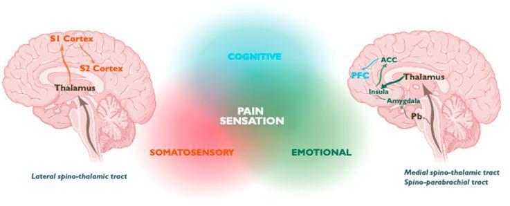
This medical illustration depicts the ascending neuroanatomical pathways for pain perception in the human brain, categorized into three functional domains: Somatosensory (red), Emotional (green), and Cognitive (cyan). Two sagittal brain diagrams flank a central 'PAIN SENSATION' Venn-style diagram. The left diagram illustrates the Lateral Spino-thalamic tract, showing nociceptive signals ascending through the thalamus to the primary (S1) and secondary (S2) somatosensory cortices. The right diagram depicts the Medial Spino-thalamic and Spino-parabrachial tracts. These pathways transmit signals from the parabrachial nucleus (Pb) and thalamus to the amygdala, insula, anterior cingulate cortex (ACC), and prefrontal cortex (PFC). Arrows represent the directional flow of neural information, highlighting the multi-dimensional nature of pain processing. This visual is designed for advanced medical education in neurology and pain management, emphasizing the distinction between sensory-discriminative and affective-cognitive processing components of the pain matrix.
This medical illustration depicts the ascending neuroanatomical pathways for pain perception in the human brain, categorized into three functional domains: Somatosensory (red), Emotional (green), and Cognitive (cyan). Two sagittal brain diagrams flank a central 'PAIN SENSATION' Venn-style diagram. The left diagram illustrates the Lateral Spino-thalamic tract, showing nociceptive signals ascending through the thalamus to the primary (S1) and secondary (S2) somatosensory cortices. The right diagram depicts the Medial Spino-thalamic and Spino-parabrachial tracts. These pathways transmit signals from the parabrachial nucleus (Pb) and thalamus to the amygdala, insula, anterior cingulate cortex (ACC), and prefrontal cortex (PFC). Arrows represent the directional flow of neural information, highlighting the multi-dimensional nature of pain processing. This visual is designed for advanced medical education in neurology and pain management, emphasizing the distinction between sensory-discriminative and affective-cognitive processing components of the pain matrix.

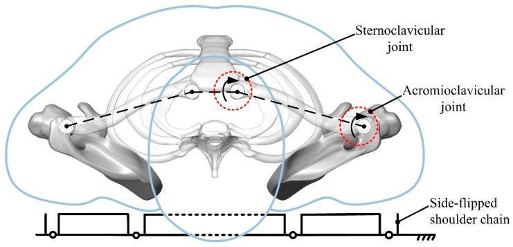
This anatomical and kinematic diagram depicts an equivalent kinematic model of the human shoulder and upper torso in a supine position, likely for rehabilitation robotics or biomechanical modeling. The visual includes a simplified skeletal rendering showing the rib cage, sternum, bilateral clavicles, and scapulae. Key anatomical landmarks are highlighted with red circular dashed lines and arrows to indicate rotational degrees of freedom: the sternoclavicular joint (connecting the clavicle to the sternum) and the acromioclavicular joint (connecting the clavicle to the scapula). Superimposed on the anatomy is a dashed line representing the functional linkage of the shoulder girdle. At the base, a schematic 'Side-flipped shoulder chain' is illustrated as a mechanical linkage system with revolute joints and grounded segments, representing a rehabilitation mechanism's interaction with the body. This diagram is used to analyze natural rollover motion trajectories and design assistive devices for physical therapy or patient positioning.

This anatomical and kinematic diagram depicts an equivalent kinematic model of the human shoulder and upper torso in a supine position, likely for rehabilitation robotics or biomechanical modeling. The visual includes a simplified skeletal rendering showing the rib cage, sternum, bilateral clavicles, and scapulae. Key anatomical landmarks are highlighted with red circular dashed lines and arrows to indicate rotational degrees of freedom: the sternoclavicular joint (connecting the clavicle to the sternum) and the acromioclavicular joint (connecting the clavicle to the scapula). Superimposed on the anatomy is a dashed line representing the functional linkage of the shoulder girdle. At the base, a schematic 'Side-flipped shoulder chain' is illustrated as a mechanical linkage system with revolute joints and grounded segments, representing a rehabilitation mechanism's interaction with the body. This diagram is used to analyze natural rollover motion trajectories and design assistive devices for physical therapy or patient positioning.

Here is a thorough breakdown of what could be causing your side rib pain above the waist, along with what to watch out for.

Pain in the Side of Your Ribs Above the Waist

Where exactly is the pain?

The location matters a lot. "Side above the waist" typically refers to the lateral chest wall / flank area, roughly where your lower ribs are on either side. Here are the most common causes, from most to least likely:

Most Likely Causes

1. Muscle Strain (Intercostal Strain) - Most Common

The intercostal muscles sit between your ribs. Straining them from twisting, lifting, coughing, or even sleeping awkwardly causes sharp or aching pain on one side. The pain often gets worse when you breathe deeply, twist, or press on the area.
  • Usually sharp, localized pain
  • Worsens with movement or deep breathing
  • Tender when pressed

2. Costochondritis

Inflammation of the cartilage connecting your ribs to the breastbone. Harrison's Principles of Internal Medicine notes that costochondritis is a relatively common cause of chest wall pain and can cause tenderness at the costochondral junctions (also called Tietze's syndrome).

3. Kidney Problems (Flank Pain)

The kidneys sit just below and behind your lower ribs. Pain from the kidney - such as from a kidney stone or infection (pyelonephritis) - classically appears as a dull ache or sharp pain in the flank/side above the waist, often reaching around to the front.
  • Kidney stone: sudden, severe, colicky pain; may radiate to groin; blood in urine
  • Kidney infection: dull ache + fever, nausea, pain on pressing the back below the ribs

4. Pleurisy (Pleural inflammation)

Inflammation of the lining around the lungs causes a sharp, "knife-like" pain that gets worse when you breathe in or cough. It is usually on one side. Harrison's describes it as "typically a knife-like pain worsened by inspiration or coughing."

5. Herpes Zoster (Shingles) - Early Stage

Before the rash appears, shingles can cause burning, shooting pain in a band around one side of your chest/ribs. If you later notice a blistering rash appearing on the same side, this is the diagnosis.

6. Slipped Rib Syndrome

The lower ribs (8-10) can temporarily slip out of position, causing sharp lateral rib pain. This is more common than usually recognized and is worsened by certain movements.

7. Gastrointestinal Causes

  • Right side: Liver, gallbladder (gallstones cause pain under right ribs, can radiate to the side/back)
  • Left side: Spleen problems, stomach issues, splenic flexure gas

8. Pulmonary Embolism (blood clot in lung) - Less common but serious

Small pulmonary emboli can cause lateral, pleuritic chest/rib pain. This is associated with breathlessness, rapid heart rate, and sometimes leg swelling.

Red Flag Symptoms - Seek Emergency Care Immediately

Go to an emergency room or call emergency services if your rib pain comes with any of these:
Warning SignPossible Cause
Sudden tearing/ripping painAortic dissection
Difficulty breathing, dizzinessPulmonary embolism, pneumothorax
Fever + painful urinationKidney infection
Pain spreading to jaw, arm, or shoulderHeart attack
Swelling or bruising over ribs after injuryRib fracture
Coughing up bloodLung clot or tumor

Questions to Help Narrow It Down

  • Which side? (Left vs. right changes the possibilities significantly)
  • Did it come on suddenly or gradually?
  • Does it get worse when breathing in deeply? (suggests pleurisy or lung issue)
  • Is there any back pain, fever, or change in urination? (suggests kidney)
  • Did you recently strain yourself, cough a lot, or sleep awkwardly? (suggests muscle)
  • Any rash appearing? (suggests shingles)

What to Do

  • If pain is mild and linked to movement: rest, avoid straining, try ibuprofen or paracetamol (if not contraindicated), apply a warm pack
  • If pain is severe, constant, or getting worse: see a doctor today
  • If any red flag symptoms are present: go to the emergency room immediately
Please remember this information is for guidance only and does not replace an in-person medical evaluation. A doctor can do a physical exam, order imaging or blood tests, and make a proper diagnosis.
This is a shared conversation. Sign in to Orris to start your own chat.