
Give me a detailed explanation of all f these questions *Intestinal Nematodes* - Ascaris, Hookworm, Strongyloids [imp topics - 5m] 1. learn the lifecycle, pathogenesis, c/f, lab diagnosis (stool culture findings), Rx and prevention for each 2. Eggs of Ascaris (fertilized, decorticated and unfertilized) - describe and draw - 3M 3. Know about Loeffler's syndrome; & charcot-Leyden Crystals seen in sputum and stool samples (non-specific ascaris finding) 4. Chandler's Index in case of Hookworms, Old world & new world hook worm classification.; ground itch + serpiginous tracks. 5. Quantity of blood loss for ankylostoma & necator ( note - more blood loss is seen in case of ankylostoma) 6. Hookworm infection a/w with Fe deficiency anemia & PEM 7. Hookworm's - Kato Katz technique = egg counting technique (MCQ); hookworm egg - describe Sepsis is imp , especially interms of clinical microbiology. it may come as a 10M, you never know ;) 1. Definition (bacteremia, sepsis, septicemia, septic shock) 2. qSOFA criteria for diagnosis 3. Most common Organisms 4. Lab diagnosis - know about how paired blood c/s is done and its significance to perform before administering broad spectrum antibiotics. 5. Extras - good to know about the sepsis 1 hour bundle. 6. Know about blood culture media [monophasic, biphasic/castaneda, BacT/ALERT bottle] 7. wrt Rx know about - a) AST methods [refer to General microbiology chapter] - b) read through Hospital infection Control Section [HAI care bundles, sterilization, BMW, Antimicrobial susceptible testing
Ascaris lumbricoides egg fertilized unfertilized decorticated microscopy
This figure presents a comparative biological and biochemical analysis of Branchiostoma belcheri eggs before and after fertilization, serving as a model for chordate egg coat (EC) and zona pellucida (ZP) protein evolution. Panels A and B are light microscopy images illustrating morphological changes: the unfertilized egg (A) measures approximately 145.77 μm in diameter with a tight egg coat, whereas the fertilized egg (B) displays significant elevation of the egg coat, expanding the total diameter to approximately 400 μm while the central cytoplasm remains constant. Panels C and D show SDS-PAGE gel electrophoresis of protein extracts from unfertilized and fertilized egg coats, respectively. Panel C reveals a high density of protein bands ranging from 30 kDa to over 100 kDa, with 11 marked slices used for LC-MS/MS analysis. Panel D demonstrates a simplified protein profile post-fertilization, with a dominant band clustered around 55 kDa and distinct minor bands at higher molecular weights (e.g., 110-170 kDa). This comparison highlights the structural remodeling and protein biochemical shifts associated with the cortical granule reaction and egg coat maturation in basal chordates.
This composite of fluorescence microscopy images illustrates the distribution and co-localization of the active Arp2/3 complex (pArp2) in amphioxus eggs from unfertilized to late fertilized stages. Section (a) shows the orientation of the egg using the polar body (pb) and vegetal tuft-like structure (tu). Section (b) is a time-series showing pArp2 (green) immunopositive signals shifting from a ubiquitous cortical distribution in unfertilized eggs to a localized concentration on one side of the egg 35 minutes post-fertilization, migrating via the animal pole. Section (c) demonstrates the co-localization of pArp2 (red) and F-actin (green), where the merged image (yellow) indicates overlapping signals in the late fertilized cortical region. Section (d) presents co-localization between pArp2 and tubulin (microtubules) in lateral and vegetal views. A prominent tuft-like structure (tu) at the vegetal pole shows strong overlap of pArp2 and tubulin. These visual data highlight the role of cortical cytoskeletal remodeling in establishing embryonic asymmetry and axis determination during early development. Scale bar represents 100 µm.
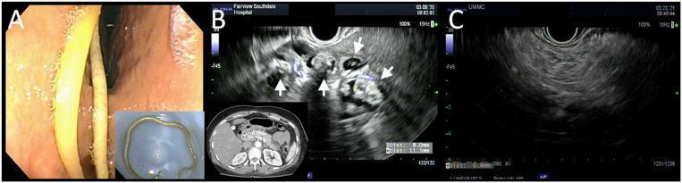
A multi-panel clinical graphic demonstrating gastric ascariasis and associated complications. Panel A shows a white-light endoscopic view of the gastric antrum containing a long, slender, pale yellowish cylindrical roundworm, identified as Ascaris lumbricoides. An inset displays the extracted intact worm. Panel B features an endoscopic ultrasound (EUS) image alongside a transverse CT slice of the abdomen. The EUS reveals a complex, heterogeneous region adjacent to the pancreatic tail containing multiple hypoechoic, roughly circular cystic structures (indicated by white arrows), representing egg collections or localized inflammation. The associated CT scan shows diffuse edematous enlargement of the pancreas consistent with acute interstitial pancreatitis. Panel C shows a follow-up EUS image after anthelmintic treatment with albendazole, demonstrating complete resolution of the cystic structures and return to more homogeneous pancreatic parenchyma. This sequence illustrates the diagnostic utility of EUS in identifying parasitic etiologies for acute pancreatitis and monitoring treatment response.
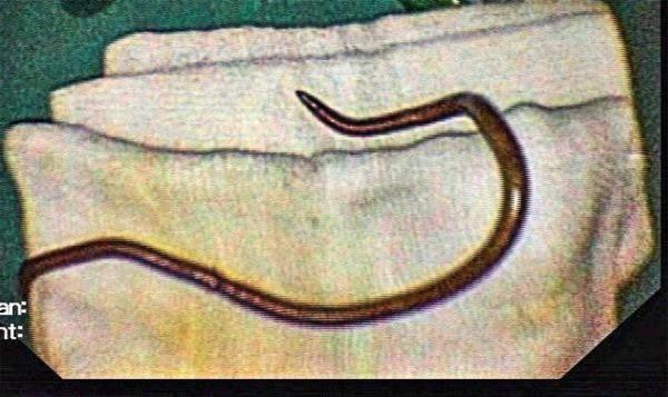
This clinical photograph displays a specimen of Ascaris lumbricoides, a human parasitic roundworm, following extraction via Endoscopic Retrograde Cholangio-Pancreaticography (ERCP). The specimen is a single, elongated, cylindrical nematode with a smooth, tan-to-brownish exterior. One visible extremity tapers to a sharp, curved point, which is characteristic of the adult worm's morphology. The organism is placed on sterile white surgical gauze, illustrating the outcome of a procedural intervention for biliary ascariasis. This image demonstrates the clinical manifestation of hepatobiliary parasitism where a nematode has migrated into the common bile duct, potentially causing obstructive jaundice or biliary colic. The educational focus is on the macroscopic identification of common helminthic infections and the visual confirmation of parasitic extraction in gastroenterology and tropical medicine.
Strongyloides stercoralis lifecycle rhabditiform filariform larvae
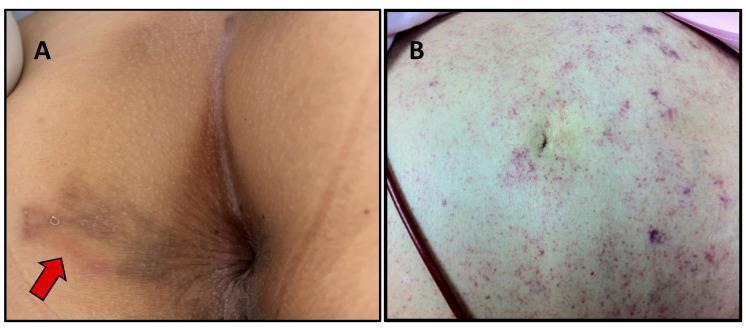
This composite clinical photograph displays two distinct dermatological manifestations of Strongyloides stercoralis infection. Image A shows larva currens in the perianal region, characterized by a serpiginous, erythematous, and slightly raised linear track (indicated by a red arrow). This pathognomonic lesion represents the rapid intradermal migration of filariform larvae. Image B displays a periumbilical 'thumbprint' purpura in a patient with disseminated strongyloidiasis. The abdominal skin exhibits extensive, non-blanching purpuric macules and petechiae, appearing as clusters of reddish-purple spots that are most concentrated in the periumbilical area. These lesions result from larvae migrating through the vessel walls into the dermis. The educational focus is on distinguishing the localized cutaneous signs of chronic infection from the systemic vascular signs associated with severe, disseminated hyperinfection syndrome in immunocompromised hosts.
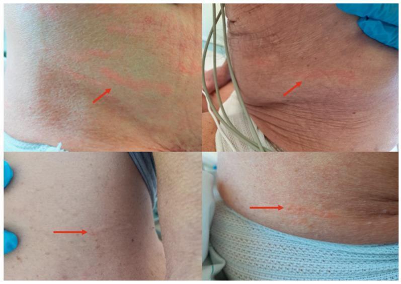
Content Type: Clinical Photograph. This four-panel composite image displays a patient's inferior abdomen and flank, showcasing pathognomonic dermatological signs of disseminated Strongyloides stercoralis hyperinfection. The primary findings are multiple, raised, erythematous, serpiginous lesions known as larva currens. These linear, wavy tracks represent the rapid cutaneous migration of filariform larvae. Red arrows highlight the specific morphology and orientation of the lesions, which appear as slightly edematous, urticarial-like streaks across the wrinkled skin of an older adult. Contextual elements include medical monitoring cables and gloved hands of healthcare providers, indicating an inpatient clinical setting. Educational Focus: Recognition of the distinctive 'racing larva' morphology and its clinical significance as a hallmark of systemic strongyloidiasis, particularly in immunosuppressed patients. Specialty: Infectious Disease, Dermatology, Tropical Medicine.
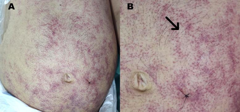
Two-panel clinical photograph (A and B) demonstrating a pathognomonic purpuric rash on the abdomen and flank of an immunocompromised patient with disseminated strongyloidiasis. Figure A provides a wide view of the torso, showing an extensive distribution of non-blanching, violaceous macules and small papules primarily concentrated in the periumbilical region, lower abdomen, and extending toward the flank. Figure B is a high-magnification view of the periumbilical area, highlighting specific morphological patterns. A black arrow indicates a classic 'thumbprint sign,' characterized by serpiginous and linear purpuric tracks caused by the subcutaneous migration of Strongyloides stercoralis filariform larvae (larva currens). The lesions appear as petechiae and ecchymoses with a distinctive reticulated or serpiginous arrangement. This finding is a critical clinical marker for Strongyloides hyperinfection syndrome or disseminated disease, often seen in the context of HIV infection or corticosteroid therapy.
hookworm egg Ancylostoma Necator thin shell embryonated
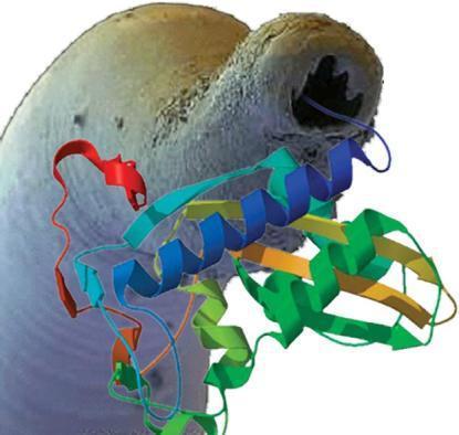
This composite educational image features a Scanning Electron Micrograph (SEM) of a hookworm specimen superimposed with a three-dimensional protein ribbon diagram. The anatomical background shows the anterior end of a parasitic nematode, likely Ancylostoma or Necator species, highlighting the buccal capsule with visible tooth-like structures or cutting plates used for mucosal attachment and blood-feeding. The specimen displays a granular, textured grey and tan surface. Overlaid is a multi-colored ribbon model representing a recombinant protein, such as those secreted by the parasite (e.g., Ancylostoma-secreted proteins). The protein structure illustrates tertiary folding patterns including alpha-helices (blue and green) and beta-pleated sheets (orange and yellow) interconnected by loops. This visual is designed to illustrate concepts in parasitology and vaccine development, specifically targeting hookworm-induced iron deficiency anemia by utilizing secreted recombinant proteins as immunogens to disrupt the parasite's life cycle or feeding mechanisms in humans.
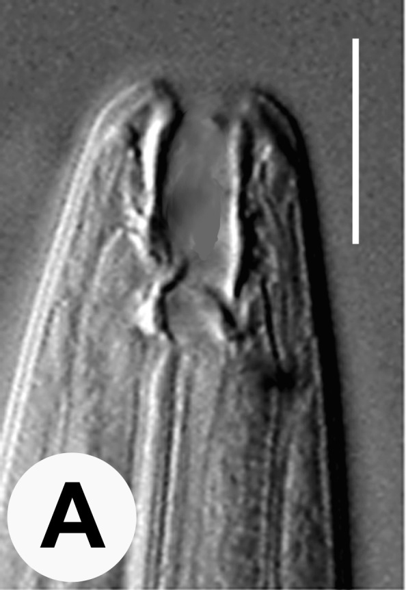
**Modality:** Differential interference contrast (DIC) light microscopy. **Subject:** Anterior end of a nematode, likely an *Ancylostoma* or *Necator* species (hookworm), viewed from a lateral perspective. **Key Anatomical Features:** * **Buccal Capsule:** A prominent, deep, and sclerotized buccal cavity is visible at the anterior terminus. * **Ventral Cutting Elements:** The image demonstrates the characteristic armature of the oral aperture, showing thickened, tooth-like or plate-like structures (cutting teeth) used for mucosal attachment. * **Esophageal Junction:** The posterior base of the buccal capsule transitions into the muscular esophagus, with visible longitudinal striations in the esophageal wall. * **Cephalic Structure:** The cuticle appears smooth with subtle longitudinal markings. **Clinical Significance:** This microscopic view is a primary diagnostic feature for identifying parasitic helminths in clinical samples (e.g., stool concentrates or tissue biopsies). The morphology of the buccal capsule—specifically the shape and number of ventral teeth—is the key taxonomic differentiator between hookworm species that cause human ancylostomiasis and necatoriasis. **Scale:** A white vertical scale bar is present in the upper right quadrant for morphometric reference.
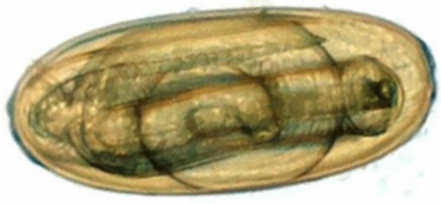
This diagnostic micrograph displays an embryonated egg of the nematode Nematodirus battus, a clinically significant helminth. The image captures the parasite in a late developmental stage, specifically featuring a well-developed third-stage larva (L3) coiled within the egg shell. The egg is characterized by a distinctive large, oval shape and a smooth, translucent outer shell. Internally, the larva is clearly visible as a complex, elongated, and folded structure with a segmented, textured appearance, occupying a significant portion of the internal volume. This 'larvated' stage is a critical diagnostic marker in parasitology, indicating maturity and readiness for hatching. Such images are used in medical and veterinary parasitology to identify species-specific egg morphology and to assess the life cycle progression of strongyle-type parasites in environmental or clinical specimens.
sepsis blood culture bottle BacT ALERT Castaneda biphasic
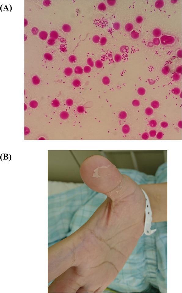
The composite image contains two panels relevant to a clinical case of sepsis. (A) Light microscopy (Gram stain, 1000x magnification) of a blood culture smear. It shows numerous small, pink-stained, Gram-negative rod-shaped bacteria (coccobacilli) scattered individually and in clusters among circular pink-stained host cells, characteristic of Yersinia pseudotuberculosis. (B) A clinical photograph of a patient's hand demonstrating distal cutaneous desquamation. The thumb displays characteristic peeling and flaking of the superficial epidermis, revealing raw, erythematous underlying skin at the fingertip. This visual finding is a classic clinical sign of Far East scarlet-like fever (FESLF) or toxic shock-like syndromes. Collectively, these images illustrate the diagnostic progression from microbiological identification of a pathogen to the manifestation of dermatological sequelae in systemic infection.
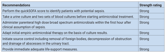
<table><thead><tr><th>Recommendations</th><th>Strength rating</th></tr></thead><tbody><tr><td>Perform the quickSOFA score to identify patients with potential sepsis.</td><td>Strong</td></tr><tr><td>Take a urine culture and two sets of blood cultures before starting antimicrobial treatment.</td><td>Strong</td></tr><tr><td>Administer parenteral high dose broad spectrum antimicrobials within the first hour after clinical assumption of sepsis.</td><td>Strong</td></tr><tr><td>Adapt initial empiric antimicrobial therapy on the basis of culture results.</td><td>Strong</td></tr><tr><td>Initiate source control including removal of foreign bodies, decompression of obstruction and drainage of abscesses in the urinary tract.</td><td>Strong</td></tr><tr><td>Provide immediate adequate life-support measures.</td><td>Strong</td></tr></tbody></table>

Educational infographic highlighting the global and domestic impact of sepsis, set against a background illustration of red blood cells in the bloodstream. The central text describes sepsis as 'the Equal Opportunity Killer'. Key epidemiological data points include: 258,000 Americans die from sepsis annually; it is the third leading cause of death in the United States after heart disease and cancer; 1.6 million cases occur in the U.S. every year; and more than 5 million children die from sepsis worldwide annually. Additionally, the graphic notes a public awareness gap, stating that only 55% of Americans have ever heard the word 'sepsis'. The visual uses iconography like the map of the United States, a globe, and human figures to contextualize the statistics. This infographic serves as a public health awareness tool to emphasize the high mortality and prevalence of this critical medical condition.
Charcot Leyden crystals sputum stool eosinophil
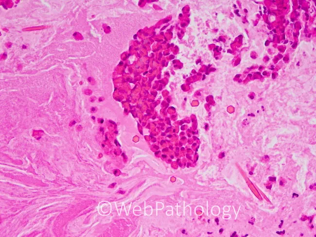
This slide shows a para-nasal sinus mucin specimen examined by brightfield light microscopy after Hematoxylin and Eosin (H&E) staining. The specimen is thick, rubbery, gelatinous, with a greenish-brown coloration typical of allergic mucin. Microscopically, the material is largely amorphous and chondroid-like, may display laminated architecture, and contains clusters of inflammatory cells within a proteinaceous matrix. The inflammatory infiltrate is eosinophil-predominant, with scattered neutrophils, plasma cells, and histiocytes. Charcot-Leyden crystals are frequently present, reflecting eosinophil breakdown, together with desquamated respiratory epithelial cells. Rare fungal hyphae are visible at the upper left and lower right corners of the field, consistent with a minority fungal component without evidence of tissue invasion. The surrounding sinus mucosa shows inflammatory exudate and mucinous debris. Collectively these features support allergic fungal rhinosinusitis (AFRS) with eosinophilic mucin rather than invasive fungal rhinosinusitis or bacterial rhinosinusitis. Diagnostic significance includes recognition of AFRS by eosinophilic mucin with Charcot-Leyden crystals and sparse hyphae, aiding differential diagnosis against chronic rhinosinusitis, fungal ball, and invasive disease. Clinically, findings correlate with chronic nasal obstruction, allergic rhinitis, and asthma; educational and research relevance spans rhinology, otolaryngology, pathology, and radiology. Morphologic features include laminated mucin, eosinophil-rich debris, and occasional contact with respiratory epithelium. Important for education.
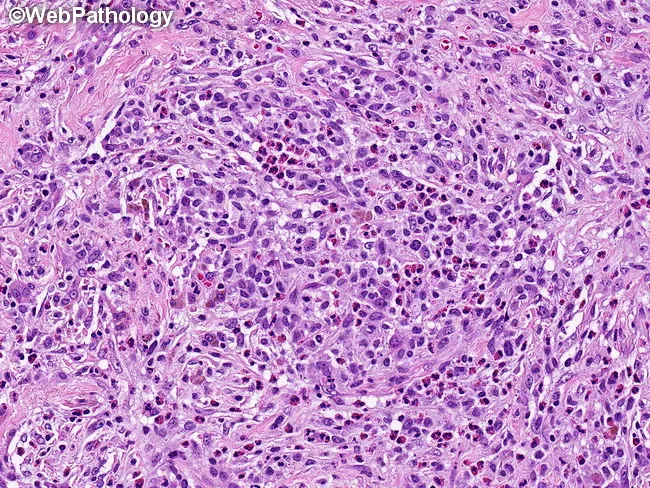
Imaging modality: Histopathology, light microscopy of hematoxylin and eosin (H&E) stained lung tissue. Specimen derived from pulmonary parenchyma via biopsy. Microscopic field reveals cellular interstitial infiltrates containing numerous Langerhans cells with vesicular, grooved nuclei and moderate to abundant eosinophilic cytoplasm, accompanied by eosinophils, lymphocytes, plasma cells, pigment-laden macrophages and occasional neutrophils. Early lesions are cellular with abundant Langerhans cells and eosinophils and minimal fibrosis; later stages show progressive fibrosis and foamy macrophages with reduced Langerhans cell density and fewer eosinophils. Langerhans cells may form cohesive clusters within airspaces and interstitium, sometimes surrounding necrotic debris; Charcot-Leyden crystals may be present in eosinophil-rich microabscesses. The histology may resemble granulomatous inflammation but lacks classic granulomas; immunophenotype is typically CD1a and CD207 (Langerin) positive in diagnostic settings. Diagnostic significance: this pattern is characteristic of pulmonary langerhans cell histiocytosis (PLCH), which commonly associates with smoking and can progress to cystic lung disease; differential includes eosinophilic pneumonia, hypersensitivity pneumonitis, granulomatous infections, and smoking-related interstitial fibrosis. Clinical correlation: correlate with smoking history, respiratory symptoms, and imaging; confirm with immunohistochemistry; assess for complications such as pneumothorax; management focuses on smoking cessation and, in selected cases, targeted therapies. Additional molecular testing may guide prognosis and targeted anti-LCH therapy in cases.
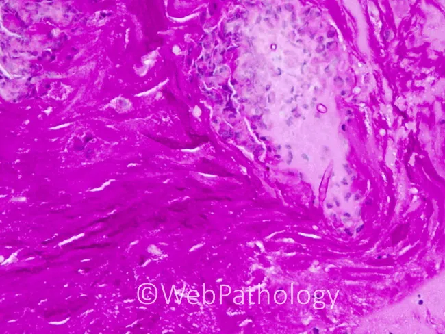
Modality and technique: Light microscopy of a paranasal sinus mucin biopsy stained with Periodic acid–Schiff (PAS). The specimen shows thick, eosinophil-rich allergic mucin containing discrete, pigmented fungal hyphae consistent with dematiaceous organisms. Hyphae are septate with acute-angled branching, noninvasive within the mucus rather than invading sinus mucosa. The pigment imparts a brown-black coloration to the hyphae, typical of Bipolaris species. No tissue invasion or necrosis is evident. The surrounding stroma demonstrates an inflammatory milieu with eosinophils, Charcot-Leyden crystals, and mucus with sparse inflammatory cells. The microbiologic appearance is consistent with allergic fungal sinusitis (AFS) rather than invasive mycosis. The case highlights Bipolaris as the causative agent among dematiaceous fungi such as Bipolaris, Exserohilum, Curvularia, Alternaria, and Drechslera; yet histology of allergic mucin remains similar irrespective of organism. For diagnosis, identification relies on fungal morphology in mucus and culture/molecular confirmation if available. Clinically, this pattern correlates with chronic rhinosinusitis with nasal polyposis and hypersensitivity reactions. Differential includes Aspergillus and other dematiaceous fungi. Recognizing dematiaceous hyphae within eosinophilic mucin guides management by guiding surgical debridement and antifungal therapy decisions, and informs prognosis regarding recurrence risk. This documentation supports educational training, targeted research, and standardized reporting of noninvasive fungal mucin infections in sinuses.
PART 1 - INTESTINAL NEMATODES
1. ASCARIS LUMBRICOIDES
Lifecycle
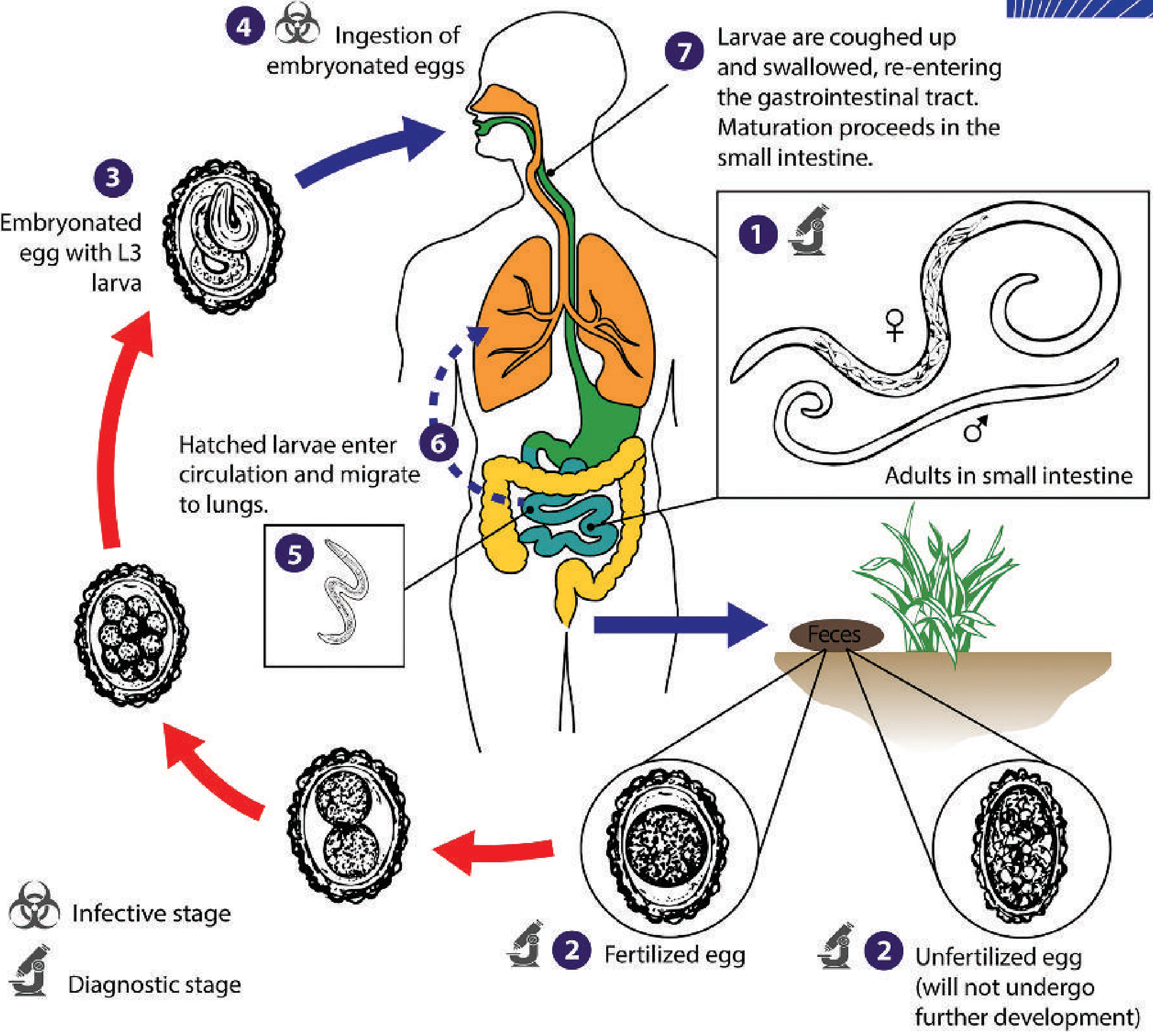
- Adults in small intestine - Female (20-35 cm, straight tail) and male (15-31 cm, curled tail). Both sexes have 3 characteristic anterior "lips."
- Egg shedding - A gravid female lays up to 200,000 eggs/day, shed in stool as fertilized or unfertilized eggs. Unfertilized eggs will NOT develop further.
- Maturation in soil - Fertilized eggs require warm, moist soil (18 days to several weeks). Eggs are extremely hardy - survive extreme temperatures, persist for months in feces/sewage.
- Ingestion - Humans ingest embryonated eggs via contaminated food/water.
- Hatching - Eggs hatch in small intestine, releasing L3 larvae.
- Lung migration - Larvae penetrate intestinal mucosa → portal circulation → lungs → penetrate alveoli → ascend bronchial tree → swallowed → return to small intestine as adults.
- Maturation - Full cycle from egg to adult takes approximately 2 months.
Pathogenesis & Clinical Features
| Phase | Mechanism | Clinical Finding |
|---|---|---|
| Larval migration (lungs) | Larvae + eosinophilic inflammation | Loeffler syndrome (see below) |
| Intestinal phase - light infection | Small worm burden | Asymptomatic or mild abdominal pain/diarrhea |
| Intestinal phase - heavy infection | Mechanical obstruction, worm mass | Intestinal obstruction (especially in children) |
| Ectopic migration | Worms invade bile duct, appendix, liver | Biliary colic, obstructive jaundice, pancreatitis |
Lab Diagnosis - Stool Findings
- Direct/concentrated stool wet mount - demonstration of characteristic eggs. Detection is very probable even with a single worm (200,000 eggs/day).
- Egg quantitation (Kato-Katz technique): <20 eggs per slide (in 2 mg feces) = light infection; >100 eggs per slide = heavy infection.
- Pulmonary phase - larvae and eosinophils may be found in sputum.
- Adult worms occasionally passed in stool or vomited, or seen on endoscopy. On abdominal X-ray, adults may be visible; cholangiogram may show worms in biliary tract.
- Blood - peripheral eosinophilia.
Treatment
- Albendazole (drug of choice) or Mebendazole or Ivermectin
- Important: In mixed infections (Ascaris + other helminths, Giardia, or E. histolytica), always treat Ascaris FIRST to avoid provoking worm migration.
- Anthelmintics are given before elective surgery in endemic areas.
Prevention
- Improved sanitation and waste treatment
- Avoid using human feces as fertilizer
- Wash hands, wash produce
- Mass drug administration (MDA) in endemic areas
2. ASCARIS EGGS - Description and Diagram [3M topic]
Fertilized Egg (most common in stool)
- Shape: Round to slightly oval
- Size: 55-75 μm long × 35-50 μm wide
- Shell: Thick shell with outer irregular, bumpy, yellow-brown mamillated (albuminous) coat
- Contents: Unembryonated cell mass (dark granular mass), does not fill entire egg
- Significance: This is the infective form - develops in soil
Unfertilized Egg (produced by solitary females)
- Shape: More elongate, narrow
- Size: Larger - up to 85-90 μm long (longer than fertilized)
- Shell: Thinner shell, irregular mamillations
- Contents: Irregularly sized granules/globules filling the entire egg; disorganized internal contents - NO organized cell mass
- Significance: Will NOT undergo further development. NOT infective.
Decorticated Egg (fertilized or unfertilized that has lost its mamillated coat)
- Appearance: Smooth outer surface - the mamillated layer has been lost
- Shell: Still has a thick shell (fertilized type)
- Differentiation: Must be differentiated from hookworm eggs - Ascaris decorticated egg has a THICKER shell; hookworm eggs have a thin shell
- Superficially resembles hookworm eggs - key differentiating feature is shell thickness
Quick Memory Diagram
FERTILIZED EGG                 UNFERTILIZED EGG              DECORTICATED EGG
 ___________                    ____________                   ___________
/~~~~~~~~~~~\    ← mamillations /~~~~~~~~~~~~\                 /           \
|  [  cell  ]|   55-75 µm long |[granules fill]| 85-90µm long | [cell mass] |
|  [  mass  ]|   thick shell   |[entire lumen  ]| thin shell  | [remains]   |
\~~~~~~~~~~~/ ← round/oval     \~~~~~~~~~~~~/               \___________/
                                 elongated                  smooth shell
                                                           (no bumps)
3. LOEFFLER'S SYNDROME & CHARCOT-LEYDEN CRYSTALS
Loeffler's Syndrome (Pulmonary Phase of Ascariasis)
- Definition: Transient pulmonary eosinophilic infiltration caused by larval migration through the lungs
- Also occurs in: Hookworm and Strongyloides (same lung migration mechanism)
- Mechanism: Larvae + host eosinophilic response → inflammation in alveolar capillary bed
- Clinical Features:
- Dry cough, wheezing, breathlessness
- Low-grade fever
- Peripheral eosinophilia (very characteristic)
- Bilateral diffuse, mottled pulmonary infiltrates on CXR (transient - "migratory/fleeting infiltrates")
- Sputum findings: Larvae + eosinophils visible
- Important: More common in previously sensitized individuals. Self-limiting.
Charcot-Leyden Crystals
- What they are: Hexagonal, bipyramidal, needle-shaped/spindle-shaped crystals derived from the breakdown of eosinophil granules (specifically lysophospholipase protein)
- Appearance: Elongated, colorless, pointed at both ends (like a double-pointed needle)
- Where found:
- Sputum - in Loeffler's syndrome (during lung migration phase)
- Stool - in intestinal phase with heavy worm burden
- Non-specific finding - NOT diagnostic only for Ascaris; found in ANY eosinophil-rich process (asthma, allergic bronchopulmonary aspergillosis, eosinophilic gastroenteritis)
- Significance: Indicates eosinophilic tissue reaction - strong clue for parasitic infection
4. HOOKWORMS
Old World vs. New World Classification
| Feature | Ancylostoma duodenale (Old World) | Necator americanus (New World) |
|---|---|---|
| Mouthparts | Teeth (cutting teeth) | Cutting plates |
| Geographic distribution | N. Africa, Middle East, S. Europe | Americas (predominant) |
| Blood loss/worm/day | 0.15-0.25 mL | 0.03 mL |
| Transmission | Skin penetration AND oral/transmammary | Skin penetration only |
Note: In practice, the Old/New World classification is oversimplified - both species overlap in distribution worldwide. A. ceylanicum is the dominant cause in Southeast Asia.
Lifecycle
- Eggs passed in stool - passed in feces, develop rapidly in warm moist soil
- Rhabditiform (L1) larvae hatch, develop over ~7 days into infective filariform (L3) stage
- Skin penetration - L3 larvae penetrate intact skin (between toes, soles of feet - "ground itch")
- Lung migration - larvae enter circulation → lungs → alveoli → bronchial tree → swallowed
- Intestinal maturation - adults attach to mucosa of small intestine using buccal capsule
- Blood feeding - adults feed on host blood for 1-18 years
- Females lay up to 200,000 eggs/day
Pathogenesis & Clinical Features
| Site | Condition | Features |
|---|---|---|
| Skin | Ground itch | Pruritic, erythematous rash, blister formation at site of larval penetration |
| Lungs | Loeffler syndrome | (same as Ascaris - eosinophilic infiltration during migration) |
| Intestine | Iron-deficiency anemia | Chronic blood loss → hypochromic microcytic anemia (Fe deficiency) |
| Systemic | PEM (Protein-Energy Malnutrition) | Heavy infection → growth stunting, developmental delay in children |
- Caused by zoonotic hookworms: A. caninum, A. braziliense, Uncinaria stenocephala
- Larvae penetrate human skin but CANNOT develop further
- Produce intensely pruritic, erythematous serpiginous (winding/serpentine) tracks - larvae migrate in the dermis
- This is called Creeping Dermatitis
- Self-limiting (4-8 weeks), but albendazole or ivermectin shortens course
Hookworm and Fe Deficiency Anemia + PEM
- Mechanism of anemia: Adults attach to intestinal mucosa and suck blood. A. duodenale causes more blood loss (0.15-0.25 mL/worm/day) vs Necator (0.03 mL/worm/day). Chronic loss → Fe-deficiency anemia (hypochromic, microcytic).
- Mechanism of PEM: Blood contains protein; chronic protein loss + reduced appetite/absorption with heavy worm burden leads to protein-energy malnutrition, growth stunting, cognitive impairment.
- Severity correlates with worm burden - egg count per gram of stool correlates with clinical severity.
- Treatment: Antiparasitic (albendazole/mebendazole/pyrantel pamoate) + iron supplementation
Chandler's Index
- Definition: A measure of the worm burden (intensity of hookworm infection) based on egg count per gram of stool
- Basis: The number of eggs per gram of stool correlates directly with the number of adult worms present
- Clinical use: Helps determine when to initiate therapy in endemic areas
- Also used to monitor response to therapy
Kato-Katz Technique (MCQ favorite)
- What it is: A quantitative/semi-quantitative stool egg counting technique used to diagnose and measure intensity of soil-transmitted helminth infections
- Principle: A standardized amount of stool (41.7 mg) is pressed through a stainless steel screen, placed on a slide, covered with a glycerol-soaked cellophane strip, cleared, and examined under microscopy
- Result expressed as: Eggs Per Gram (EPG) of feces
- Uses:
- Diagnosis of hookworm, Ascaris, Trichuris
- Measure worm burden (intensity of infection)
- Monitor treatment response
- Epidemiological surveys
- NOT part of standard O&P (ova and parasite) examination - requires specific request
Hookworm Egg - Description
- Shape: Oval
- Size: 55-76 μm long × 36-40 μm wide
- Shell: Thin shell (key feature - distinguishes from Ascaris)
- Contents: Partially embryonated when passed (2-8 cell stage visible inside)
- Color: Colorless, transparent
- Important: In unpreserved specimens not examined promptly - eggs may hatch releasing rhabditiform larvae, which must be differentiated from Strongyloides larvae (hookworm rhabditiform larvae have a longer buccal canal and inconspicuous genital primordium vs Strongyloides which has short buccal canal + prominent genital primordium)
Hookworm Treatment & Prevention
- Rx: Albendazole, Mebendazole, or Pyrantel pamoate + iron supplementation
- Prevention: Footwear (prevents skin penetration), improved sanitation, avoid walking barefoot in endemic areas, MDA programs
5. STRONGYLOIDES STERCORALIS
Lifecycle (most complex of the three - unique features)
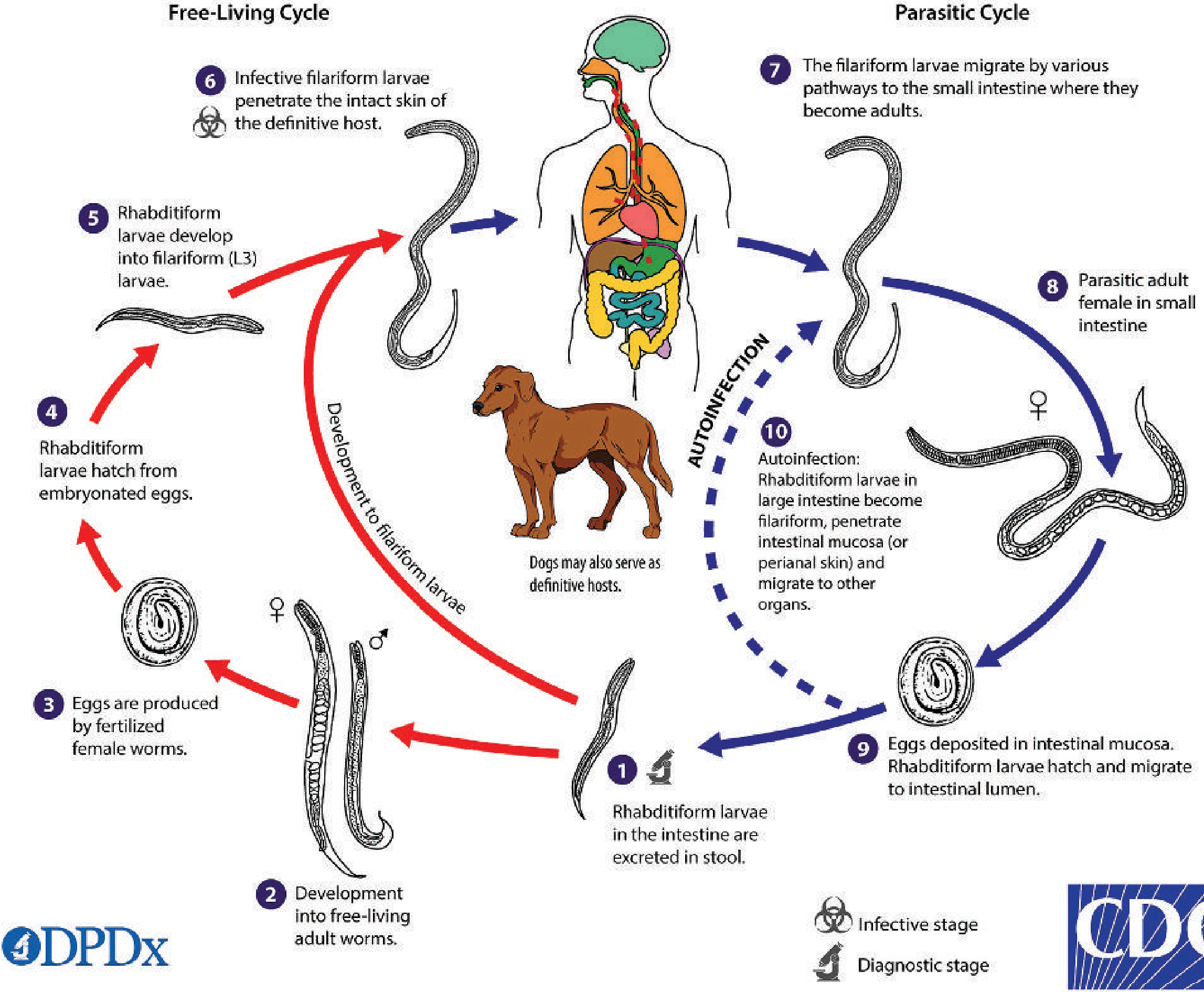
- Rhabditiform (L1) larvae shed in stool
- Develop into free-living adult male and female worms in soil
- Adults mate; female produces eggs
- Eggs hatch → rhabditiform → develop into infective filariform (L3) larvae
- L3 larvae penetrate intact skin of new host
- L3 (filariform) larvae penetrate skin → circulation → lungs → bronchial tree → swallowed → small intestine
- Only parthenogenetic females (no males) establish infection in intestinal mucosa
- Female is embedded in intestinal mucosa and deposits eggs (which HATCH in intestine)
- Rhabditiform larvae are shed in stool (NOTE: LARVAE seen in stool, NOT eggs - this is the key diagnostic difference from other helminths!)
- Some rhabditiform larvae in the large intestine transform to filariform (L3) larvae
- These penetrate intestinal mucosa (or perianal skin) without leaving the host
- → Lungs → re-establish infection - completing the cycle internally
- This allows lifelong infection even without re-exposure
- In immunocompromised patients (HIV, corticosteroids, transplant) → Hyperinfection Syndrome (massive larvae throughout body) → can be fatal
- Hyperinfection is accompanied by gram-negative bacteremia (larvae carry gut bacteria through the intestinal wall)
Pathogenesis & Clinical Features
| Mild/Chronic Infection | Severe/Hyperinfection |
|---|---|
| Asymptomatic or mild diarrhea | Severe diarrhea, malabsorption |
| Larva currens (skin) | Disseminated strongyloidiasis |
| Eosinophilia | Meningitis, pneumonia (from larvae carrying bacteria) |
| Mild pulmonary symptoms | Life-threatening sepsis |
Lab Diagnosis
- LARVAE in stool (NOT eggs - this is the hallmark) - rhabditiform larvae are the diagnostic stage
- Stool wet mount: Low sensitivity (30%). Multiple stool specimens (up to 7) on different days needed
- Stool agar culture (Harada-Mori technique) - preferred concentration/culture method; increases sensitivity greatly by allowing larvae to crawl on agar surface
- Baermann technique - another concentration method (uses warm water to attract larvae)
- Rhabditiform larva morphology: 180-380 μm long; short buccal canal, esophagus runs 1/3 the larval length, prominent genital primordium (distinguishes from hookworm larvae)
- Serology (ELISA) - useful in low-burden infections; may cross-react with other helminths
- Duodenal aspirate/string test - increases sensitivity
- Filariform larvae occasionally in sputum during lung migration phase
Treatment & Prevention
- Rx: Ivermectin (drug of choice) - 2 doses 2 weeks apart; also albendazole
- Immunocompromised: Ivermectin + reduce immunosuppression + broad-spectrum antibiotics for disseminated infection
- Prevention: Footwear, improved sanitation, screen immunocompromised patients before starting steroids/chemotherapy
PART 2 - SEPSIS (Clinical Microbiology)
1. DEFINITIONS (Rosen's Emergency Medicine / Sepsis-3)
| Term | Definition |
|---|---|
| Bacteremia (Fungemia) | Presence of viable bacteria (fungi) in the blood, as evidenced by positive blood cultures. Bacteremia does NOT require SIRS. |
| SIRS (Systemic Inflammatory Response Syndrome) | ≥2 of: Temp >38°C or <35°C; HR >90 bpm; RR >20 breaths/min or PaCO2 <32 mmHg; WBC >12,000 or <4,000/dL or >10% bands |
| Sepsis (original 1992 definition) | SIRS + proven or suspected microbial source |
| Sepsis-3 (2016) | Life-threatening organ dysfunction caused by a dysregulated host response to infection. Organ dysfunction defined by SOFA score increase ≥2. |
| Septicemia | Older term for bacteremia with clinical manifestations of systemic infection (largely replaced by "sepsis") |
| Severe Sepsis | Sepsis + organ dysfunction (older term; merged into "sepsis" in Sepsis-3) |
| Septic Shock | Sepsis + hemodynamic compromise: vasopressor requirement to maintain MAP ≥65 mmHg AND serum lactate >2 mmol/L DESPITE adequate fluid resuscitation. Mortality 35-54%. |
| MODS | Dysfunction of more than one organ requiring intervention to maintain homeostasis |
2. qSOFA (Quick Sequential Organ Failure Assessment)
- Respiratory rate ≥22 breaths/min
- Altered mental status (any new confusion, GCS <15)
- Systolic BP ≤100 mmHg
- qSOFA ≥2 = heightened risk of poor outcomes/mortality; suspect sepsis and escalate care
- Used to SCREEN, NOT diagnose sepsis
- Less sensitive but more specific than SIRS criteria
- Particularly useful in ED and ward settings
3. Most Common Organisms in Sepsis
| Category | Organisms | Notes |
|---|---|---|
| Gram-positive (25-50%) | S. aureus, S. pneumoniae, Enterococcus spp., CoNS | Especially with IV catheters, prosthetics |
| Gram-negative (30-60%) | E. coli, Klebsiella, Pseudomonas, Enterobacter | UTI, pneumonia, abdominal source |
| Fungi (2-10%) | Candida spp. | Immunocompromised, prolonged ICU |
4. Lab Diagnosis - Blood Culture (Paired Blood Culture)
Why Blood Cultures?
- To identify the causative organism and guide targeted antibiotic therapy
- Results not helpful acutely (take 24-72 hours) but guide de-escalation from broad-spectrum
- MUST be collected BEFORE starting antibiotics - delay of even 1 hour after starting antibiotics significantly reduces yield
How Paired Blood Cultures Are Done
| Parameter | Details |
|---|---|
| Volume per set | 8-10 mL adult per bottle (aerobic + anaerobic = 1 set) |
| Number of sets | 2 sets (2 aerobic + 2 anaerobic bottles) = paired |
| Sites | Different venipuncture sites (e.g., right arm + left arm) or peripheral vein + central line |
| Timing | Drawn simultaneously or within 10 minutes |
| Before antibiotics | ALWAYS collected before administration of broad-spectrum antibiotics |
- Helps distinguish true bacteremia from contamination
- If the SAME organism grows in BOTH sets → TRUE bacteremia
- If only ONE set positive (especially with a skin commensal like CoNS) → likely CONTAMINATION
- Increases sensitivity (single culture misses ~20% of cases)
- Allows differentiation of catheter-related vs peripheral bacteremia (if CVC blood culture turns positive faster = differential time-to-positivity)
Blood Culture Media
| Type | Description | Use |
|---|---|---|
| Monophasic (standard) | One phase only - liquid broth (e.g., tryptic soy broth). Standard automated bottles. | Routine aerobic and anaerobic cultures |
| Biphasic / Castaneda bottle | Contains BOTH solid agar slant on one side + liquid broth. Flask is tilted to allow broth to wash over agar. Colonies grow on agar without subculture needed. | Fastidious organisms, Brucella, fungemia |
| BacT/ALERT bottle | Automated, continuous monitoring. Colorimetric sensor at base of bottle detects CO2 production by growing bacteria. Signals when positive. | Modern standard automated blood culture system |
- Collect paired sets → inoculate aerobic + anaerobic bottles → load into automated system (BacT/ALERT or BACTEC) → system flags when CO2 sensor changes color (positive) → Gram stain → subculture → identification + AST
5. Sepsis 1-Hour Bundle (Surviving Sepsis Campaign)
- Measure lactate (if lactate >2 mmol/L, recheck after fluid resuscitation)
- Obtain blood cultures (before antibiotics - at least 2 sets)
- Administer broad-spectrum IV antibiotics (empirical, based on suspected source)
- IV fluid resuscitation - 30 mL/kg crystalloid (for hypotension or lactate ≥4 mmol/L)
- Vasopressors if hypotensive during/after fluids to maintain MAP ≥65 mmHg (Norepinephrine is first-line)
6. Antimicrobial Susceptibility Testing (AST) Methods
Purpose
Methods
- Antibiotic-impregnated paper disks placed on Mueller-Hinton agar plate inoculated with organism
- After overnight incubation, measure zone of inhibition (in mm)
- Interpret as Susceptible (S), Intermediate (I), or Resistant (R) using CLSI/EUCAST breakpoints
- Simple, cheap, widely used - but qualitative, not quantitative
- Serial 2-fold dilutions of antibiotic in broth, inoculated with organism
- After incubation, the Minimum Inhibitory Concentration (MIC) = lowest concentration with no visible growth
- Gold standard - gives exact MIC
- Used for reference, automated systems (Vitek 2, Phoenix, MicroScan)
- Plastic strip with gradient of antibiotic concentration applied to agar plate
- Ellipse-shaped inhibition zone forms; read MIC at intersection of ellipse with strip
- Combines ease of disk diffusion with quantitative MIC value
- Inoculate a card with pre-loaded antibiotics; automated reader gives ID + MIC in 4-8 hours
- Most modern hospital labs use this
Key Terms
- MIC (Minimum Inhibitory Concentration): Lowest concentration that inhibits visible growth
- MBC (Minimum Bactericidal Concentration): Lowest concentration that kills 99.9% of organisms
- CLSI / EUCAST: Organizations that publish breakpoints for S/I/R interpretation
7. Hospital Infection Control - Key Points
Healthcare-Associated Infections (HAI) Care Bundles
- Hand hygiene
- Maximal barrier precautions during insertion
- Chlorhexidine skin antisepsis
- Optimal catheter site selection (avoid femoral)
- Daily review of line necessity and remove promptly
- Head of bed elevation 30-45°
- Daily sedation vacation and readiness to extubate assessment
- Peptic ulcer disease prophylaxis
- Deep vein thrombosis prophylaxis
- Oral care with chlorhexidine
Sterilization vs Disinfection vs Antisepsis
| Level | Method | Destroys |
|---|---|---|
| Sterilization | Autoclave (121°C, 15 min, 15 psi); Ethylene oxide; Dry heat; Gamma radiation | All microbial forms including spores |
| High-level disinfection | Glutaraldehyde, OPA, H2O2 vapor | All except high numbers of spores |
| Low-level disinfection | 70% alcohol, chlorhexidine | Vegetative bacteria, some viruses |
| Antisepsis | Chlorhexidine, povidone-iodine (skin) | Surface decontamination |
Biomedical Waste (BMW) Categories (India)
| Category | Color | Examples |
|---|---|---|
| Yellow | Yellow bag/container | Anatomical waste, body fluids, microbiological waste |
| Red | Red bag | Contaminated waste (soiled gloves, tubing) - autoclave then shredder |
| White | White puncture-proof | Sharps (needles, blades, glass) |
| Blue | Blue/cardboard | Glassware, metallic implants |
Antimicrobial Stewardship
- Goal: Optimize antibiotic therapy, reduce resistance, minimize collateral damage
- Core strategies: De-escalation (step down from broad to narrow spectrum once cultures return), antibiotic timeouts, restriction policies, PK/PD optimization
- Blood cultures BEFORE antibiotics is a fundamental stewardship principle
QUICK SUMMARY TABLE - Intestinal Nematodes
| Feature | Ascaris | Hookworm (Ancylostoma/Necator) | Strongyloides |
|---|---|---|---|
| Infective stage | Embryonated egg | Filariform (L3) larva | Filariform (L3) larva |
| Route of infection | Oral (ingestion) | Percutaneous (skin) | Percutaneous (+ autoinfection) |
| Lung migration | YES | YES | YES |
| Loeffler syndrome | YES | YES | YES |
| Diagnostic stage | Eggs in stool | Eggs in stool | LARVAE in stool |
| Culture method | Direct/Kato-Katz | Direct/Kato-Katz | Agar plate culture (Harada-Mori), Baermann |
| Main pathology | Obstruction, ectopic migration | Iron-deficiency anemia, PEM | Autoinfection, hyperinfection in immunocomp. |
| Treatment | Albendazole/Mebendazole | Albendazole/Mebendazole + Iron | Ivermectin (DOC) |
| Blood loss/worm | None significant | A. duodenale: 0.15-0.25 mL/day; Necator: 0.03 mL/day | None |
| Unique feature | 200K eggs/day; hardy eggs | Ground itch; Chandler's Index; Kato-Katz | Autoinfection; only LARVAE in stool; parthenogenesis |