
विवत्रीमक answer for the question, enumerate opening of right and left atrium of heart from biliary cholesia exam point of view only and in textbook language.
Reading File
Searching sources...
Finding Sources
Finding Sources
Reading File
Reading File
Reading File
Reading File
Now let me also get the left atrium section from Gray's Anatomy for Students and also fetch the relevant diagrams for visual clarity:
Finding Sources
Reading File
Loading Image
I now have comprehensive textbook content. Here is the complete, vivat exam-oriented answer:
Openings of the Right and Left Atrium of the Heart
(Exam-oriented | Textbook Language)
A. OPENINGS OF THE RIGHT ATRIUM
The right atrium receives blood from the systemic circulation and cardiac walls through the following openings:
1. Opening of the Superior Vena Cava (SVC)
- Situated at the upper posterior part of the right atrium.
- Opens downward and anteriorly.
- Has no valve guarding its opening.
- Returns deoxygenated blood from the upper half of the body (above the diaphragm).
2. Opening of the Inferior Vena Cava (IVC)
- Situated at the lowest point of the right atrium, at its junction with the floor.
- Guarded by a crescent-shaped (semilunar) valve known as the Valve of the Inferior Vena Cava (Eustachian valve).
- During fetal life, this valve is large and directs oxygenated blood from the IVC through the foramen ovale into the left atrium, bypassing the non-functional lungs.
- After birth, the foramen ovale closes and a depression called the Fossa Ovalis is found at this site, bordered by a prominent margin - the Limbus Fossae Ovalis.
- Returns deoxygenated blood from the lower half of the body (below the diaphragm).
3. Opening of the Coronary Sinus
- Situated medial to the opening of the IVC, in the lower posterior part of the right atrium.
- Guarded by a valvular fold - the Valve of the Coronary Sinus (Thebesian valve).
- Returns the greater portion of deoxygenated blood from the walls of the heart itself (cardiac veins).
4. Opening of the Right Atrioventricular (AV) Orifice
- Located anteriorly, directed forward and medially.
- Transmits blood from the right atrium into the right ventricle.
- Guarded by the Tricuspid Valve (right atrioventricular valve), which closes during ventricular contraction (systole) to prevent backflow.
5. Openings of the Venae Cordis Minimae (Foramina of the Smallest Cardiac Veins / Thebesian Veins)
- Numerous minute openings scattered throughout the walls of the right atrium.
- These tiny cardiac veins drain myocardium directly into the right atrial cavity.
- They have no valves.
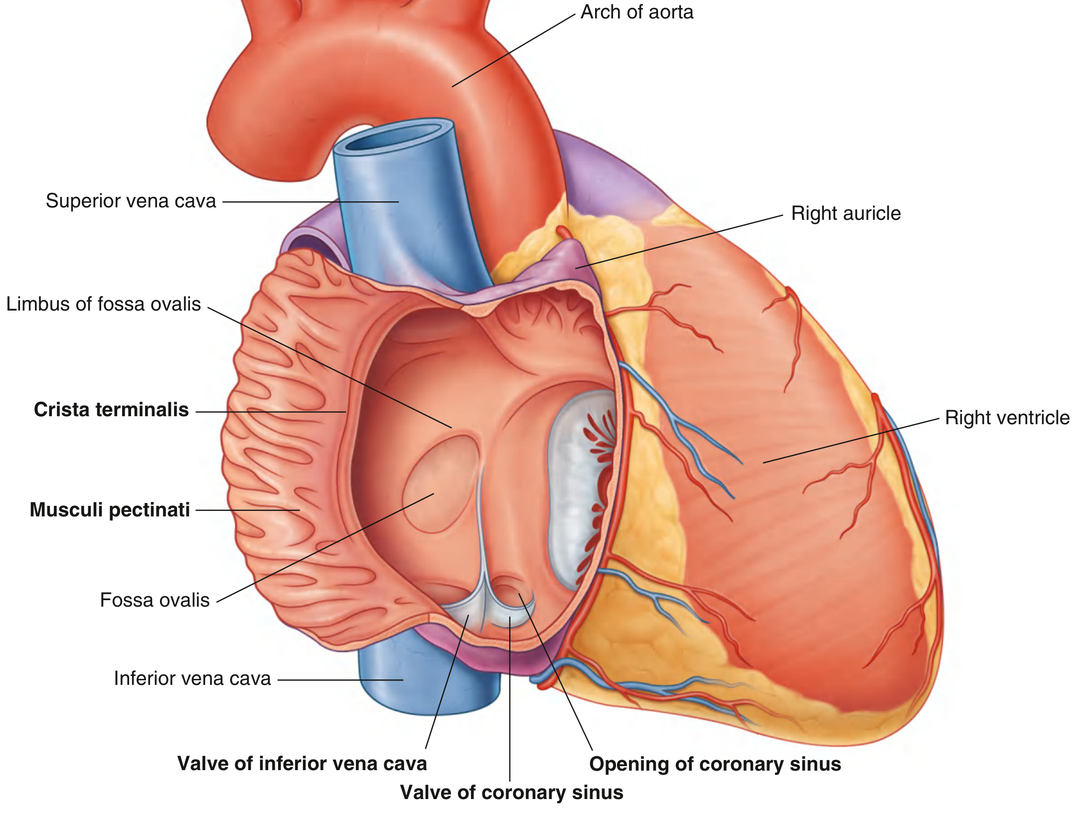
Fig. Internal View of Right Atrium showing: Superior Vena Cava, Inferior Vena Cava, Valve of Inferior Vena Cava, Opening of Coronary Sinus, Valve of Coronary Sinus, Fossa Ovalis, Limbus of Fossa Ovalis, Crista Terminalis, Musculi Pectinati.
B. OPENINGS OF THE LEFT ATRIUM
The left atrium forms most of the base (posterior surface) of the heart. Its interior is predominantly smooth-walled. It receives oxygenated blood from the lungs and transmits it to the left ventricle.
1. Openings of the Four Pulmonary Veins
- Generally four pulmonary veins open into the left atrium - two from each lung (right pulmonary veins and left pulmonary veins).
- They open into the upper portion (posterior, smooth-walled part) of the left atrium.
- There are no valves at the openings of the pulmonary veins.
- This part of the left atrium has smooth walls derived embryologically from the proximal parts of the pulmonary veins that are incorporated into the left atrium during development.
2. Opening of the Left Atrioventricular (AV) Orifice (Mitral Orifice)
- Transmits blood from the left atrium into the left ventricle.
- Guarded by the Mitral Valve (left atrioventricular valve / bicuspid valve), which has two cusps - the anterior (aortic) cusp and the posterior (mural) cusp.
- Closes during ventricular contraction (systole) to prevent backflow into the left atrium.
3. Valve of the Foramen Ovale
- Found near the interatrial septum, which forms part of the anterior wall of the left atrium.
- The thin area or depression in the septum is the Valve of the Foramen Ovale, which corresponds to the floor of the Fossa Ovalis seen from the right atrium.
- During development, this valve prevents blood from passing from the left atrium back to the right atrium.
- In some adults, the valve may not be completely fused, leaving a "probe-patent" foramen ovale - a potential passage between the two atria.
SUMMARY TABLE (Exam Quick Reference)
| Opening | Location | Valve Present | Notes |
|---|---|---|---|
| Superior Vena Cava | Upper posterior RA | No valve | Returns blood from upper body |
| Inferior Vena Cava | Lower posterior RA | Eustachian valve | Directs blood through foramen ovale in fetus |
| Coronary Sinus | Medial to IVC, RA | Thebesian valve | Drains cardiac walls |
| Right AV Orifice | Anterior RA | Tricuspid valve | RA to RV passage |
| Venae Cordis Minimae | Scattered in RA walls | No valve | Drain myocardium directly |
| 4 Pulmonary Veins | Upper posterior LA | No valves | Return oxygenated blood from lungs |
| Left AV Orifice (Mitral) | Anterior LA | Mitral (Bicuspid) valve | LA to LV passage |
| Valve of Foramen Ovale | Interatrial septum, LA | - | Fetal remnant; may be probe-patent in adults |
Sources: - Gray's Anatomy for Students, pp. 231-234 | - Color Atlas of Human Anatomy Vol. 2 (Internal Organs), pp. 43-46
This is a shared conversation. Sign in to Orris to start your own chat.