
Describe mandibular nerve course and branches
mandibular nerve branches diagram anatomy
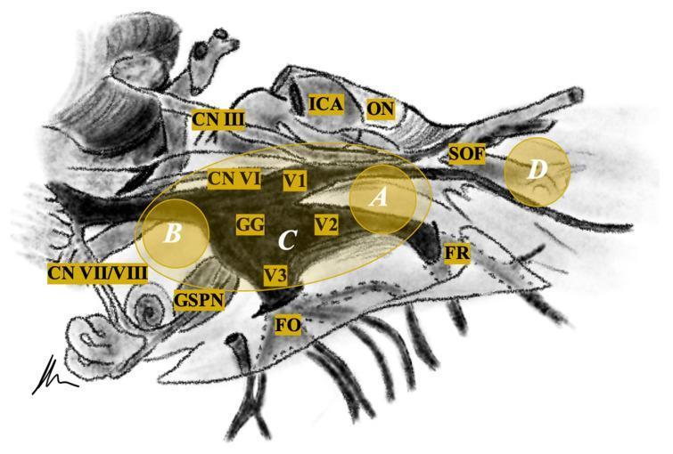
This medical schematic diagram illustrates the neuroanatomical relationships of the trigeminal nerve (CN V) and its surroundings, specifically designed for tumor localization and classification. The central feature is the Gasserian Ganglion (GG), which branches into three divisions: the Ophthalmic (V1) extending toward the Superior Orbital Fissure (SOF), the Maxillary (V2) toward the Foramen Rotundum (FR), and the Mandibular (V3) toward the Foramen Ovale (FO). Adjacent neurovascular structures are labeled, including the Internal Carotid Artery (ICA), Optic Nerve (ON), and Cranial Nerves III, VI, and VII/VIII complex. The Greater Superficial Petrosal Nerve (GSPN) is also indicated inferior to the ganglion. Overlaid on the anatomy are shaded yellow regions and white letters (A, B, C, D) representing a clinical classification system for trigeminal schwannomas: Type A (middle fossa), Type B (posterior fossa), Type C (dumbbell-shaped involving both fossa), and Type D (extracranial/extradural). This diagram serves as a surgical planning guide for neurosurgeons and medical students focusing on skull base pathology.
This composite educational resource illustrates the reduction of a mandibular condyle fracture using a specialized retractor system. Image (a) is an anatomical diagram utilizing a skeletal model to demonstrate the mechanical principle: the retractor tips are inserted into the joint space between the zygomatic arch and the condylar process to pull down the condyle and create space for reduction. Image (b) is an intraoperative clinical photograph showing the same procedure in a live surgical field. An S-shaped preauricular incision is retracted with stay sutures, exposing the underlying soft tissue, facial nerve branches, and the condylar head. The metallic retractor is positioned deep within the surgical site, demonstrating its clinical application for managing dislocated or fractured condylar segments. The comparison highlights the transition from theoretical biomechanics to real-world surgical anatomy, specifically for oral and maxillofacial surgery training.
This composite educational image features two diagnostic CT scans and one anatomical diagram focusing on the right parotid gland region. The first and second panels are axial CT slices of the head and neck. The left-most panel (current admission) demonstrates a hyperdense sialolith (red arrow) within the right Stensen's duct, accompanied by a dilated ductal segment (yellow arrow) and soft tissue swelling within the right parotid gland compared to the contralateral side. The middle panel shows a prior CT from six months earlier, highlighting the same sialolith (red arrow) in a more posterior position and smaller size, illustrating disease progression and anterior displacement. The third panel is an anatomical line drawing of a lateral profile of the head, showing the parotid gland and the branching patterns of the facial nerve (CN VII), including the temporal, zygomatic, buccal, mandibular, and cervical branches. This figure serves as a comparative study of sialolithiasis, demonstrating diagnostic imaging of ductal obstruction and the relevant regional anatomy for surgical or interventional planning in otolaryngology.
Mandibular Nerve (V3) - Course and Branches
Overview
- Gray's Anatomy for Students, p. 1123
Course
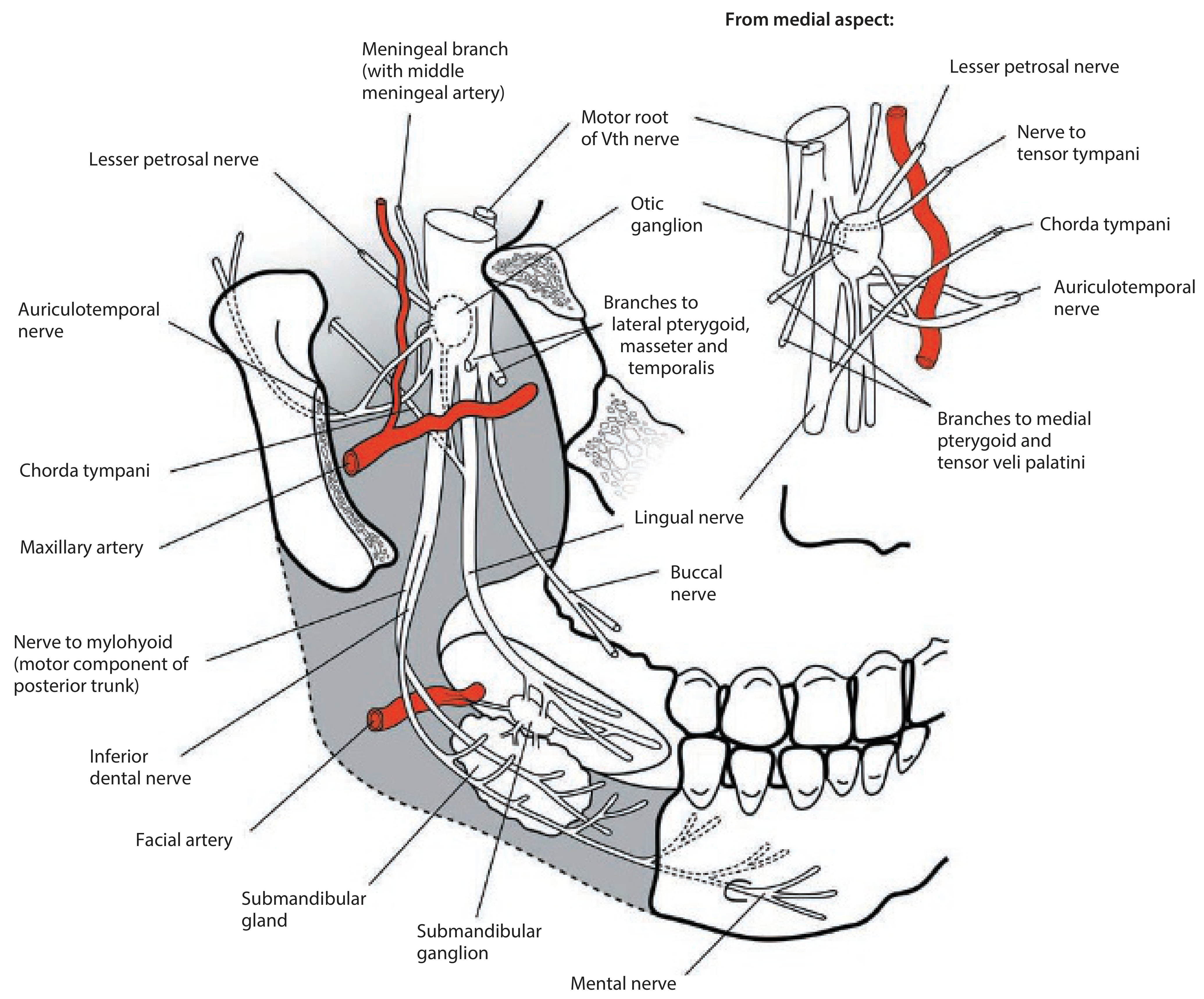
- Origin: The sensory part arises from the trigeminal ganglion in the middle cranial fossa. The small motor root passes medial to the ganglion.
- Exit from skull: Both roots pass through the foramen ovale into the infratemporal fossa, where they immediately unite to form the mixed mandibular nerve trunk.
- Initial position: The united trunk lies deep to the upper head of the lateral pterygoid muscle, between the tensor veli palatini (medially) and the lateral pterygoid (laterally).
- Division: After a very short course, the trunk gives off two immediate branches and then divides into anterior and posterior trunks.
Immediate Branches (before division)
1. Meningeal Branch (Nervus Spinosus)
- Ascends back into the skull through the foramen spinosum with the middle meningeal artery
- Sensory to dura mater of the middle cranial fossa and mastoid air cells
2. Nerve to Medial Pterygoid
- Descends to supply the medial pterygoid muscle (motor)
- Gives off two small twigs:
- Nerve to tensor veli palatini
- Nerve to tensor tympani (ascends to the bony canal of the middle ear)
Anterior Trunk (predominantly motor)
| Branch | Type | Distribution |
|---|---|---|
| Buccal nerve | Sensory (main branch) | Skin over buccinator, mucosa of cheek, buccal gingivae of lower molars |
| Masseteric nerve | Motor | Passes through mandibular notch to supply masseter |
| Deep temporal nerves (usually 2) | Motor | Curve around infratemporal crest to supply temporalis from its deep surface |
| Nerve to lateral pterygoid | Motor | May arise from anterior trunk or from buccal nerve; enters lateral pterygoid directly |
- Gray's Anatomy for Students, pp. 1124-1125
Posterior Trunk (predominantly sensory)
1. Auriculotemporal Nerve
- Origin: Two roots that encircle the middle meningeal artery (i.e., arise around the artery as it ascends to the foramen spinosum)
- Course: Passes between tensor veli palatini and the upper head of lateral pterygoid, then between the sphenomandibular ligament and the neck of the mandible, curves behind the temporomandibular joint, and ascends deep to the parotid gland
- Sensory supply:
- Skin over the temple (superficial temporal branches)
- Auricle, external acoustic meatus, and tympanic membrane
- Temporomandibular joint
- Special role: Carries postganglionic parasympathetic fibers from the otic ganglion (which relays signals from the glossopharyngeal nerve via the lesser petrosal nerve) to the parotid gland - this is why parotid surgery can cause Frey's syndrome (gustatory sweating)
2. Lingual Nerve
- Course: Descends between tensor veli palatini and lateral pterygoid, then crosses the lateral surface of medial pterygoid; enters the oral cavity between the superior pharyngeal constrictor and the posterior mylohyoid attachment; grooves the medial surface of the mandible just below the last molar; then passes onto the lateral surface of hyoglossus into the tongue
- Joined high in the infratemporal fossa by the chorda tympani (branch of VII), which adds:
- Taste from anterior 2/3 of tongue
- Preganglionic parasympathetic fibers (synapse in the submandibular ganglion) to supply the submandibular and sublingual salivary glands
- Sensory supply:
- General sensation from anterior 2/3 of tongue
- Oral mucosa on floor of mouth
- Lingual gingivae of lower teeth
- Clinical note: The lingual nerve is vulnerable during lower molar (wisdom tooth) extraction
3. Inferior Alveolar Nerve (Inferior Dental Nerve)
-
Course: Descends on the lateral surface of medial pterygoid, passes between the sphenomandibular ligament and the neck of the mandible, and enters the mandibular foramen to travel through the mandibular canal
-
Before entering the foramen, gives off:
- Nerve to mylohyoid - the sole motor branch of the posterior trunk; runs in the mylohyoid groove on the medial surface of the mandible; supplies mylohyoid and the anterior belly of digastric
-
Within the canal: Gives dental branches to all lower molar and premolar teeth
-
Terminal branches at the mental foramen:
- Mental nerve - exits the mental foramen; sensory to skin and mucosa of the lower lip and chin
- Incisive nerve - continues within the bone to supply lower canine and incisor teeth
-
Gray's Anatomy for Students, pp. 1125-1126; Scott-Brown's Otorhinolaryngology Vol 2, pp. 1407-1408
Summary: Motor Muscles Supplied by V3
| Muscle | Branch |
|---|---|
| Temporalis | Deep temporal nerves (anterior trunk) |
| Masseter | Masseteric nerve (anterior trunk) |
| Lateral pterygoid | Nerve to lateral pterygoid (anterior trunk) |
| Medial pterygoid | Nerve to medial pterygoid (main trunk) |
| Tensor veli palatini | Via nerve to medial pterygoid |
| Tensor tympani | Via nerve to medial pterygoid |
| Mylohyoid | Mylohyoid nerve (from inferior alveolar, posterior trunk) |
| Anterior belly of digastric | Mylohyoid nerve (from inferior alveolar, posterior trunk) |
Otic Ganglion - Associated Parasympathetic Ganglion
-
Input: Preganglionic fibers from CN IX (via the lesser petrosal nerve)
-
Output: Postganglionic fibers travel on the auriculotemporal nerve to reach the parotid gland
-
The lesser petrosal nerve exits through the foramen ovale (or foramen innominatum) to reach the ganglion
-
Scott-Brown's Otorhinolaryngology Vol 2, p. 1248