
pathophysiology of bronchiectasis
bronchiectasis pathophysiology diagram airway dilation
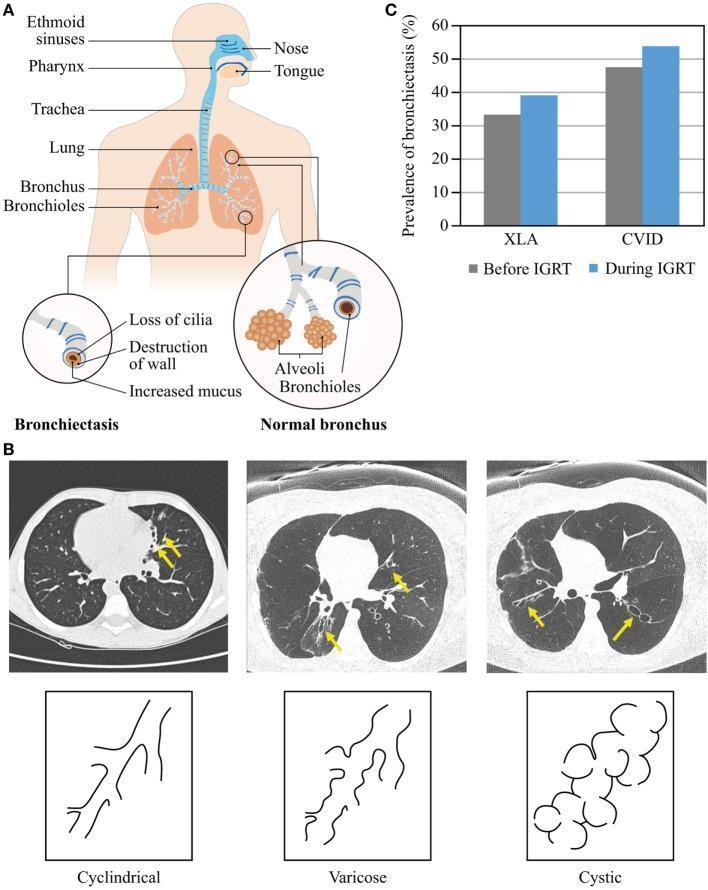
This composite educational image illustrates the pathophysiology and types of bronchiectasis in the context of primary antibody deficiencies. Panel A contains an anatomical diagram showing the respiratory system from the nasal cavity to the bronchioles. It provides a comparative cross-sectional schematic between a normal bronchus and one affected by bronchiectasis, highlighting histopathological changes including loss of cilia, destruction of the bronchial wall, and increased mucus production. Panel B presents three axial High-Resolution Computed Tomography (HRCT) lung windows with corresponding line drawings illustrating the morphological classification of bronchiectasis: Cylindrical (uniform, tubular dilation), Varicose (irregular, beaded appearance with alternating dilation and constriction), and Cystic (sac-like dilations often appearing in clusters). Panel C is a bar graph showing the prevalence of bronchiectasis in patients with X-linked agammaglobulinemia (XLA) and common variable immunodeficiency (CVID) before and during immunoglobulin replacement therapy (IGRT), demonstrating an increase in prevalence with disease duration despite treatment. This resource is intended for medical students and clinicians focusing on pulmonology and immunology.
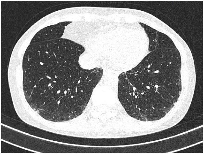
High-resolution computed tomography (HRCT) of the chest in axial view demonstrating bilateral bronchiectasis in a patient with rheumatoid arthritis. The imaging shows classic signs of airway pathology, specifically the 'signet-ring sign,' where dilated bronchi are seen adjacent to smaller pulmonary arteries, indicating a broncho-arterial ratio significantly greater than 1. The bronchial walls are visibly thickened and exhibit a lack of normal tapering as they extend toward the lung periphery. These dilated, thick-walled airways are distributed throughout the lung parenchyma, appearing as circular or oval lucencies. In addition to the bronchiectatic changes, there is evidence of mild interstitial thickening and subtle patchy opacities. This diagnostic image illustrates a common extra-articular manifestation of rheumatoid arthritis, highlighting chronic inflammatory damage to the lower airways resulting in permanent bronchial dilation and structural remodeling.
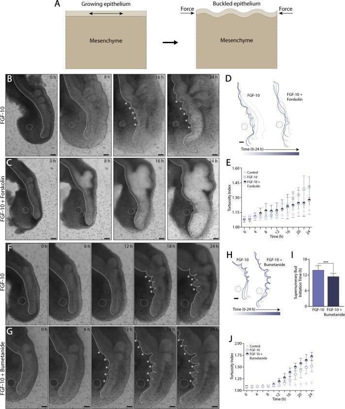
This composite educational image illustrates the mechanical principles of lung airway branching using ex vivo embryonic lung explants. Panel A presents a pathophysiology diagram of 'buckling morphogenesis,' showing how epithelial growth constrained by mesenchyme leads to folding and bud formation under compressive force. Panels B, C, F, and G are bright-field time-lapse clinical photographs showing lung explants over a 24-hour period. Each explant contains an FGF-10-loaded bead (indicated by white dashed circles) placed near the ventral epithelium. White asterisks denote supernumerary buds. Comparison highlights include: - FGF-10 only (B, F): Shows moderate ectopic budding along the ventral epithelium. - FGF-10 + Forskolin (C): An agonist of fluid secretion that causes airway dilation and stretching, suppressing bud formation. - FGF-10 + Bumetanide (G): An inhibitor of fluid secretion that leads to airway collapse and increased, earlier, and more expansive supernumerary budding. Graphs (E, I, J) and contour maps (D, H) quantify the 'tortuosity index' and 'supernumerary bud initiation time,' demonstrating that altered luminal pressure and epithelial tension directly modulate branching morphogenesis in developmental biology.
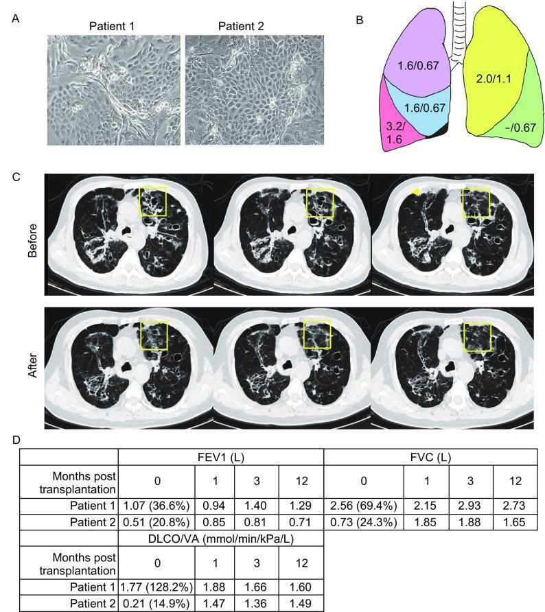
This composite educational image illustrates an autologous SOX9+ basal cell (BC) transplantation study for bronchiectasis. Panel A displays phase-contrast microscopy of cultured SOX9+ BCs from two patients, showing distinct morphologies: Patient 1 exhibits elongated, spindle-like shapes with visible intercellular spaces, while Patient 2 shows a dense, confluent epithelial-like monolayer. Panel B is an anatomical diagram of the lungs, color-coded by lobe, indicating the cell dosage (×10^7) delivered per lobe for each patient. Panel C presents consecutive axial high-resolution computed tomography (HRCT) scans of Patient 2 before and one year after transplantation. Yellow squares highlight a region of severe bronchiectasis; the 'After' images demonstrate regional recovery, characterized by a reduction in cystic dilation and decreased bronchial wall thickening. Panel D contains data tables tracking pulmonary function (FEV1, FVC, and DLCO/VA) from baseline to 12 months post-transplantation. These clinical findings collectively suggest that SOX9+ BC transplantation may facilitate structural and functional repair of damaged airway tissue in chronic obstructive lung disease.
bronchiectasis pathophysiology
Pathophysiology of Bronchiectasis
1. Definition and Basic Concept
"Two intertwined processes contribute to bronchiectasis: obstruction and chronic infection. Either may be the initiator."
- Robbins & Kumar Basic Pathology
2. Mechanical Mechanisms of Airway Dilation
a. Traction Bronchiectasis
b. Pulsion Bronchiectasis
c. Weakness of Airway Walls (Structural)
- Mounier-Kuhn syndrome - congenital tracheobronchomegaly from atrophy of elastic fibers
- Williams-Campbell syndrome - absence of cartilaginous rings in segmental/subsegmental bronchi
- Marfan syndrome
- Relapsing polychondritis
3. The "Vicious Cycle" / "Vicious Vortex" (Cole's Hypothesis)
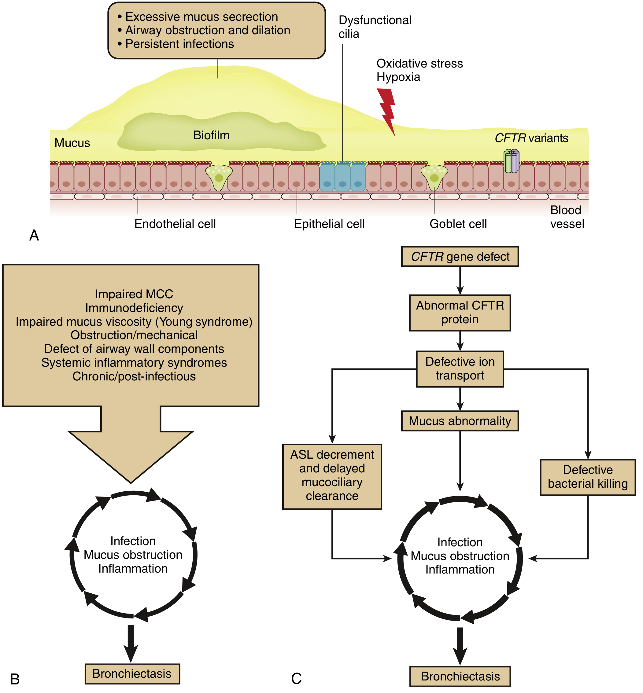
- Airway epithelial and ciliary dysfunction + mucus hypersecretion - impairs mucociliary clearance (MCC)
- Chronic bacterial infection - induces further mucus hypersecretion and colonization (biofilm formation by Pseudomonas aeruginosa, H. influenzae, NTM)
- Inflammation - causes permanent airway injury and further dilation
- Resulting bronchiectatic airways - which are poor in MCC, perpetuating infection, inflammation, and further epithelial/ciliary dysfunction
4. Cellular and Molecular Mechanisms
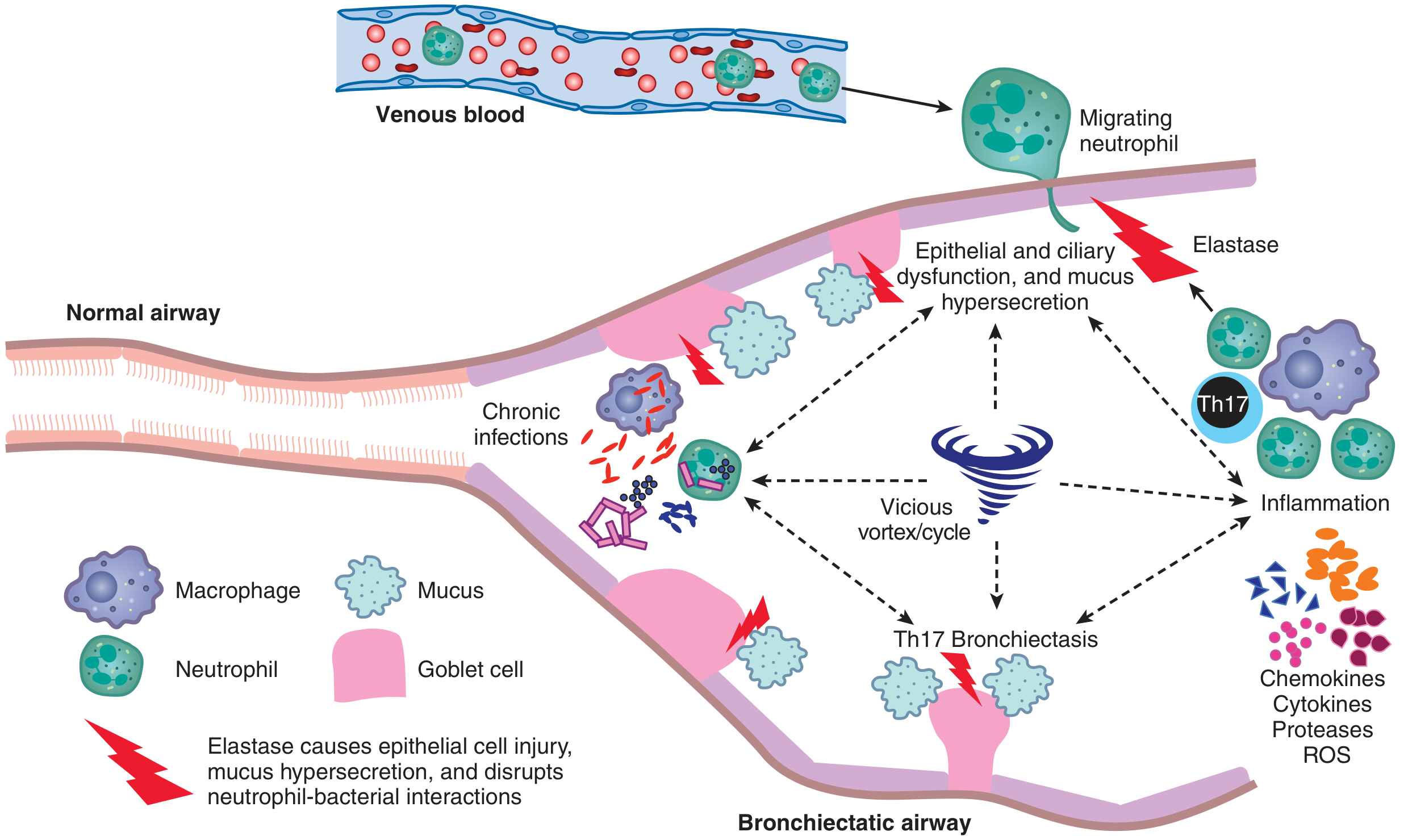
Neutrophil-Dominated Inflammation
- IL-8 (CXCL8) - the primary neutrophil chemokine, produced by bacteria (via glycoproteins) and airway epithelial cells
- Macrophage inflammatory protein-2 (MIP-2)
- TNF-alpha
- Th17 cells - amplify neutrophilic recruitment
Protease-Antiprotease Imbalance
- Neutrophil elastase (HNE) - the key destructive enzyme; damages bronchial epithelium, causes mucus hypersecretion, disrupts normal neutrophil-bacterial interactions, and degrades structural proteins (elastin, collagen)
- Matrix metalloproteinases (MMPs) - further break down the airway wall matrix
- Reactive oxygen species (ROS) - oxidative stress contributes to epithelial injury
Microbial Biofilm Formation
Goblet Cell Hyperplasia
5. Impaired Mucociliary Clearance (MCC) - The Upstream Defect
| Cause | Mechanism |
|---|---|
| Primary ciliary dyskinesia (PCD) | Inherited cilia ultrastructural defects (dynein arms) - complete failure of ciliary beat |
| Cystic fibrosis | CFTR mutation → defective Cl⁻/Na⁺ transport → depleted airway surface liquid (ASL) → dehydrated, viscid mucus that cannot be cleared |
| Young syndrome | Abnormally viscid mucus impairing ciliary transport |
| Post-infectious | Ciliary loss/dysfunction following viral or bacterial infection (influenza, adenovirus, Bordetella pertussis) |
6. Initiating Causes (Etiologic Classification)
| Category | Examples |
|---|---|
| Bronchial obstruction | Foreign body, tumor, mucus impaction, ABPA |
| Post-infectious | TB (major cause in endemic areas), S. aureus, Klebsiella, NTM, post-COVID-19 |
| Congenital/hereditary | Cystic fibrosis, PCD, Kartagener syndrome, immunoglobulin deficiencies, alpha-1 antitrypsin deficiency |
| Immunodeficiency | Common variable immunodeficiency (CVID), X-linked agammaglobulinemia (XLA) |
| Structural airway weakness | Mounier-Kuhn, Williams-Campbell, Marfan, relapsing polychondritis |
| Autoimmune/inflammatory | Rheumatoid arthritis (30% prevalence on CT), inflammatory bowel disease |
| Traction | Sarcoidosis, pulmonary fibrosis |
7. Morphologic Consequences
Macroscopic
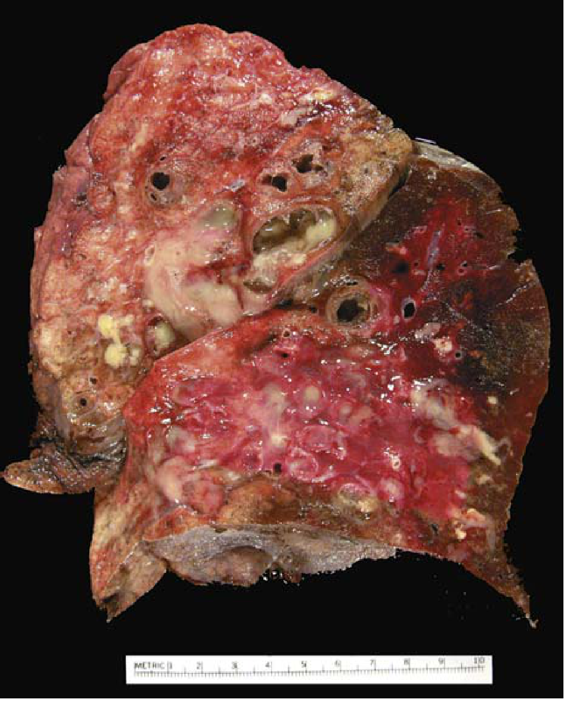
- Lower lobes predominantly affected bilaterally (most vertical airways)
- Airways dilated up to 4x normal diameter, visible grossly near the pleural surface
- Three morphologic types: cylindrical (uniform, tubular), varicose (beaded, irregular), cystic/saccular (sac-like clusters)
Microscopic
- Active disease: Intense acute + chronic inflammatory exudate in bronchial/bronchiolar walls, desquamation of lining epithelium, ulceration
- Healing/chronic: Fibrosis of bronchial and peribronchial walls, peribronchiolar fibrosis
- End-stage: Necrosis can destroy the bronchial wall entirely, producing abscess cavities
- The injury usually cannot be fully repaired, so abnormal dilation and scarring persist even after the active infection resolves.
8. Physiological Consequences
| Consequence | Mechanism |
|---|---|
| Chronic productive cough | Excess mucopurulent secretions; cough is the primary compensatory clearance mechanism |
| Obstructive ventilatory defect | Airway wall thickening, luminal secretions, bronchoconstriction |
| Hypoxemia | V/Q mismatch from airway obstruction and mucus plugging |
| Hypercapnia | In advanced disease with severe airflow limitation |
| Pulmonary hypertension & cor pulmonale | Chronic hypoxic vasoconstriction; vascular remodeling |
| Hemoptysis | Erosion of bronchial vessels by inflammation; systemic bronchial artery hypertrophy |
| Systemic amyloidosis | Rare, from chronic systemic inflammation (AA amyloid) |
| Brain abscess | Hematogenous spread (rare with modern treatment) |
Summary
- Robbins & Kumar Basic Pathology, Chapter 11
- Murray & Nadel's Textbook of Respiratory Medicine, 2-Volume Set, Chapter 69 (Pathophysiology section, Figs. 69.1-69.3)
- Fishman's Pulmonary Diseases and Disorders, 2-Volume Set, Chapter on Bronchiectasis
- Recent systematic reviews (PMID 40662885, 42342264, 41534709) continue to support the anti-inflammatory and mucoactive therapeutic directions derived from this pathophysiologic model.