Epiglottis in image view

Reading File
Finding Sources
Searching Images

epiglottis laryngoscopic view

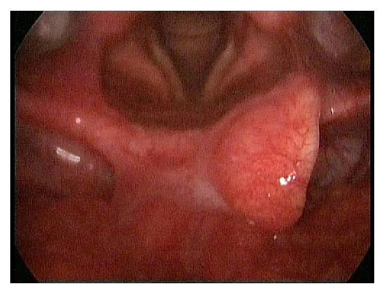
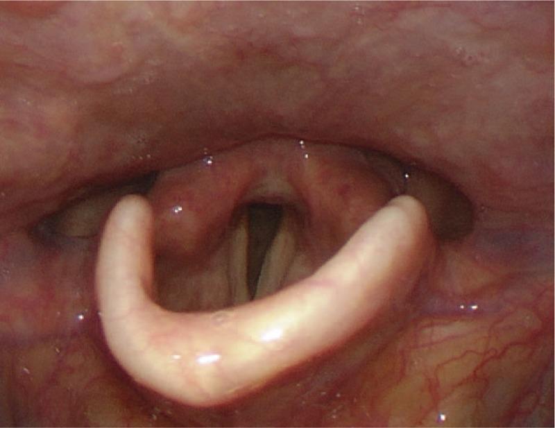
This endoscopic clinical photograph shows a laryngoscopic view of the upper airway, specifically focusing on the epiglottis and the laryngeal inlet. The image demonstrates the anatomical appearance six months following the surgical resection of a large vallecular cyst. The epiglottis exhibits a reddish-pink mucosal color with a slightly irregular surface texture and visible superficial vascular patterns. There is evidence of permanent structural modification to the right side of the epiglottis, likely due to preoperative pressure necrosis from the cyst and subsequent surgical excision. The surrounding pharyngeal tissues and vallecula appear healthy with no evidence of recurrent masses, active inflammation, or significant edema. The laryngeal inlet and vocal fold region are visible in the background, appearing patent and clear of obstruction. This image serves as a clinical example of postoperative healing following the removal of an epiglottic vallecular cyst using CO2 laser microsurgery.
This endoscopic clinical photograph shows a laryngoscopic view of the upper airway, specifically focusing on the epiglottis and the laryngeal inlet. The image demonstrates the anatomical appearance six months following the surgical resection of a large vallecular cyst. The epiglottis exhibits a reddish-pink mucosal color with a slightly irregular surface texture and visible superficial vascular patterns. There is evidence of permanent structural modification to the right side of the epiglottis, likely due to preoperative pressure necrosis from the cyst and subsequent surgical excision. The surrounding pharyngeal tissues and vallecula appear healthy with no evidence of recurrent masses, active inflammation, or significant edema. The laryngeal inlet and vocal fold region are visible in the background, appearing patent and clear of obstruction. This image serves as a clinical example of postoperative healing following the removal of an epiglottic vallecular cyst using CO2 laser microsurgery.

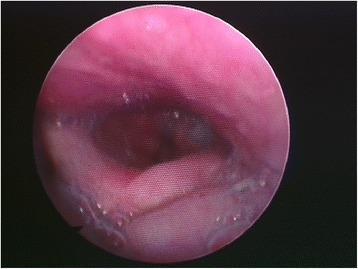
This diagnostic image is a laryngoscopic view of the oropharynx, specifically demonstrating tongue and epiglottal incompetence. The image captures the base of the tongue and the epiglottis, which exhibits abnormal positioning and hypotonia. The epiglottis appears flattened and more horizontal than expected, losing its typical upright, flexible orientation. Notable clinical findings include the pooling of saliva and secretions within the vallecula—the anatomical space between the tongue base and the epiglottis—indicating an impaired swallowing mechanism or poor clearance of secretions. This visual is characteristic of cranial nerve paralysis affecting pharyngeal and lingual motor function, leading to dysphagia and pharyngoepiglottic muscle hypotonia. The mucosal surfaces appear moist and erythematous, with visible fluid accumulation that poses a risk for aspiration. This clinical photograph is highly relevant for otolaryngology and neurology education regarding post-surgical nerve injury and its impact on upper airway and esophageal protection.
This diagnostic image is a laryngoscopic view of the oropharynx, specifically demonstrating tongue and epiglottal incompetence. The image captures the base of the tongue and the epiglottis, which exhibits abnormal positioning and hypotonia. The epiglottis appears flattened and more horizontal than expected, losing its typical upright, flexible orientation. Notable clinical findings include the pooling of saliva and secretions within the vallecula—the anatomical space between the tongue base and the epiglottis—indicating an impaired swallowing mechanism or poor clearance of secretions. This visual is characteristic of cranial nerve paralysis affecting pharyngeal and lingual motor function, leading to dysphagia and pharyngoepiglottic muscle hypotonia. The mucosal surfaces appear moist and erythematous, with visible fluid accumulation that poses a risk for aspiration. This clinical photograph is highly relevant for otolaryngology and neurology education regarding post-surgical nerve injury and its impact on upper airway and esophageal protection.

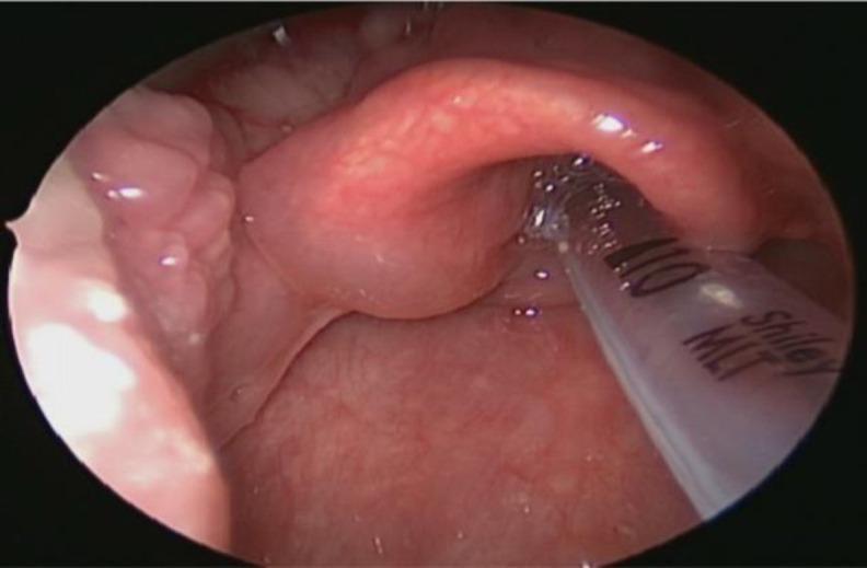
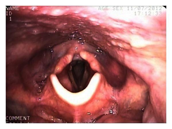
This endoscopic clinical photograph displays a direct laryngoscopic view of the supraglottic laryngeal structures. The primary finding is significant asymmetrical edema involving the left aspect of the epiglottis, affecting both the lingual and laryngeal surfaces. The epiglottis appears markedly swollen, pinkish-red, and displays a smooth but thickened surface texture. The neighboring arytenoid regions also show signs of reactive edema. In the right foreground, a clear medical device, identified as a Shiley MLT (Microlaryngeal Tube) with the number '110' visible, is positioned within the laryngeal inlet, partially obscuring the glottic aperture. This image is used to demonstrate supraglottic pathology, such as that seen in reactive plasmacytosis or localized laryngeal inflammation, highlighting the importance of direct visualization for assessing airway patency and tissue morphology in Otolaryngology.
This endoscopic clinical photograph displays a direct laryngoscopic view of the supraglottic laryngeal structures. The primary finding is significant asymmetrical edema involving the left aspect of the epiglottis, affecting both the lingual and laryngeal surfaces. The epiglottis appears markedly swollen, pinkish-red, and displays a smooth but thickened surface texture. The neighboring arytenoid regions also show signs of reactive edema. In the right foreground, a clear medical device, identified as a Shiley MLT (Microlaryngeal Tube) with the number '110' visible, is positioned within the laryngeal inlet, partially obscuring the glottic aperture. This image is used to demonstrate supraglottic pathology, such as that seen in reactive plasmacytosis or localized laryngeal inflammation, highlighting the importance of direct visualization for assessing airway patency and tissue morphology in Otolaryngology.

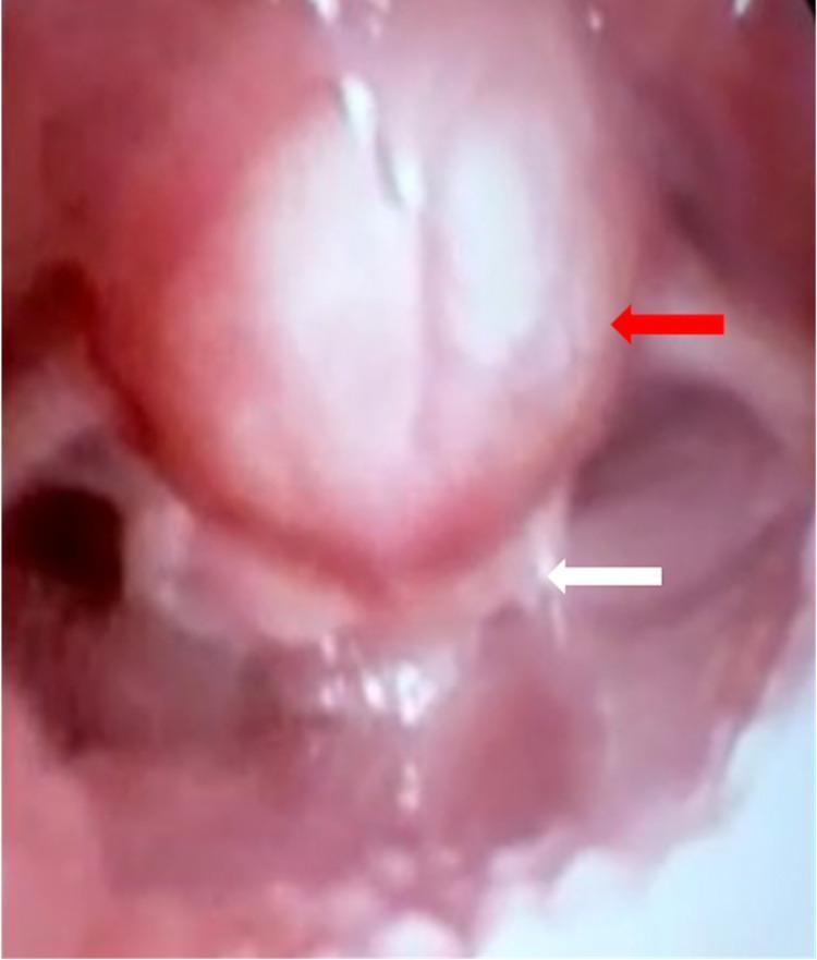
This clinical photograph displays a flexible laryngoscopic view of the upper airway, highlighting a large vallecular cyst. The diagnostic image shows a prominent, spherical, and smooth-surfaced cystic mass (indicated by a red arrow) originating from the vallecula at the base of the tongue. The cyst exhibits a pinkish-red hue consistent with normal mucosal coverage but is under tension, causing significant posterior displacement and compression of the epiglottis (indicated by a white arrow). The epiglottis appears pale and distorted due to the extrinsic pressure. The mass occupies a substantial portion of the oropharyngeal space, resulting in severe narrowing and near-complete occlusion of the laryngeal inlet. This visual demonstrates the pathophysiology of airway obstruction caused by benign epiglottic or vallecular lesions, highlighting the clinical significance of laryngoscopy in diagnosing obstructive laryngeal masses.
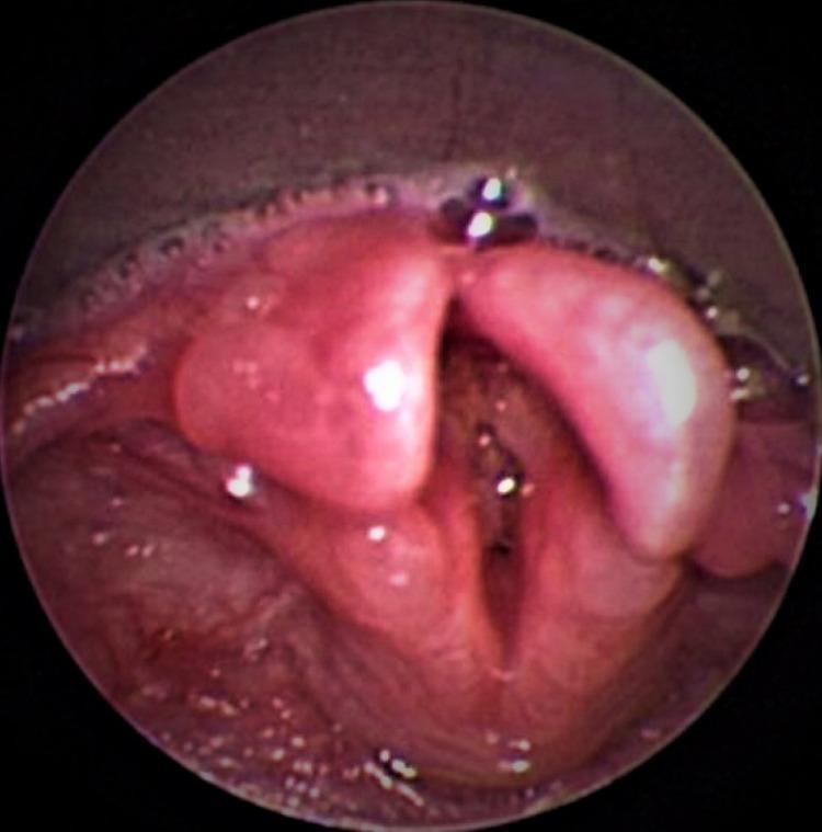
This clinical photograph displays a flexible laryngoscopic view of the upper airway, highlighting a large vallecular cyst. The diagnostic image shows a prominent, spherical, and smooth-surfaced cystic mass (indicated by a red arrow) originating from the vallecula at the base of the tongue. The cyst exhibits a pinkish-red hue consistent with normal mucosal coverage but is under tension, causing significant posterior displacement and compression of the epiglottis (indicated by a white arrow). The epiglottis appears pale and distorted due to the extrinsic pressure. The mass occupies a substantial portion of the oropharyngeal space, resulting in severe narrowing and near-complete occlusion of the laryngeal inlet. This visual demonstrates the pathophysiology of airway obstruction caused by benign epiglottic or vallecular lesions, highlighting the clinical significance of laryngoscopy in diagnosing obstructive laryngeal masses.

This clinical photograph displays a fiberoptic laryngoscopic view of a neonatal upper airway. The primary diagnostic finding is a bifid epiglottis, characterized by a complete midline cleft that divides the epiglottic cartilage into two distinct lateral lobes. These lobes are situated at the base of the tongue, superior to the glottic opening. The laryngeal mucosa appears healthy with a normal pinkish hue and moist surface, with some light reflections indicating the presence of physiological secretions. Inferior to the malformed epiglottis, the vocal cords and arytenoid cartilages are partially visualized. This congenital anomaly is a rare structural defect often associated with broader syndromic presentations, such as Pallister-Hall syndrome. The image illustrates the anatomical cause of inspiratory stridor or irregular breathing patterns in the pediatric population and serves as a critical diagnostic marker for otorhinolaryngologists and pediatricians.
This clinical photograph displays a fiberoptic laryngoscopic view of a neonatal upper airway. The primary diagnostic finding is a bifid epiglottis, characterized by a complete midline cleft that divides the epiglottic cartilage into two distinct lateral lobes. These lobes are situated at the base of the tongue, superior to the glottic opening. The laryngeal mucosa appears healthy with a normal pinkish hue and moist surface, with some light reflections indicating the presence of physiological secretions. Inferior to the malformed epiglottis, the vocal cords and arytenoid cartilages are partially visualized. This congenital anomaly is a rare structural defect often associated with broader syndromic presentations, such as Pallister-Hall syndrome. The image illustrates the anatomical cause of inspiratory stridor or irregular breathing patterns in the pediatric population and serves as a critical diagnostic marker for otorhinolaryngologists and pediatricians.

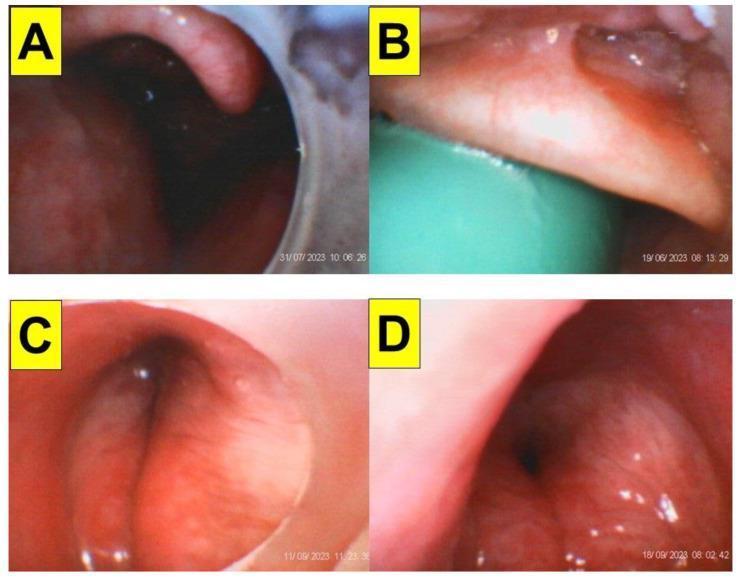
This composite clinical endoscopic image displays four panels (A-D) showing the upper airway anatomy and laryngeal structures of obese patients during video styletubation. Panel A demonstrates significant narrowing due to a collapsed lateral pharyngeal wall and a concave, omega-shaped epiglottis. Panel B illustrates an anterior-posterior collapsed epiglottis being managed with a suction-airway device. Panel C reveals edematous and hypertrophic bilateral vestibular folds, obstructing the glottic view. Panel D identifies chronic hypertrophic laryngitis characterized by prominent bilateral arytenoid edema and generalized mucosal congestion. Collectively, these images illustrate the diagnostic challenges of managing difficult airways in patients with high Body Mass Index (BMI), specifically highlighting soft tissue redundancy, airway collapse, and laryngeal edema that can impede successful tracheal intubation. The images are representative of the anesthesiology specialty, specifically focused on bariatric airway management and the educational identification of laryngoscopic landmarks under pathological conditions.
This composite clinical endoscopic image displays four panels (A-D) showing the upper airway anatomy and laryngeal structures of obese patients during video styletubation. Panel A demonstrates significant narrowing due to a collapsed lateral pharyngeal wall and a concave, omega-shaped epiglottis. Panel B illustrates an anterior-posterior collapsed epiglottis being managed with a suction-airway device. Panel C reveals edematous and hypertrophic bilateral vestibular folds, obstructing the glottic view. Panel D identifies chronic hypertrophic laryngitis characterized by prominent bilateral arytenoid edema and generalized mucosal congestion. Collectively, these images illustrate the diagnostic challenges of managing difficult airways in patients with high Body Mass Index (BMI), specifically highlighting soft tissue redundancy, airway collapse, and laryngeal edema that can impede successful tracheal intubation. The images are representative of the anesthesiology specialty, specifically focused on bariatric airway management and the educational identification of laryngoscopic landmarks under pathological conditions.

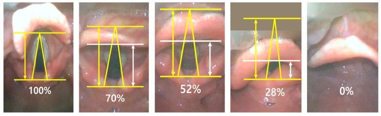
This set of five diagnostic images showcases video laryngoscopic views of the human larynx, illustrating the 'Percentage of Glottic Opening' (POGO) scoring system used during endotracheal intubation. The series progresses from a clear view of the glottis to complete obstruction. Anatomical structures visible include the epiglottis, vocal cords, anterior commissure, and arytenoid cartilages. Yellow superimposed lines delineate the full potential glottic span from the interarytenoid notch to the anterior commissure. In the partial views (70%, 52%, and 28%), white horizontal lines and double-headed arrows quantify the visible portion of the glottis relative to the total span. The 100% score indicates full visibility of the vocal cords, whereas the 0% score shows the glottic opening completely obscured by the epiglottis. This comparison chart serves as an educational tool for anesthesiology and emergency medicine to assess and communicate the difficulty of laryngeal exposure and airway management.
This set of five diagnostic images showcases video laryngoscopic views of the human larynx, illustrating the 'Percentage of Glottic Opening' (POGO) scoring system used during endotracheal intubation. The series progresses from a clear view of the glottis to complete obstruction. Anatomical structures visible include the epiglottis, vocal cords, anterior commissure, and arytenoid cartilages. Yellow superimposed lines delineate the full potential glottic span from the interarytenoid notch to the anterior commissure. In the partial views (70%, 52%, and 28%), white horizontal lines and double-headed arrows quantify the visible portion of the glottis relative to the total span. The 100% score indicates full visibility of the vocal cords, whereas the 0% score shows the glottic opening completely obscured by the epiglottis. This comparison chart serves as an educational tool for anesthesiology and emergency medicine to assess and communicate the difficulty of laryngeal exposure and airway management.

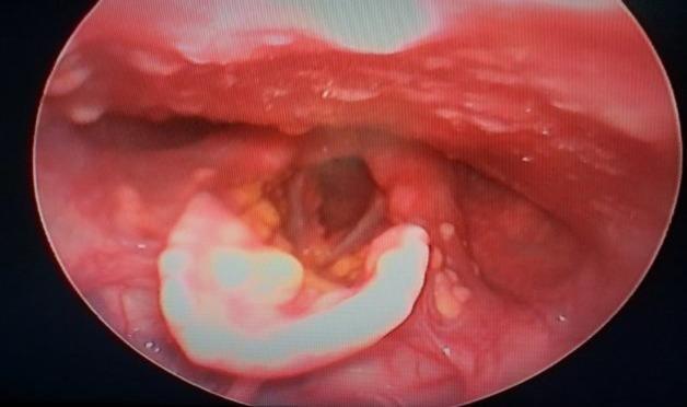
This clinical image shows a laryngoscopic view of the larynx, demonstrating multiple yellow to reddish-yellow papules and nodules across various laryngeal surfaces. The lesions are prominent on the laryngeal surface of the epiglottis, bilateral aryepiglottic folds, and the interarytenoid region. Subglottic involvement is also visible. The morphology of these lesions ranges from discrete, small papules to larger, raised nodules with a characteristic xanthomatous (yellowish) appearance. Such findings are indicative of systemic disorders with mucosal infiltration, such as xanthomatosis or non-Langerhans cell histiocytosis (e.g., xanthoma disseminatum), often correlating with clinical symptoms like hoarseness and visible cutaneous lesions. The image serves as a diagnostic reference for identifying laryngeal manifestations of infiltrative histiocytic diseases in an otolaryngology or dermatology context.

This clinical image shows a laryngoscopic view of the larynx, demonstrating multiple yellow to reddish-yellow papules and nodules across various laryngeal surfaces. The lesions are prominent on the laryngeal surface of the epiglottis, bilateral aryepiglottic folds, and the interarytenoid region. Subglottic involvement is also visible. The morphology of these lesions ranges from discrete, small papules to larger, raised nodules with a characteristic xanthomatous (yellowish) appearance. Such findings are indicative of systemic disorders with mucosal infiltration, such as xanthomatosis or non-Langerhans cell histiocytosis (e.g., xanthoma disseminatum), often correlating with clinical symptoms like hoarseness and visible cutaneous lesions. The image serves as a diagnostic reference for identifying laryngeal manifestations of infiltrative histiocytic diseases in an otolaryngology or dermatology context.

Reading File
Searching Images

normal larynx laryngoscopy epiglottis vocal cords aryepiglottic fold piriform fossa

Searching Images

epiglottis posterior view cartilages of larynx
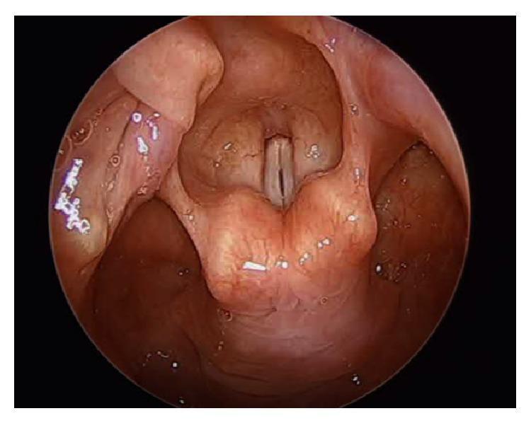
This diagnostic clinical photograph, captured via intraoperative endoscopy, provides a supraglottic view of the human larynx. The primary anatomical structures visible include the true vocal folds (vocal cords), the arytenoid cartilages, and the epiglottis superiorly. The vocal folds appear as symmetric, pale, whitish-gray bands meeting at the anterior commissure, with a narrow vertical glottic chink visible between them. The surrounding laryngeal mucosa and the aryepiglottic folds exhibit a healthy pinkish-red hue with no gross evidence of ulceration or significant edema. The arytenoid cartilages are prominently visible as rounded mucosal-covered structures at the posterior aspect of the glottis. Small, white, punctate specular reflections across the mucosal surfaces are artifacts from the endoscopic light source. The image demonstrates a normal laryngeal configuration during a surgical procedure, likely prior to or during a laryngeal intervention such as an arytenoidectomy, as indicated by the medialized position of the vocal folds and the surgical context.
This diagnostic clinical photograph, captured via intraoperative endoscopy, provides a supraglottic view of the human larynx. The primary anatomical structures visible include the true vocal folds (vocal cords), the arytenoid cartilages, and the epiglottis superiorly. The vocal folds appear as symmetric, pale, whitish-gray bands meeting at the anterior commissure, with a narrow vertical glottic chink visible between them. The surrounding laryngeal mucosa and the aryepiglottic folds exhibit a healthy pinkish-red hue with no gross evidence of ulceration or significant edema. The arytenoid cartilages are prominently visible as rounded mucosal-covered structures at the posterior aspect of the glottis. Small, white, punctate specular reflections across the mucosal surfaces are artifacts from the endoscopic light source. The image demonstrates a normal laryngeal configuration during a surgical procedure, likely prior to or during a laryngeal intervention such as an arytenoidectomy, as indicated by the medialized position of the vocal folds and the surgical context.

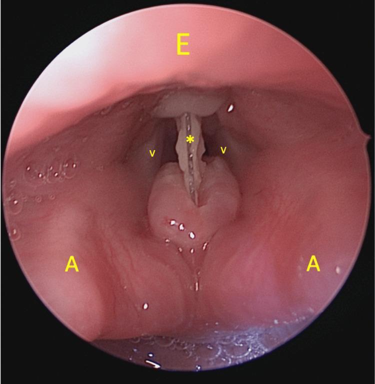
This clinical endoscopic photograph captures a direct laryngoscopy view of the upper airway, demonstrating a laryngeal foreign body obstruction. The visual is labeled with anatomical markers: the epiglottis (E) is seen superiorly as a smooth, pinkish structure; the true vocal folds (V) are visible as paler, longitudinal bands lateral to the midline; and the arytenoid cartilages (A) appear as prominent, rounded soft tissue masses in the posterior larynx. A rigid, light-colored foreign body (marked with an asterisk *) is lodged vertically at the level of the supraglottis (false vocal folds), traversing the anterior-to-posterior dimension of the laryngeal inlet. This image is used in medical education to illustrate an emergency airway scenario, specifically the localization of supraglottic foreign bodies and the relevant laryngeal landmarks for endoscopic evaluation and retrieval.
This clinical endoscopic photograph captures a direct laryngoscopy view of the upper airway, demonstrating a laryngeal foreign body obstruction. The visual is labeled with anatomical markers: the epiglottis (E) is seen superiorly as a smooth, pinkish structure; the true vocal folds (V) are visible as paler, longitudinal bands lateral to the midline; and the arytenoid cartilages (A) appear as prominent, rounded soft tissue masses in the posterior larynx. A rigid, light-colored foreign body (marked with an asterisk *) is lodged vertically at the level of the supraglottis (false vocal folds), traversing the anterior-to-posterior dimension of the laryngeal inlet. This image is used in medical education to illustrate an emergency airway scenario, specifically the localization of supraglottic foreign bodies and the relevant laryngeal landmarks for endoscopic evaluation and retrieval.

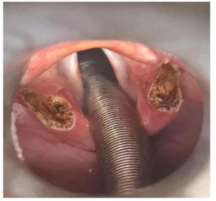
This clinical photograph captures an intraoperative view of the larynx during a supraglottoplasty, specifically demonstrating the immediate result of bilateral cuneiform tubercle removal. The view, obtained via suspension laryngoscopy, shows two distinct, rough-textured surgical defects (voids) located within the aryepiglottic folds where the redundant cuneiform cartilages were excised. These surgical sites exhibit signs of thermal alteration and tissue removal, appearing brownish and irregular compared to the surrounding pink mucosal tissue. An armored, metallic-appearing laser-resistant endotracheal tube is centrally positioned through the glottis, secured in the posterior commissure to maintain the airway and provide protection during the use of a CO2 laser. The epiglottis is visible superiorly, and the operative field shows the widening of the supraglottic space. This procedure is typically performed to treat conditions such as severe laryngomalacia or exercise-induced laryngeal obstruction (EILO) by reducing supraglottic tissue collapse.
This clinical photograph captures an intraoperative view of the larynx during a supraglottoplasty, specifically demonstrating the immediate result of bilateral cuneiform tubercle removal. The view, obtained via suspension laryngoscopy, shows two distinct, rough-textured surgical defects (voids) located within the aryepiglottic folds where the redundant cuneiform cartilages were excised. These surgical sites exhibit signs of thermal alteration and tissue removal, appearing brownish and irregular compared to the surrounding pink mucosal tissue. An armored, metallic-appearing laser-resistant endotracheal tube is centrally positioned through the glottis, secured in the posterior commissure to maintain the airway and provide protection during the use of a CO2 laser. The epiglottis is visible superiorly, and the operative field shows the widening of the supraglottic space. This procedure is typically performed to treat conditions such as severe laryngomalacia or exercise-induced laryngeal obstruction (EILO) by reducing supraglottic tissue collapse.

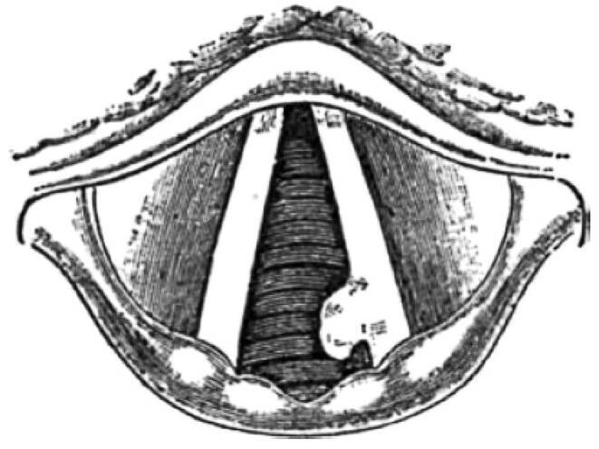
This historical anatomical illustration provides an indirect laryngoscopic view of the larynx, specifically the glottic region. Key anatomical structures visible include the bilateral vocal cords (true vocal folds) meeting at the anterior commissure, with the epiglottis positioned superiorly and the arytenoid cartilages at the posterior aspect. A pathological finding is clearly depicted on the left vocal cord: a prominent, irregular, lobulated mass is situated in the posterior third of the glottis. The growth is nodular in appearance and significantly distorts the smooth edge of the vocal fold, encroaching upon the laryngeal airway (rima glottidis). This drawing serves as a clinical documentation of a glottic tumor, illustrating the localized morphological changes and tissue proliferation characteristic of laryngeal malignancy or advanced dysplasia. The visual focus is the demonstration of the lesion's site, size, and its impact on glottic symmetry, which are critical findings for the diagnosis of persistent hoarseness and laryngeal cancer.
This historical anatomical illustration provides an indirect laryngoscopic view of the larynx, specifically the glottic region. Key anatomical structures visible include the bilateral vocal cords (true vocal folds) meeting at the anterior commissure, with the epiglottis positioned superiorly and the arytenoid cartilages at the posterior aspect. A pathological finding is clearly depicted on the left vocal cord: a prominent, irregular, lobulated mass is situated in the posterior third of the glottis. The growth is nodular in appearance and significantly distorts the smooth edge of the vocal fold, encroaching upon the laryngeal airway (rima glottidis). This drawing serves as a clinical documentation of a glottic tumor, illustrating the localized morphological changes and tissue proliferation characteristic of laryngeal malignancy or advanced dysplasia. The visual focus is the demonstration of the lesion's site, size, and its impact on glottic symmetry, which are critical findings for the diagnosis of persistent hoarseness and laryngeal cancer.

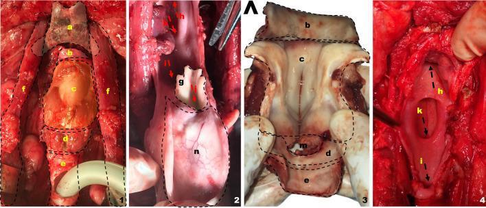
A series of four clinical photographs demonstrating the surgical anatomy of a total laryngectomy on a porcine model, used as a surrogate for human surgical training. Panel 1 shows a superior view of the exposed larynx with the hyoid bone (a), thyrohyoid membrane (b), thyroid cartilage (c), cricoid cartilage (d), and trachea (e) in a cranial-to-caudal sequence; the prelaryngeal musculature (f) is retracted laterally. Panel 2 highlights the everted epiglottis (n), vocal rudiment (g), and glottic space (j), indicating the anatomical orientation toward the oropharynx (h) and esophagus (i). Panel 3 provides a detailed anatomical view of the thyroid (c) and cricoid (d) cartilages, the cricothyroid membrane (m), and the thyrohyoid membrane (b). Panel 4 illustrates the posterior surgical bed after laryngeal removal, revealing the esophageal diverticulum (k) and the relationship between the oropharyngeal inlet (h) and the esophageal direction (i). The images serve to teach laryngeal dissection techniques and comparative anatomy for head and neck surgical procedures.
A series of four clinical photographs demonstrating the surgical anatomy of a total laryngectomy on a porcine model, used as a surrogate for human surgical training. Panel 1 shows a superior view of the exposed larynx with the hyoid bone (a), thyrohyoid membrane (b), thyroid cartilage (c), cricoid cartilage (d), and trachea (e) in a cranial-to-caudal sequence; the prelaryngeal musculature (f) is retracted laterally. Panel 2 highlights the everted epiglottis (n), vocal rudiment (g), and glottic space (j), indicating the anatomical orientation toward the oropharynx (h) and esophagus (i). Panel 3 provides a detailed anatomical view of the thyroid (c) and cricoid (d) cartilages, the cricothyroid membrane (m), and the thyrohyoid membrane (b). Panel 4 illustrates the posterior surgical bed after laryngeal removal, revealing the esophageal diverticulum (k) and the relationship between the oropharyngeal inlet (h) and the esophageal direction (i). The images serve to teach laryngeal dissection techniques and comparative anatomy for head and neck surgical procedures.

This clinical endoscopic photograph captures a rigid laryngoscopic view of the larynx, highlighting the vocal folds and glottic opening. The image demonstrates a right vocal fold palsy where the right vocal fold is medially fixed in a paramedian position. In contrast, the left vocal fold exhibits compensatory movement, crossing the midline to approximate the fixed right fold during attempted phonation. The vocal folds appear smooth and pearly white with fine superficial vasculature. Anatomical landmarks including the epiglottis in the foreground, the arytenoid cartilages, and the posterior commissure are clearly visible. The overall mucosal surface appears healthy without evidence of acute inflammation, edema, or space-occupying lesions. This diagnostic image is significant for otolaryngology education, illustrating the visual signs of unilateral vocal cord paralysis and the resulting glottic configuration. It serves as an educational resource for medical students and residents to recognize laryngeal asymmetry and the mechanical compensation of the contralateral fold in postoperative clinical scenarios.

This clinical endoscopic photograph captures a rigid laryngoscopic view of the larynx, highlighting the vocal folds and glottic opening. The image demonstrates a right vocal fold palsy where the right vocal fold is medially fixed in a paramedian position. In contrast, the left vocal fold exhibits compensatory movement, crossing the midline to approximate the fixed right fold during attempted phonation. The vocal folds appear smooth and pearly white with fine superficial vasculature. Anatomical landmarks including the epiglottis in the foreground, the arytenoid cartilages, and the posterior commissure are clearly visible. The overall mucosal surface appears healthy without evidence of acute inflammation, edema, or space-occupying lesions. This diagnostic image is significant for otolaryngology education, illustrating the visual signs of unilateral vocal cord paralysis and the resulting glottic configuration. It serves as an educational resource for medical students and residents to recognize laryngeal asymmetry and the mechanical compensation of the contralateral fold in postoperative clinical scenarios.

Searching Images

normal endoscopic view of larynx epiglottis and vocal folds
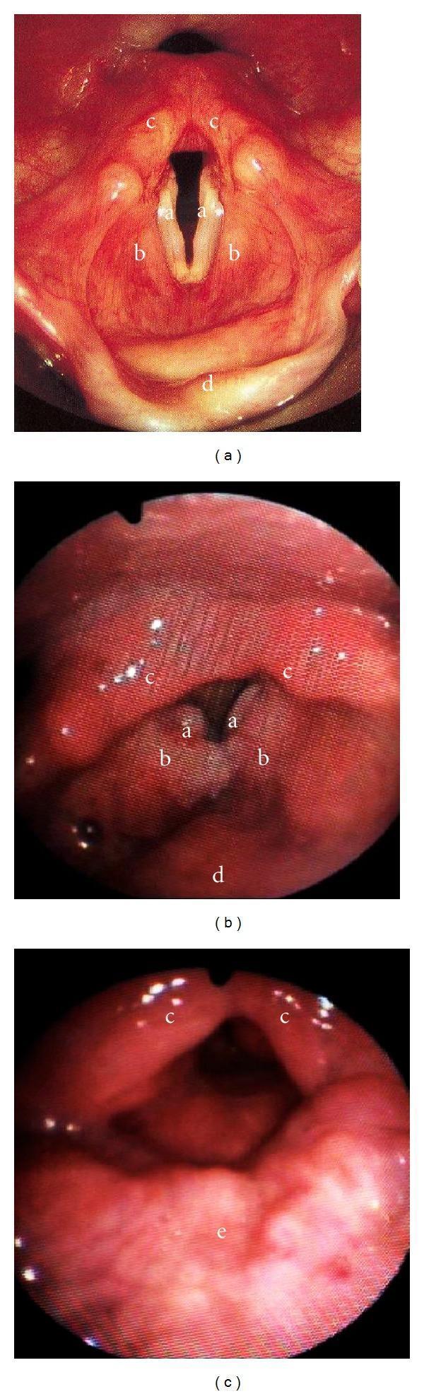
This composite of three fiberoptic endoscopic images demonstrates the comparative surgical and pathological anatomy of the larynx. Image (a) depicts a normal larynx during respiration, clearly identifying the true vocal folds (a), false vocal folds (b), arytenoids (c), and the epiglottis (d) forming a patent glottis. Image (b) shows a larynx with glottic tumors, where exophytic growths are visible on the true vocal folds (a), potentially impairing sphincteric closure and airway protection. Image (c) shows the post-surgical anatomy following a supracricoid laryngectomy with cricohyoidopexy. This view illustrates the complete absence of the vocal folds and epiglottis. The remaining functional anatomy consists of the arytenoids (c) positioned against the tongue base (e), which serves as the primary mechanism for airway protection during swallowing. This comparison is used to teach laryngeal oncology, the physiological consequences of partial laryngectomy, and the mechanisms of post-operative aspiration risk in otolaryngology and speech-language pathology.
This composite of three fiberoptic endoscopic images demonstrates the comparative surgical and pathological anatomy of the larynx. Image (a) depicts a normal larynx during respiration, clearly identifying the true vocal folds (a), false vocal folds (b), arytenoids (c), and the epiglottis (d) forming a patent glottis. Image (b) shows a larynx with glottic tumors, where exophytic growths are visible on the true vocal folds (a), potentially impairing sphincteric closure and airway protection. Image (c) shows the post-surgical anatomy following a supracricoid laryngectomy with cricohyoidopexy. This view illustrates the complete absence of the vocal folds and epiglottis. The remaining functional anatomy consists of the arytenoids (c) positioned against the tongue base (e), which serves as the primary mechanism for airway protection during swallowing. This comparison is used to teach laryngeal oncology, the physiological consequences of partial laryngectomy, and the mechanisms of post-operative aspiration risk in otolaryngology and speech-language pathology.

This clinical photograph displays an endoscopic view of the human larynx, specifically a fiberoptic nasoendoscopy. The image highlights the glottis and surrounding supraglottic structures. Centrally, the true vocal folds (vocal cords) appear as bright, pearly-white, crescent-shaped bands. The edges of the folds are smooth and symmetrical, framing the dark, patent rima glottidis (airway opening). Above the vocal folds, the vestibular folds (false vocal cords) and the laryngeal vestibule are visible, exhibiting a healthy, reddish-pink mucosal lining with normal vascularity. The epiglottis is partially visible at the top of the frame. Small droplets of clear mucus are scattered across the mucosal surface, which is a normal finding. This image serves as an educational example of normal post-traumatic recovery, demonstrating preserved laryngeal anatomy and unobstructed airway patency following a penetrating neck injury.
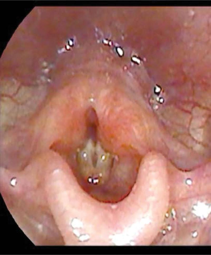
This clinical photograph displays an endoscopic view of the human larynx, specifically a fiberoptic nasoendoscopy. The image highlights the glottis and surrounding supraglottic structures. Centrally, the true vocal folds (vocal cords) appear as bright, pearly-white, crescent-shaped bands. The edges of the folds are smooth and symmetrical, framing the dark, patent rima glottidis (airway opening). Above the vocal folds, the vestibular folds (false vocal cords) and the laryngeal vestibule are visible, exhibiting a healthy, reddish-pink mucosal lining with normal vascularity. The epiglottis is partially visible at the top of the frame. Small droplets of clear mucus are scattered across the mucosal surface, which is a normal finding. This image serves as an educational example of normal post-traumatic recovery, demonstrating preserved laryngeal anatomy and unobstructed airway patency following a penetrating neck injury.

This clinical photograph displays a fiberscopic view of the human larynx during a laryngoscopic examination. The image provides a superior view of the glottis and surrounding supraglottic structures. Key anatomical features include the epiglottis in the foreground, the true vocal folds (vocal cords), and the arytenoid cartilages. The mucosa appears pink and healthy with normal vascularity and no obvious masses, ulcerations, or acute inflammatory exudates. The true vocal folds are positioned medially near the midline, showing a narrow glottic aperture. In the clinical context of bilateral vocal cord paralysis (BVCP), this still image represents the characteristic adducted or paramedian position of the folds that fails to abduct during inspiration, leading to clinical symptoms such as stridor. The educational focus is on the endoscopic recognition of laryngeal anatomy and the clinical presentation of neurogenic airway obstruction where structural integrity is preserved but functional mobility is lost.

This clinical photograph displays a fiberscopic view of the human larynx during a laryngoscopic examination. The image provides a superior view of the glottis and surrounding supraglottic structures. Key anatomical features include the epiglottis in the foreground, the true vocal folds (vocal cords), and the arytenoid cartilages. The mucosa appears pink and healthy with normal vascularity and no obvious masses, ulcerations, or acute inflammatory exudates. The true vocal folds are positioned medially near the midline, showing a narrow glottic aperture. In the clinical context of bilateral vocal cord paralysis (BVCP), this still image represents the characteristic adducted or paramedian position of the folds that fails to abduct during inspiration, leading to clinical symptoms such as stridor. The educational focus is on the endoscopic recognition of laryngeal anatomy and the clinical presentation of neurogenic airway obstruction where structural integrity is preserved but functional mobility is lost.

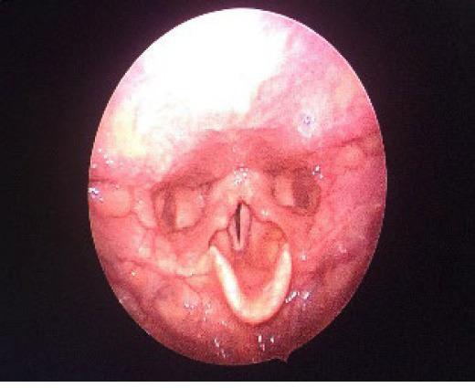
This diagnostic clinical photograph, captured via intraoperative endoscopy, provides a supraglottic view of the human larynx. The primary anatomical structures visible include the true vocal folds (vocal cords), the arytenoid cartilages, and the epiglottis superiorly. The vocal folds appear as symmetric, pale, whitish-gray bands meeting at the anterior commissure, with a narrow vertical glottic chink visible between them. The surrounding laryngeal mucosa and the aryepiglottic folds exhibit a healthy pinkish-red hue with no gross evidence of ulceration or significant edema. The arytenoid cartilages are prominently visible as rounded mucosal-covered structures at the posterior aspect of the glottis. Small, white, punctate specular reflections across the mucosal surfaces are artifacts from the endoscopic light source. The image demonstrates a normal laryngeal configuration during a surgical procedure, likely prior to or during a laryngeal intervention such as an arytenoidectomy, as indicated by the medialized position of the vocal folds and the surgical context.
This diagnostic clinical photograph, captured via intraoperative endoscopy, provides a supraglottic view of the human larynx. The primary anatomical structures visible include the true vocal folds (vocal cords), the arytenoid cartilages, and the epiglottis superiorly. The vocal folds appear as symmetric, pale, whitish-gray bands meeting at the anterior commissure, with a narrow vertical glottic chink visible between them. The surrounding laryngeal mucosa and the aryepiglottic folds exhibit a healthy pinkish-red hue with no gross evidence of ulceration or significant edema. The arytenoid cartilages are prominently visible as rounded mucosal-covered structures at the posterior aspect of the glottis. Small, white, punctate specular reflections across the mucosal surfaces are artifacts from the endoscopic light source. The image demonstrates a normal laryngeal configuration during a surgical procedure, likely prior to or during a laryngeal intervention such as an arytenoidectomy, as indicated by the medialized position of the vocal folds and the surgical context.

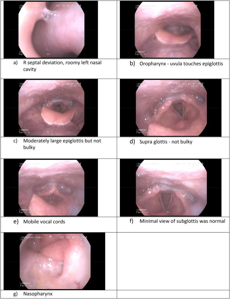
A series of seven endoscopic still frames (labeled a-g) obtained via flexible nasendoscopy, documenting an upper airway assessment. (a) Nasal cavity view showing right septal deviation and a patent left nasal passage. (g) A view of the nasopharynx appearing adequate without obvious mucosal lesions. (b) Oropharyngeal view demonstrating a high larynx with a minimal distance between the uvula/soft palate and the epiglottis. (c, d) Supraglottic views showing a moderately large but non-bulky epiglottis and clear aryepiglottic folds. (e, f) Laryngeal views illustrating symmetric, mobile vocal cords in an abducted position, providing a limited but normal view of the subglottis. These images are clinically relevant for evaluating airway anatomy and potential difficulty during intubation, particularly in patients with suspected mucopolysaccharidosis or skeletal dysplasias where high laryngeal positioning and anatomical crowding are common.
A series of seven endoscopic still frames (labeled a-g) obtained via flexible nasendoscopy, documenting an upper airway assessment. (a) Nasal cavity view showing right septal deviation and a patent left nasal passage. (g) A view of the nasopharynx appearing adequate without obvious mucosal lesions. (b) Oropharyngeal view demonstrating a high larynx with a minimal distance between the uvula/soft palate and the epiglottis. (c, d) Supraglottic views showing a moderately large but non-bulky epiglottis and clear aryepiglottic folds. (e, f) Laryngeal views illustrating symmetric, mobile vocal cords in an abducted position, providing a limited but normal view of the subglottis. These images are clinically relevant for evaluating airway anatomy and potential difficulty during intubation, particularly in patients with suspected mucopolysaccharidosis or skeletal dysplasias where high laryngeal positioning and anatomical crowding are common.

This diagnostic image is a flexible fiberoptic laryngoscopy frame showing a superior endoscopic view of the human larynx. The laryngeal inlet is patent and exhibits anatomical symmetry, representing a normal post-operative state following the excision of a supraglottic schwannoma. The true vocal cords are visible, appearing pearly white with smooth edges and no evidence of nodules or polyps. The glottic aperture is open, demonstrating normal patency. The supraglottic structures, including the epiglottis, aryepiglottic folds, and the bilateral arytenoid complexes, show healthy pink mucosa without signs of inflammation, edema, or residual mass. The surrounding pharyngeal mucosa and piriform sinuses appear clear. This image serves as an educational reference for normal laryngeal anatomy and successful recovery in otorhinolaryngology, specifically in the context of head and neck oncology follow-up.

This diagnostic image is a flexible fiberoptic laryngoscopy frame showing a superior endoscopic view of the human larynx. The laryngeal inlet is patent and exhibits anatomical symmetry, representing a normal post-operative state following the excision of a supraglottic schwannoma. The true vocal cords are visible, appearing pearly white with smooth edges and no evidence of nodules or polyps. The glottic aperture is open, demonstrating normal patency. The supraglottic structures, including the epiglottis, aryepiglottic folds, and the bilateral arytenoid complexes, show healthy pink mucosa without signs of inflammation, edema, or residual mass. The surrounding pharyngeal mucosa and piriform sinuses appear clear. This image serves as an educational reference for normal laryngeal anatomy and successful recovery in otorhinolaryngology, specifically in the context of head and neck oncology follow-up.

Epiglottis in image view

Anatomy: The epiglottis is a leaf-shaped elastic cartilage. Its stem attaches by the thyro-epiglottic ligament to the back of the thyroid cartilage, in the midline about halfway between the laryngeal prominence and the inferior thyroid notch. It projects posterosuperiorly, and its upper margin lies behind the pharyngeal part of the tongue. The lower half of its posterior surface is raised slightly to form the epiglottic tubercle. - Gray's Anatomy for Students, Section "Epiglottis" (Fig. 8.215)

1. Gross anatomy (sagittal and superior dissection)
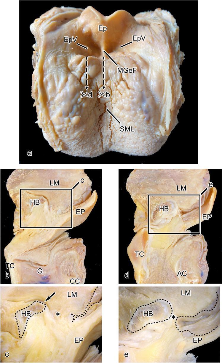
Dissection showing tongue base, epiglottis, valleculae and sagittal sections
  • Panel (a) is a superior view of the tongue base. The median glossoepiglottic fold runs from the tongue to the epiglottis, with a vallecula on each side.
  • The sagittal sections show the epiglottis in relation to the hyoid bone, thyroid cartilage and cricoid cartilage.
  • The pre-epiglottic space sits between the epiglottis and the hyoid.

2. Normal endoscopic (laryngoscopic) view

Normal larynx: true vocal folds, false vocal folds, arytenoids, epiglottis
Panel (a) shows a normal larynx in respiration. Seen from the front of the view to the back, the structures are:
  • the epiglottis at the top of the frame
  • the false (vestibular) folds
  • the true vocal folds
  • the arytenoids posteriorly
Supraglottic view with epiglottis, aryepiglottic folds, arytenoids and vocal folds
The epiglottis is at the top. The aryepiglottic folds run from it to the arytenoids, and the pale true vocal folds meet at the anterior commissure.

3. Pathology as seen on the epiglottis

Vallecular cyst pushing the epiglottis back and narrowing the airway
Large vallecular cyst compressing the epiglottis
Bifid epiglottis in a neonate. A midline cleft splits the cartilage into two lobes.
Bifid epiglottis, fiberoptic view
Omega-shaped or collapsed epiglottis in the airway of an obese patient. This is relevant for intubation.
Obese airway: omega-shaped epiglottis and collapse

Key points

  • Function: It folds back over the laryngeal inlet during swallowing to protect the airway.
  • Histology: It is elastic cartilage. The textbook histology atlas includes an H&E and orcein stained section, ×250.
  • Clinical: In video laryngoscopy, the POGO score (percentage of glottic opening) is limited by how much the epiglottis covers the glottis. Acute epiglottitis shows a swollen, cherry-red epiglottis.
I can pull the elastic cartilage histology slide or an epiglottitis image next if that would help. I did not run the recent-evidence PubMed check for this anatomy question.

Cartoon photo

Generating Image

A cartoon-style labeled illustration of the epiglottis and larynx, showing the epiglottis as a leaf-shaped flap above the glottis, tongue base, vallecula, hyoid bone, thyroid cartilage, and the open versus closed position during swallowing
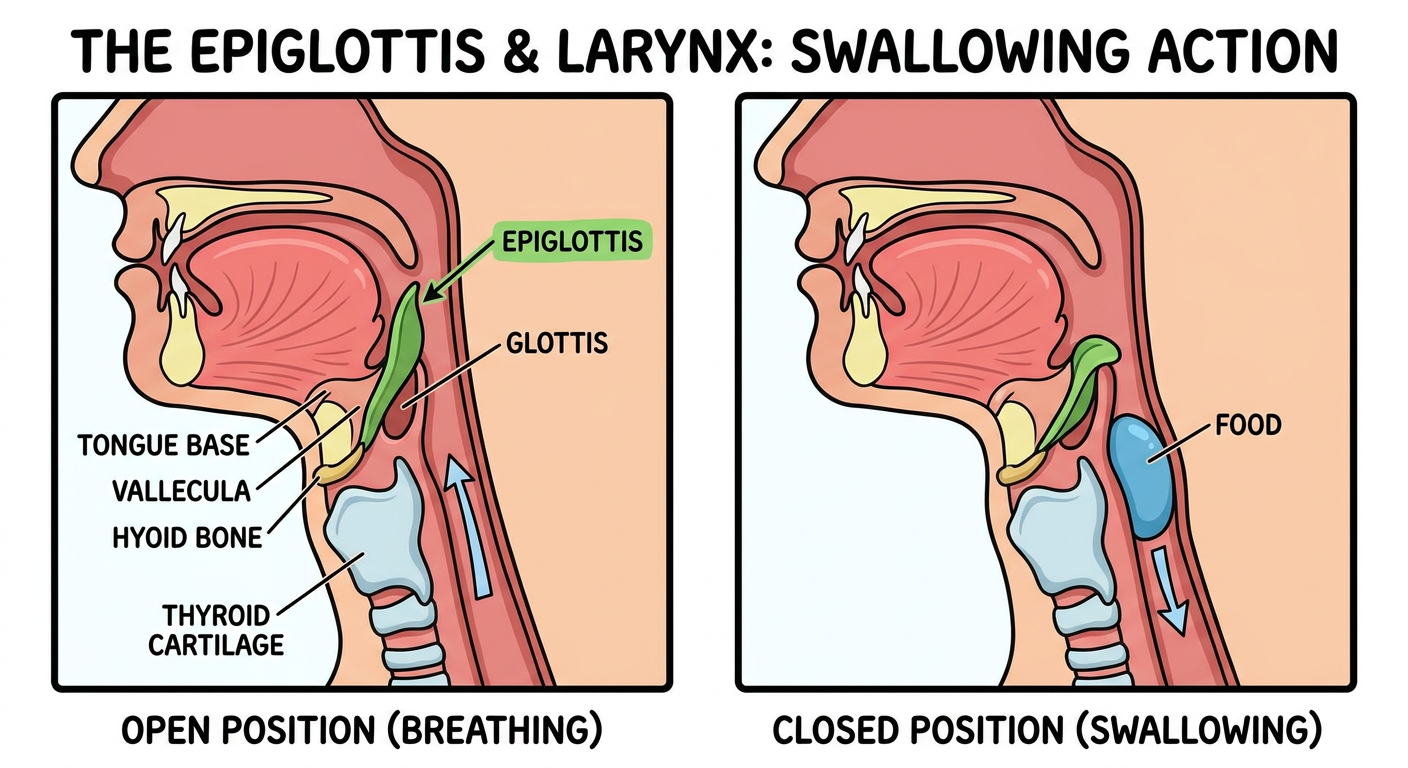
Cartoon illustration of the epiglottis and larynx
This is a cartoon of the epiglottis. It shows the leaf-shaped flap above the glottis, with the tongue base, vallecula, hyoid bone and thyroid cartilage around it. It also shows the open position (breathing) against the closed position (swallowing).
Generated cartoons can have labeling or proportion errors. For accurate anatomy, use the dissection and endoscopic images above.
I can make a simpler version, such as a single swallowing-sequence strip, or one in a different style.
This is a shared conversation. Sign in to Orris to start your own chat.