
Senile
What aspect of 'Senile' are you asking about?
age-related changes elderly aging body systems anatomy
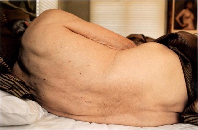
This clinical photograph captures a lateral, slightly curled view of an elderly individual's posterior torso and upper arm while lying in a bed. The image serves as a visual study of geriatric skin and age-related cutaneous findings. Key visible features include multiple benign-appearing skin lesions, such as melanocytic nevi (birthmarks) and potentially seborrheic keratoses, distributed across the back. The skin exhibits signs of chronological aging, including decreased elasticity (laxity), fine wrinkling on the elbow, and visible thinning of the dermis, making superficial bluish veins faintly visible on the arm. The positioning of the body highlights the natural curvature of the spine. The photograph is taken in a domestic setting with natural light, providing high-contrast visibility between the texture of the aging skin and the surrounding smooth bedding. This image is educationally relevant for geriatric medicine and dermatology, illustrating common age-related skin changes and the distribution of common cutaneous lesions in an elderly patient.
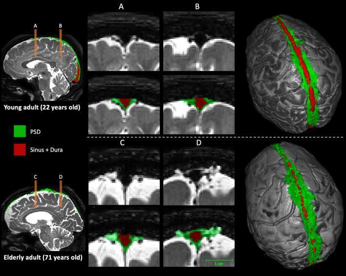
This diagnostic comparison image illustrates the age-related expansion of the parasagittal dural (PSD) space using T2-weighted MRI and 3D segmentation. The figure contrasts a young adult (22 years old) with an elderly adult (71 years old). High-resolution coronal slices focus on two neuroanatomical regions: the posterior aspect of the frontal lobe (A, C) and the anterior aspect of the parietal lobe (B, D). Color-coded label maps identify the PSD space in green and the superior sagittal sinus and contributing veins in red. In the elderly adult, there is a marked increase in the green-labeled PSD volume compared to the young adult, particularly evident in the anterior parietal region. The right-hand column presents 3D surface reconstructions of the brain, demonstrating a more extensive and wider distribution of the green PSD space along the longitudinal fissure in the older adult. These findings demonstrate the morphological changes of the parasagittal space associated with normal aging and its potential clinical significance in cerebrospinal fluid (CSF) dynamics and glymphatic function.
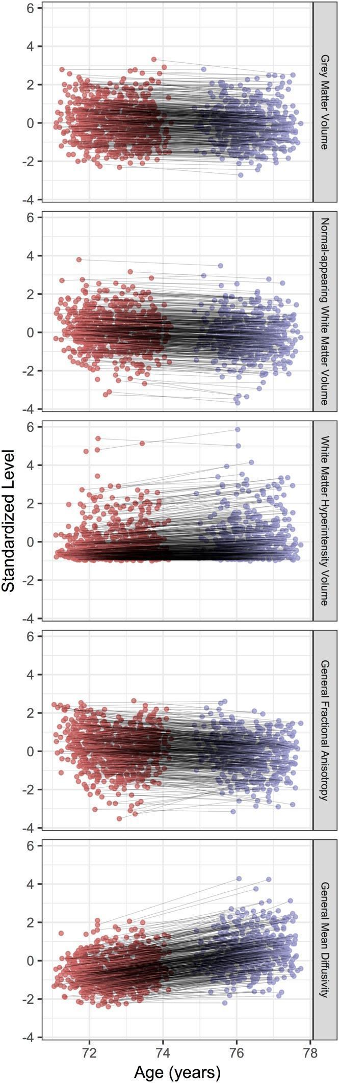
This Comparison Chart consists of five longitudinal scatter plots illustrating changes in brain structural measures in an elderly cohort between the ages of 73 (red clusters) and 76 (purple clusters). The x-axis represents age in years, and the y-axis represents the standardized level of each measure. The brain measures analyzed include Grey Matter Volume, Normal-appearing White Matter Volume, White Matter Hyperintensity Volume, General Fractional Anisotropy (FA), and General Mean Diffusivity (MD). Individual longitudinal trajectories are indicated by grey lines connecting paired data points for each participant across the two waves. The plots reveal a general downward trend (atrophy) in Grey Matter and Normal-appearing White Matter volumes, as well as a decrease in General FA, indicating reduced white matter microstructural integrity. Conversely, there is a clear upward trend in White Matter Hyperintensity Volume and General Mean Diffusivity, reflecting increased pathological vascular burden and increased water diffusion consistent with aging-related tissue degradation. This visualization demonstrates both the consistent mean shifts across the 3-year interval and the significant individual variability in the rate of neuroanatomical aging.
Senile (Age-Related) Changes - A System-by-System Overview
Quantitative Benchmarks at Age 80
| Parameter | % Decrease |
|---|---|
| Brain weight | 10-15% |
| Blood flow to brain | 20% |
| Cardiac output at rest | 35% |
| Glomerular filtration rate | 31% |
| Number of kidney glomeruli | 44% |
| Nerve fiber count | 37% |
| Nerve conduction velocity | 10% |
| Number of taste buds | 64% |
| Max O2 utilization with exercise | 60% |
| Max breathing capacity | 44% |
| Power of hand grip | 45% |
| Basal metabolic rate | 16% |
| Body water content | 18% |
1. Nervous System (Gerontologic Neurology)
- Brain weight falls 10-15% by age 80
- Cerebral blood flow decreases by ~20%
- Loss of neurons (especially in cortex, cerebellum, and substantia nigra), with gyral thinning and sulcal widening
- Accumulation of lipofuscin ("wear-and-tear" pigment) in surviving neurons
- A scattering of senile plaques (amyloid) and neurofibrillary tangles can appear in normal aging - but in far smaller numbers than in Alzheimer's disease
- Expansion of the parasagittal dural space (seen on MRI), reflecting changes in CSF dynamics and glymphatic function
- Progressive pupillary constriction (miosis)
- Decreased light reactions
- Presbyopia - loss of accommodation (lens hardening)
- Insufficiency of convergence
- Restricted upward conjugate gaze
- Diminished dark adaptation; increased glare sensitivity
- Presbycusis - progressive high-frequency hearing loss, with loss of hair cells in the organ of Corti and consequent decline in speech discrimination
- Diminished smell (anosmia) and to a lesser degree taste (64% fewer taste buds by age 80)
- Reduced motor speed, agility, fine coordination
- Slowed reaction time
- Sarcopenia - loss of muscle mass, especially legs, proximal muscles, dorsal interossei, thenar, and anterior tibial muscles
- Progressive loss of anterior horn cells contributes to denervation atrophy; denervation atrophy of the gastrocnemius is found in 80% of people over 70
- Lost muscle fibers are replaced by endomysial connective tissue and fat
- Ankle reflexes diminished in those over 70; Achilles reflexes lost in those over 80
- "Cortical release" signs (snout, palmomental) appear in up to 50% of normal people over 60
-
"Senile gait" - decreased stride length, reduced hip excursion, less toe elevation, stooped posture, slower cadence, widened base for stability
-
Adams and Victor's Principles of Neurology, 12th Ed.
2. Skin (Dermatogerontology)
- Epidermis thins, rete ridges flatten
- Decreased collagen and elastin fiber count
- Pallor, fine wrinkles, xerosis (dry skin), decreased elasticity, fragility
- Weakened antioxidant defenses lead to oxidative stress and cellular senescence
- Telomere shortening with each somatic division eventually triggers cell-cycle arrest
- UVA and UVB cause DNA damage, MMP activation, collagen degradation, and elastotic material accumulation (solar elastosis)
- Features: deep coarse wrinkles, mottled pigmentation, telangiectases, laxity, fragility, dyspigmentation
- Solar lentigines ("age spots")
- Seborrheic keratoses
- Senile angiomas (cherry angiomas)
- Xerosis and asteatotic eczema
- Pruritus
- Increased risk of both benign and malignant growths
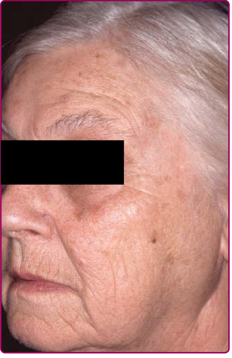
- Fitzpatrick's Dermatology, Vol. 1-2
3. Respiratory System
- Ciliary motility decreases significantly with age, impairing mucociliary clearance and increasing susceptibility to lower respiratory infections
- After age 30-40, respiratory bronchioles and alveolar ducts progressively enlarge ("ductectasia")
- Alveolar septa shorten; mean linear intercept (distance between alveolar walls) increases
- Alveolar surface area decreases by ~15% by age 70
- Surface-to-volume ratio falls
- These changes were historically called "senile emphysema" - but unlike true emphysema, there is NO destruction of alveolar septa with fusion of airspaces. The NHLBI recommends using the term "aging lung" instead and retiring "senile emphysema"
-
Max breathing capacity: -44% by age 80
-
Max ventilatory volume: -47% by age 80
-
Max O2 utilization with exercise: -60% by age 80
-
Fishman's Pulmonary Diseases and Disorders
4. Cardiovascular System
-
Cardiac output at rest falls ~35% by age 80
-
Decreased sympathetic response to stress
-
Reduced vascular compliance (arteries stiffen)
-
Impaired baroreceptor responses - predisposes to orthostatic hypotension and perioperative blood pressure lability
-
Increased prevalence of coronary artery disease, hypertension, atrial fibrillation
-
Left ventricular hypertrophy from chronic pressure overload; diastolic dysfunction is common
-
These changes place elderly patients at significantly higher perioperative cardiac risk (POISE-2 trial: age >75 was an independent risk factor for postoperative MI, HR 1.89)
-
Barash's Clinical Anesthesia, 9th Ed.
5. Musculoskeletal System
- Occurs in both sexes with advanced age (distinct from postmenopausal/Type I osteoporosis)
- Classified as "low-turnover" osteoporosis - reduced physical activity, declining sex hormones, and impaired calcium absorption all contribute
- Reduced bone mass increases fracture risk, especially hip and vertebral fractures
- Decreased physical activity compounds the problem as osteocytes respond to mechanical loading
-
Age-related loss of skeletal muscle mass and strength
-
Due to combined denervation (loss of motor neurons), disuse, and hormonal changes
-
Robbins & Cotran Pathologic Basis of Disease
6. Renal System
- Number of glomeruli falls by ~44% by age 80
- GFR declines by ~31% by age 80
- Reduced renal reserve means drugs cleared renally (e.g., aminoglycosides, NSAIDs, digoxin) require dose adjustment
- Diminished ability to concentrate urine and handle volume/electrolyte stress
7. ENT - Senile Rhinitis
-
Aging nose shows decreased nasal blood flow and diminished humidification
-
Predisposes to rhinitis in elderly; termed "senile rhinitis" in otolaryngology literature
-
Cummings Otolaryngology
8. Pharmacologic Implications
-
Altered volume of distribution (less body water, more fat)
-
Reduced cardiac output delays IV drug onset
-
Impaired renal/hepatic clearance prolongs drug half-lives
-
Reduced protein binding increases free drug fraction
-
Elderly patients are highly susceptible to adverse drug reactions and polypharmacy interactions
-
Goodman & Gilman's Pharmacological Basis of Therapeutics
Named "Senile" Conditions in Medicine
| Condition | Description |
|---|---|
| Senile plaques | Extracellular amyloid-beta deposits in brain; normal aging has a few; Alzheimer's has massive burden |
| Senile osteoporosis | Low-turnover bone loss with advanced age in both sexes |
| Senile emphysema | Outdated term for age-related alveolar duct enlargement - now called "aging lung" |
| Senile angiomas | Cherry angiomas - benign vascular proliferations common in elderly skin |
| Senile gait | Stooped, short-stride, broad-based gait of normal aging |
| Senile rhinitis | Age-related nasal mucosal dryness and rhinitis |
| Senile cataracts | Lens opacification with age (most common cause of cataracts worldwide) |
| Senile purpura | Easy bruising from dermal atrophy and capillary fragility in elderly skin |