
60 yr old female, came with c/o Rt ankle pain and swelling for 10 days, no h/o trauma, RA, SLE, thyroid, DM, neuropathy o/e ankle effusion +, medial joint line tenderness + ROM pain free, No bony tenderness over medial and lateral malleoli, serology, cbc, rbs, uric acid, RA factor was normal, xray showed mild arthritic changes in lat view , not settles with pain killers and immobilisation
Reading File
Quick Answer: This 60-year-old female with a painful, swollen ankle, no trauma, normal serology (including uric acid and RA factor), and only mild arthritic changes on X-ray, not responding to analgesics/immobilisation, is a diagnostic puzzle. Key differentials include calcium pyrophosphate deposition (CPPD/pseudogout), pigmented villonodular synovitis (PVNS), osteochondral lesion, occult fracture, or early septic arthritis. The next step is an MRI of the ankle and joint aspiration for synovial fluid analysis.
Searching sources...
Finding Sources
Finding Sources
Finding Sources
Searching PubMed
ankle monoarthritis differential diagnosis non-traumatic
Finding Sources
Finding Sources
Finding Sources
Reading Article
PMID: 40531148
Reading File
Reading File
Finding Sources
Reading File
Reading File
Searching Images
ankle joint effusion swelling MRI osteochondral lesion
Here is a structured clinical analysis of this case:
Clinical Analysis: 60F, Right Ankle Pain & Swelling, 10 Days, No Trauma
Problem Summary
- Acute/subacute monoarthritis of the right ankle
- 60-year-old female
- No trauma, no RA, no SLE, no thyroid disease, no DM, no neuropathy
- Ankle effusion + medial joint line tenderness
- ROM pain-free (important - suggests intra-articular not periarticular)
- No bony tenderness over malleoli
- Normal: CBC, serology, RBS, uric acid, RA factor
- X-ray: mild arthritic changes (lateral view)
- Not responding to analgesics + immobilisation
Key Diagnostic Clues
| Feature | Interpretation |
|---|---|
| Effusion + medial joint line tenderness | True intra-articular pathology |
| ROM pain-free | Excludes severe inflammatory or septic process; also suggests periarticular structures are not the primary issue |
| Normal uric acid | Makes gout less likely, but does NOT exclude it during an acute attack (uric acid drops during attacks) |
| Normal RA factor | Makes RA less likely, but seronegative RA/spondyloarthropathy still possible |
| Normal CBC | Against septic arthritis (though WBC can be normal early) |
| Mild arthritic change on X-ray only | Does not explain 10 days of effusion and persistent pain |
| No response to analgesics/immobilisation | Suggests an underlying cause that needs specific treatment |
Differential Diagnosis (Most to Least Likely)
1. CPPD / Pseudogout - TOP DIFFERENTIAL
- Most common crystal arthropathy at this age in females, after gout is excluded
- Presents as acute monoarthritis mimicking gout - "pseudogout"
- Uric acid is normal - consistent with CPPD
- Ankle is a recognised site (though knee and wrist are more common)
- X-ray may show chondrocalcinosis (cartilage calcification) - check carefully on the lateral view
- Diagnosis: synovial fluid aspiration - CPPD crystals are weakly positive birefringent, rhomboid-shaped under polarised microscopy
- As noted in Grainger & Allison's Diagnostic Radiology: "Pseudogout: an acute clinical condition produced by CPPD crystals within joints, resulting in acute attacks of gout-like symptoms. The diagnosis is clinical and cannot be made radiologically."
2. Gout (Acute Attack with Normal Uric Acid)
- Serum uric acid drops during an acute attack - a normal level does NOT exclude gout
- However, ankle gout is less common than first MTP involvement at this age
- Synovial fluid aspiration: monosodium urate crystals - needle-shaped, strongly negatively birefringent
3. Osteochondral Lesion of the Talus (OLT)
- Can present as ankle pain and swelling with no trauma history (up to 25% are atraumatic)
- Medial talar dome is the most common site - explains medial joint line tenderness
- X-ray often misses early lesions - MRI is required
- ROM is often preserved in early lesions
- Does NOT respond to analgesics or immobilisation if the fragment is unstable
4. Septic Arthritis (Low probability but must exclude)
- Normal CBC and serology make this less likely
- However, early septic arthritis can have a normal or mildly elevated WBC
- Synovial fluid analysis is the gold standard - WBC >50,000/mm³ with >90% PMNs + positive culture
- As per Firestein & Kelley's Rheumatology: "In acute monoarthritis, synovial fluid analysis is the most valuable laboratory test"
- Must not be missed - permanent joint destruction if untreated
5. Pigmented Villonodular Synovitis (PVNS)
- Rare, but can present as monoarticular swelling in ankle (as referenced in Campbell's Operative Orthopaedics)
- Insidious onset, recurrent bloody effusions, marked synovial thickening
- MRI is diagnostic: diffuse synovial thickening with hemosiderin deposits giving "blooming" on gradient echo sequences
6. Reactive Arthritis / Early Spondyloarthropathy
- Seronegative, asymmetric, lower limb predominant
- History of prior GI/urogenital infection? Psoriasis? Back pain?
- Consider HLA-B27 testing
7. Occult Stress Fracture / Bone Contusion
- No trauma, but insufficiency fractures can occur in older postmenopausal women (osteoporosis)
- X-ray negative early; MRI shows marrow oedema pattern
- No bony tenderness over malleoli - but midfoot/subtalar pathology possible
Immediate Next Steps
A. Synovial Fluid Aspiration (MOST IMPORTANT)
This is the single most important investigation in acute monoarthritis. Analyse for:
| Parameter | What it tells you |
|---|---|
| Cell count + differential | >50,000 WBC with PMNs = septic; 2,000-50,000 = inflammatory |
| Crystal microscopy (polarised) | CPPD (rhomboid, weakly +ve birefringent) vs Gout (needle, strongly -ve birefringent) |
| Gram stain + Culture | Septic arthritis |
| Lactate | >4.3 mmol/L supports septic arthritis |
- From Tietz Textbook of Laboratory Medicine: Synovial fluid WBC >50×10⁹/L gives a likelihood ratio of 3.59 for infection; >100×10⁹/L gives +LR of 13.2
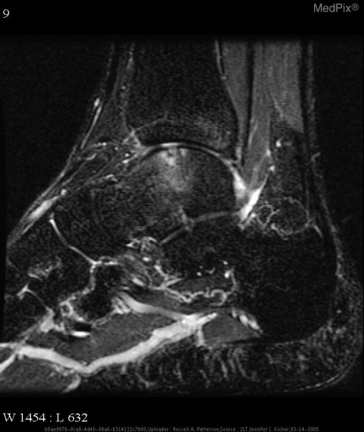
B. MRI Ankle (RIGHT)
- Detects: osteochondral lesion of talus, PVNS, occult fractures, ligamentous pathology, early bone marrow oedema
- Should be done even if aspiration gives a diagnosis, to look for any underlying structural lesion
C. Additional Blood Tests
- Serum uric acid (repeat, ideally 4-6 weeks after acute attack to catch baseline hyperuricemia)
- ESR, CRP (baseline inflammatory markers)
- HLA-B27 (if reactive/spondyloarthropathy suspected)
- PTH, calcium, phosphate, ferritin, Mg (screen for metabolic causes of CPPD - hyperparathyroidism, haemochromatosis, hypomagnesaemia)
- DEXA scan (postmenopausal female - osteoporosis risk for insufficiency fractures)
- Blood cultures if septic arthritis is still in the running
D. Repeat X-ray Review
- Look carefully for chondrocalcinosis (linear calcification in cartilage) in the ankle joint - may be subtle
- Weight-bearing AP and mortise views if not already done
Likely Diagnosis
Most probable: CPPD/Pseudogout - 60-year-old postmenopausal female, acute monoarthritis, ankle effusion, normal uric acid and serology, mild arthritic change on X-ray, not responding to simple analgesics.
Second most probable: Osteochondral Lesion of the Talus - given medial joint line tenderness, pain-free ROM, and failure to respond to immobilisation.
Management Once Diagnosis Confirmed
| Diagnosis | Treatment |
|---|---|
| CPPD/Pseudogout | Joint aspiration (therapeutic) + intra-articular steroid injection; NSAIDs/colchicine for acute attack |
| Gout | NSAIDs, colchicine, or short course steroids acutely; urate-lowering therapy later |
| Septic arthritis | Urgent IV antibiotics + surgical washout/repeated aspiration |
| OLT | Conservative (immobilisation, physiotherapy) vs arthroscopic surgery for unstable lesions |
| PVNS | Arthroscopic synovectomy |
| Insufficiency fracture | Offloading, calcium/vitamin D, bisphosphonates |
References
- Firestein & Kelley's Textbook of Rheumatology (10th Ed.) - Chapter on Early Arthritis
- Grainger & Allison's Diagnostic Radiology - CPPD Deposition Disease
- Tietz Textbook of Laboratory Medicine (7th Ed.) - Synovial Fluid Analysis
- Campbell's Operative Orthopaedics (15th Ed.) - PVNS of the Ankle
- Swisher et al., Acute Monoarthritis: Diagnosis in Adults, American Family Physician, 2025
This is a shared conversation. Sign in to Orris to start your own chat.