
If you had to prepare an MD Paediatrics level seminar on Encopresis and constipation in children, which sources will you refer to?
If you had to prepare an MD Paediatrics level seminar on Encopresis and constipation in children, which sources will you refer to?
NASPGHAN ESPGHAN guideline functional constipation children 2014 encopresis sources pediatric textbook Rome IV
pediatric functional constipation
https://pmc.ncbi.nlm.nih.gov/articles/PMC11795385
Core sources
-
ESPGHAN/NASPGHAN evidence-based guideline
- Tabbers MM, DiLorenzo C, Berger MY, et al. Evaluation and Treatment of Functional Constipation in Infants and Children. Journal of Pediatric Gastroenterology and Nutrition. 2014;58:258-274.
- This should be the main clinical source for definitions, alarm features, investigations, disimpaction, maintenance treatment, follow-up, and indications for referral.
- The full NASPGHAN guideline includes useful diagnostic and treatment algorithms.
-
Rome IV paediatric disorders
- Hyams JS, Di Lorenzo C, Saps M, et al. Childhood Functional Gastrointestinal Disorders: Child/Adolescent. Gastroenterology. 2016;150:1456-1468.
- Use it for diagnostic criteria and differentiation of:
- Functional constipation
- Functional non-retentive faecal incontinence
- Other disorders of gut-brain interaction.
- This is particularly important for a seminar slide distinguishing constipation-associated overflow incontinence from non-retentive soiling.
-
Latest ESPGHAN/NASPGHAN guideline-development update
- The 2024 ESPGHAN/NASPGHAN protocol is useful to show that the 2014 guidance is being updated using GRADE methodology and expanded coverage of faecal impaction, refractory constipation, and surgical options.
- Phrase this carefully: it is a protocol for an updated guideline, not itself the final treatment guideline.
-
NASPGHAN recommendations on paediatric refractory constipation
- Use the current NASPGHAN position paper/recommendations for children who fail standard disimpaction, maintenance therapy, toileting intervention, and adherence optimisation.
- This is useful for advanced seminar sections on anorectal manometry, colonic transit, pelvic-floor dyssynergia, transanal irrigation, antegrade continence enemas, and surgical referral.
Textbooks
-
Nelson Textbook of Pediatrics, latest edition
- Primary textbook source for epidemiology, history, examination, red flags, organic causes, Hirschsprung disease, spinal disorders, endocrine/metabolic causes, and management principles.
-
Pediatric Gastrointestinal and Liver Disease by Wyllie, Hyams, Kay, et al., latest edition
- Best in-depth specialty reference for:
- Pathophysiology of stool withholding and rectal dilatation
- Faecal impaction and overflow soiling
- Refractory constipation
- Motility investigations
- Bowel-management programmes and procedural options.
- Best in-depth specialty reference for:
-
The Harriet Lane Handbook, latest edition
- Good for ward-level practical details: medication choices, dosing frameworks, follow-up, counselling, and referral thresholds. The available paediatric text library contains dedicated constipation sections in The Harriet Lane Handbook, 23rd ed.
-
Rudolph’s Pediatrics, latest edition
- A useful alternative general paediatric source, especially for clinical approach and differential diagnosis.
Complementary sources
-
NICE guideline: Constipation in children and young people
- Useful for structured treatment pathways, family education, toilet programmes, and longer-term follow-up. Compare it with NASPGHAN/ESPGHAN rather than using it as the sole authority.
-
DSM-5-TR
- Relevant only for the terminology and psychiatric/developmental context of encopresis, particularly when discussing non-retentive faecal incontinence, comorbid behavioural disorders, psychosocial stressors, and stigma.
- Avoid presenting faecal soiling as primarily wilful or psychiatric. Most cases are constipation-associated overflow.
- ICCS terminology documents
- Helpful if including bladder-bowel dysfunction, daytime urinary symptoms, enuresis, recurrent UTI, or dysfunctional elimination syndrome.
Evidence updates to cite selectively
- The 2024 SUCCESS evidence synthesis addresses strategies for childhood chronic functional constipation, PMID 38343084.
- For severe refractory disease, a 2025 systematic review evaluates antegrade continence enemas, PMID 39389879.
- A 2026 systematic review/meta-analysis examines physiotherapeutic interventions, PMID 41961325.
A practical reference hierarchy for the seminar
- ESPGHAN/NASPGHAN guideline
- Rome IV criteria
- Nelson + Wyllie/Hyams/Kay
- Current NASPGHAN refractory constipation recommendations
- NICE and selected systematic reviews
- DSM-5-TR/ICCS only for specific terminology or comorbidity slides
Create diagrams demonstrating the anatomy and physiology first. Detailed diagrams.
A detailed, medically accurate paediatric educational infographic with three clearly labelled panels for an MD Paediatrics seminar. Panel 1: sagittal pelvic anatomy of a toilet-trained child showing descending colon, sigmoid colon, rectum, anal canal, internal anal sphincter, external anal sphincter, puborectalis sling, pelvic floor, sacrum, pudendal nerve and S2-S4 parasympathetic supply. Panel 2: normal defecation physiology: colonic mass movement into rectum, rectal distension, rectoanal inhibitory reflex causing internal sphincter relaxation, conscious sensation, voluntary external sphincter and puborectalis relaxation, abdominal pressure and stool expulsion. Panel 3: functional constipation with retentive faecal incontinence: painful stool leads to withholding, contracted external sphincter and puborectalis, progressive rectal stool accumulation and megarectum, reduced rectal sensation, liquid stool leaking around the faecal impaction as overflow soiling. Use correct directional arrows and concise educational labels. Ensure anatomical proportions are appropriate for a child and distinguish normal physiology from pathological cycle.
normal defecation physiology rectoanal inhibitory reflex pediatric functional constipation overflow fecal incontinence NASPGHAN
pediatric functional constipation physiology
Anatomy and physiology of defecation, and the mechanism of constipation-associated overflow soiling
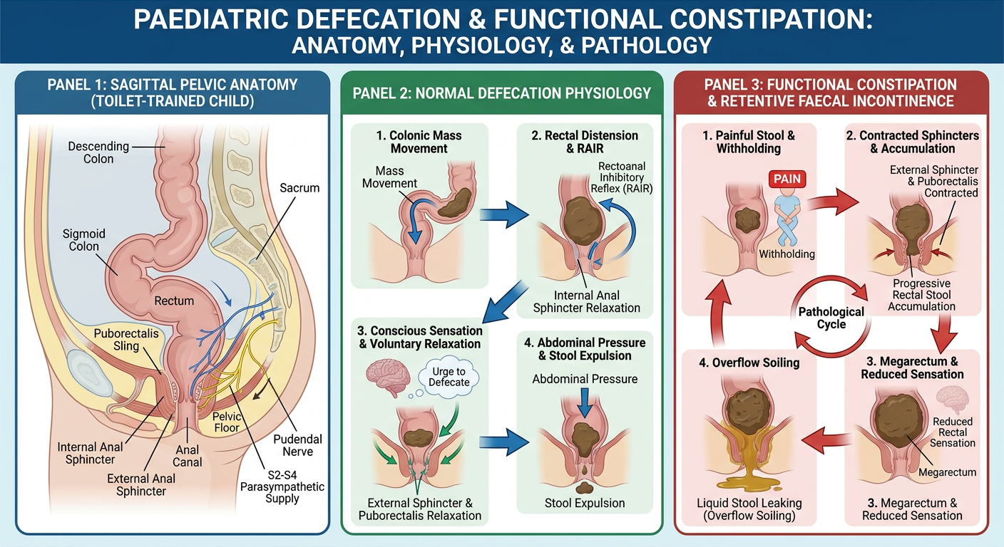
How to present this diagram
- The rectum serves as a compliant reservoir. At baseline it is usually relatively empty.
- The internal anal sphincter (IAS) is smooth muscle and involuntary. It maintains most resting anal tone.
- The external anal sphincter (EAS) is striated muscle under voluntary control, via the pudendal nerve.
- The puborectalis, part of levator ani, forms a sling around the anorectal junction and maintains the anorectal angle. Its contraction supports continence.
- Parasympathetic sacral outflow, chiefly S2-S4, promotes distal colonic and rectal activity. Somatic pudendal innervation permits conscious continence control.
- Colonic propagating contractions move stool into the rectum.
- Rectal distension activates stretch receptors and produces the urge to defecate.
- Distension evokes the rectoanal inhibitory reflex (RAIR), causing transient relaxation of the IAS.
- The child consciously assesses rectal contents and either postpones defecation by contracting the EAS/puborectalis, or proceeds to evacuate.
- Defecation requires voluntary relaxation of the EAS and puborectalis, straightening of the anorectal angle, coordinated abdominal wall contraction, and expulsion of stool.
- A painful or frightening bowel movement initiates stool withholding.
- The child contracts the EAS and puborectalis, often with characteristic retentive posturing.
- Stool remains in the rectum, becomes progressively drier and larger, and may form a faecal impaction.
- Chronic rectal distension causes increased rectal compliance and reduced awareness of rectal filling.
- Softer stool from proximal colon passes around the impacted stool and leaks out involuntarily: overflow faecal incontinence, often called soiling or encopresis.
Add labelled diagrams of the enteric nervous system and defecation reflex
Detailed medically accurate labelled teaching diagram for an MD Paediatrics seminar: enteric nervous system of the distal colon, rectum and anal canal. Show a longitudinal cutaway of colonic wall with mucosa, submucosa, circular muscle and longitudinal muscle. Clearly label the submucosal Meissner plexus and myenteric Auerbach plexus, intrinsic sensory neurons, interneurons, excitatory motor neurons releasing acetylcholine and substance P that contract smooth muscle, inhibitory motor neurons releasing nitric oxide and VIP that relax smooth muscle, enteric glia, interstitial cells of Cajal, extrinsic parasympathetic pelvic splanchnic nerves S2-S4, sympathetic hypogastric input, and pudendal somatic innervation of external anal sphincter. Include a small inset showing coordinated peristalsis: contraction behind stool and relaxation ahead. Use arrows and concise labels, with a clear educational medical illustration style.
Detailed medically accurate labelled teaching flow diagram for an MD Paediatrics seminar showing the normal defecation reflex and voluntary continence control in a toilet-trained child. Show rectal filling by colonic mass movement, rectal stretch receptors, afferent signals to sacral defecation centre S2-S4 and brain, parasympathetic efferents via pelvic splanchnic nerves, rectoanal inhibitory reflex with internal anal sphincter relaxation, sampling of rectal contents, conscious urge to defecate, external anal sphincter and puborectalis under pudendal voluntary control, and two outcome branches: defer defecation with contraction of external sphincter and puborectalis, versus defecate with relaxation of external sphincter and puborectalis, straightening of anorectal angle, abdominal straining and stool expulsion. Include a small contrast inset that notes absent RAIR in Hirschsprung disease. Use unambiguous directional arrows and concise labels.
enteric nervous system defecation reflex rectoanal inhibitory reflex pediatric review
pediatric functional constipation enteric nervous system
1. Enteric nervous system of the distal colon, rectum, and anal canal
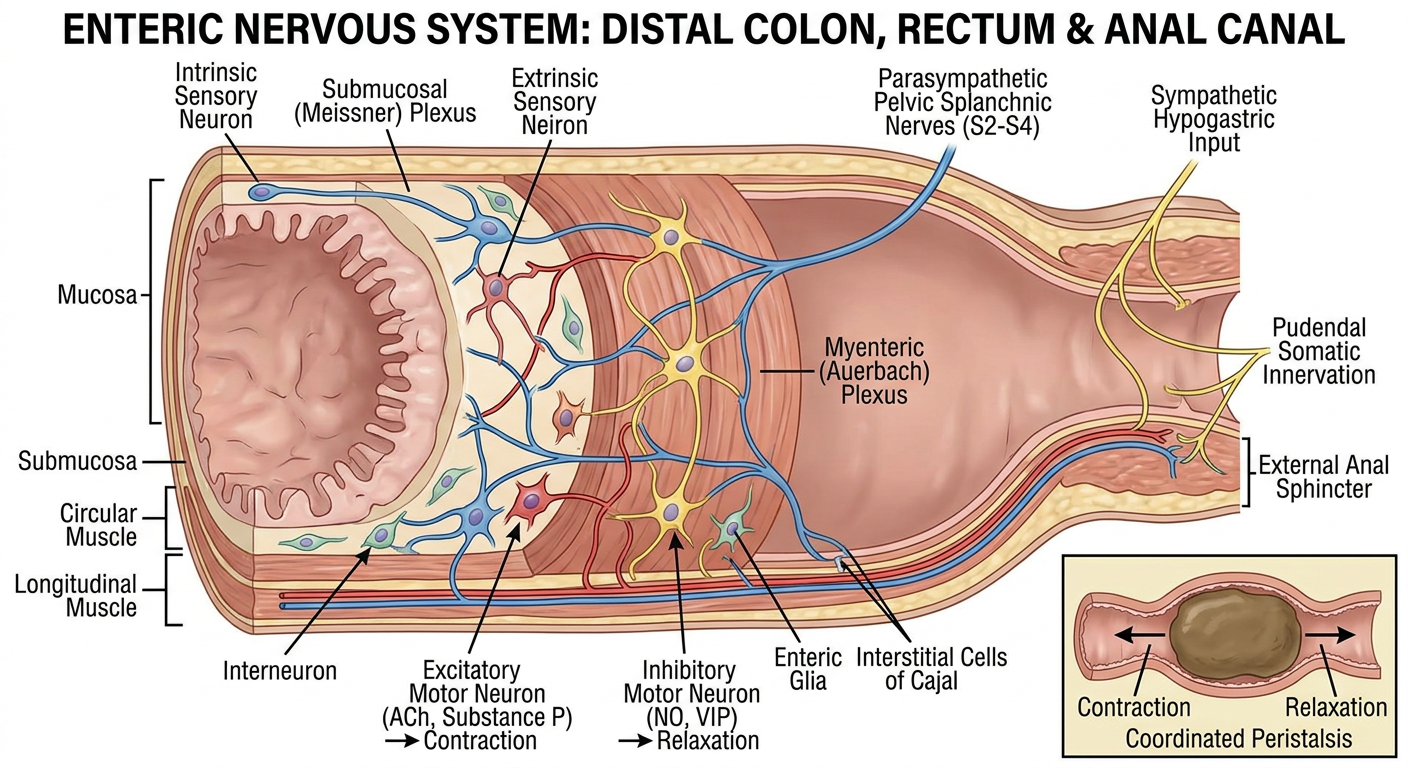
Key labels to explain
- Myenteric plexus of Auerbach: located between circular and longitudinal muscle layers. It coordinates motility, including propagated contractions and the relaxation ahead of the stool bolus.
- Submucosal plexus of Meissner: regulates mucosal secretion, blood flow, and local sensory processing.
- Intrinsic primary afferent neurons: detect mechanical distension and chemical contents in the gut.
- Excitatory motor neurons: mainly acetylcholine and substance P mediated. They produce smooth-muscle contraction behind the advancing stool.
- Inhibitory motor neurons: principally nitric oxide and VIP mediated. They relax smooth muscle ahead of the bolus and contribute to internal anal sphincter relaxation.
- Interstitial cells of Cajal: contribute to neuromuscular transmission and coordination of gastrointestinal electrical activity.
- Parasympathetic pelvic splanchnic nerves, S2-S4: facilitate distal colonic motility and the defecation reflex.
- Sympathetic hypogastric pathways: generally support storage by reducing colonic activity and increasing internal sphincter tone.
- Pudendal nerve: somatic control of the external anal sphincter and contributes to voluntary continence.
2. Defecation reflex and voluntary continence control
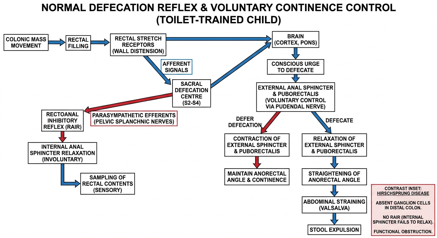
Sequence for the seminar
-
Colonic mass movement
- A high-amplitude propagated contraction transfers stool into the rectum.
-
Rectal distension
- Rectal stretch receptors are activated.
- Sensory signals reach sacral spinal centers and are perceived centrally as the urge to defecate.
-
Rectoanal inhibitory reflex
- Rectal distension induces transient relaxation of the internal anal sphincter.
- This permits “sampling” of rectal contents in the upper anal canal.
-
Continence decision
- If socially inappropriate to pass stool, the child voluntarily contracts the external anal sphincter and puborectalis through pudendal innervation.
- Rectal accommodation occurs and the urge may temporarily decline.
-
Defecation
- If conditions are appropriate, voluntary relaxation of external anal sphincter and puborectalis occurs.
- The anorectal angle straightens.
- Abdominal wall contraction, diaphragmatic descent, and increased intra-abdominal pressure assist evacuation.
-
Return to resting state
- Anal canal pressure rises again and the internal and external sphincters restore continence.