
Hypertensive disorders in pregnancy
Reading File
Now let me search the medical library for comprehensive content on hypertensive disorders in pregnancy, running parallel queries:
Finding Sources
Finding Sources
Searching PubMed
"hypertensive disorders" AND pregnancy
Excellent results. Now let me read the primary sources in parallel - the maternal-fetal medicine textbook (most comprehensive) and Robbins pathology for pathophysiology:
Reading File
Reading File
Reading File
Good. Now let me read deeper from Creasy & Resnik (the gold standard MFM textbook) for the full classification, management, and diagnosis content, plus Guyton physiology:
Reading File
Reading File
Reading File
Excellent. Now let me read more from Creasy & Resnik on diagnosis/classification and the Braunwald Heart Disease table for ACOG classification, plus continue the Goldman-Cecil treatment section:
Reading File
Reading File
Reading File
Now let me fetch the key images and read the prevention/management section from Creasy & Resnik:
Reading File
Loading Image
Loading Image
Excellent content and images gathered. Let me now get the prevention data and fetal complications section from Creasy & Resnik:
Reading File
I now have comprehensive content from multiple authoritative textbooks. Let me compile the full detailed response.
Hypertensive Disorders of Pregnancy (HDP)
Overview
Hypertensive disorders complicate approximately 5-8% of all pregnancies (up to 85 per 1000 deliveries in the United States) and represent one of the leading causes of maternal and perinatal morbidity and mortality worldwide. They encompass a spectrum ranging from mild gestational hypertension to life-threatening eclampsia and HELLP syndrome.
Classification (ACOG)
The ACOG Practice Bulletin No. 222 defines four main categories:
| Category | Definition |
|---|---|
| Gestational Hypertension | New-onset BP ≥140/90 mmHg after 20 weeks, without proteinuria or end-organ damage; resolves postpartum |
| Preeclampsia | Gestational hypertension + proteinuria (≥300 mg/24h, protein:creatinine ≥0.3, or dipstick 2+) OR new end-organ damage |
| Chronic Hypertension | HTN diagnosed before pregnancy or before 20 weeks, or persisting postpartum |
| Chronic HTN with Superimposed Preeclampsia | Preeclampsia developing in a woman with pre-existing chronic hypertension |
Note: Eclampsia = Preeclampsia + new-onset tonic-clonic, focal, or multifocal seizures in the absence of other causes.
Onset subclassification:
- Early-onset preeclampsia: <34 weeks (more severe, higher maternal/fetal risk)
- Late-onset preeclampsia: ≥34 weeks (more common)
Epidemiology and Risk Factors
- Gestational hypertension: 6-17% of nulliparous, 2-4% of parous women
- Preeclampsia: 5-8% of all pregnancies
- Eclampsia: ~1 in 2000-3000 deliveries in high-resource settings
Major risk factors (Creasy & Resnik's Maternal-Fetal Medicine, Table 45.2):
- Nulliparity (highest population-attributable fraction: 32.3%)
- Prior preeclampsia (8-fold increased risk)
- Chronic hypertension (25% risk of superimposed preeclampsia)
- Antiphospholipid syndrome
- Pregestational diabetes mellitus (20% overall; up to 70% with White's class F/R)
- Renal disease
- Connective tissue disorders (SLE)
- Multifetal gestation
- IVF conception
- Extremes of maternal age
- Non-Hispanic Black race (related more to severity than incidence)
- Obesity, family history of preeclampsia
Pathophysiology
The central mechanism is a two-stage model:
Stage 1 - Defective Placentation
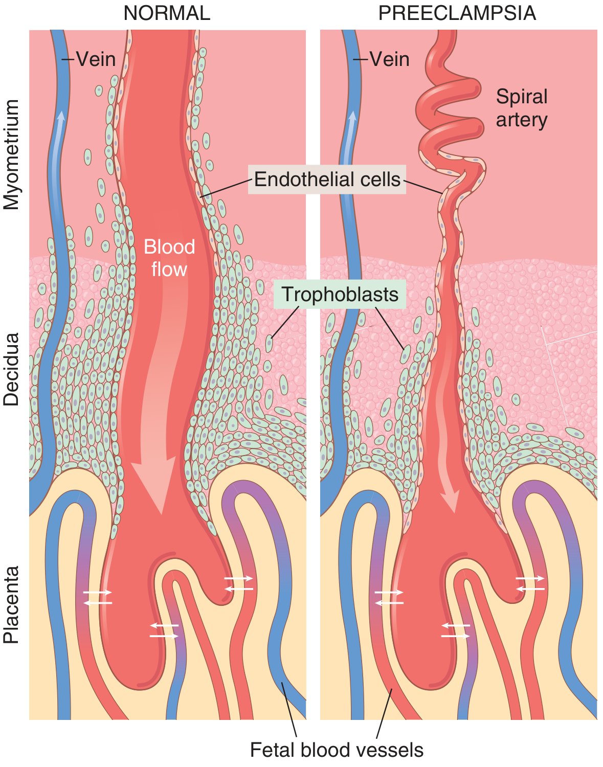
In normal pregnancy, trophoblasts invade the spiral arteries of the uterine endometrium, remodeling them into wide, low-resistance vascular sinusoids. In preeclampsia, this process is impaired - the muscloelastic walls are retained, channels remain narrow, and uteroplacental blood flow is reduced, leading to placental hypoxia.
Stage 2 - Endothelial Dysfunction
Placental ischemia triggers release of circulating factors into the maternal bloodstream:
- sFlt-1 (soluble fms-like tyrosine kinase-1) - antagonizes VEGF
- Soluble endoglin (sEng) - antagonizes TGF-β
- Inflammatory cytokines (TNF-α, IL-6)
These antiangiogenic factors cause systemic maternal endothelial dysfunction, leading to:
| Consequence | Mechanism |
|---|---|
| Hypertension | Reduced endothelial PGI₂/PGE₂ (vasodilators) + increased TXA₂ (vasoconstrictor); increased SVR |
| Proteinuria / renal injury | Glomerular endotheliosis; decreased GFR, decreased renal blood flow |
| Coagulopathy/HELLP | Endothelial activation → platelet consumption, microangiopathic hemolysis |
| Fetal growth restriction | Chronic uteroplacental hypoperfusion |
| Seizures (eclampsia) | Cerebral vasospasm, blood-brain barrier disruption, PRES (posterior reversible encephalopathy syndrome) |
| Hepatic injury | Periportal fibrin deposition; elevated AST/ALT; risk of subcapsular hematoma/rupture |
| Pulmonary edema | Increased afterload, decreased oncotic pressure, endothelial permeability |
Vascular reactivity: Preeclamptic women show exaggerated sensitivity to all endogenous pressors - angiotensin II, norepinephrine, epinephrine, and vasopressin - a finding detectable weeks before clinical hypertension develops.
Diagnosis
Preeclampsia Without Severe Features
- BP ≥140/90 mmHg on two occasions ≥4 hours apart, at or after 20 weeks
- PLUS: proteinuria ≥300 mg/24h (or protein:creatinine ≥0.3, or dipstick 2+)
- OR: any of the following end-organ manifestations even without proteinuria:
- Thrombocytopenia (<100,000/μL)
- Renal insufficiency (creatinine >1.1 mg/dL or doubling of baseline)
- Impaired liver function (AST/ALT ≥2× upper limit of normal)
- Pulmonary edema
- New-onset headache unresponsive to analgesics
- Visual disturbances
Severe Features (Any One Qualifies)
Per ACOG (also from Goldman-Cecil Medicine, Table 221-7):
| Severe Feature | Threshold |
|---|---|
| Severe hypertension | SBP ≥160 or DBP ≥110 mmHg on 2 occasions ≥4 h apart |
| Thrombocytopenia | Platelets <100,000/μL |
| Elevated liver enzymes | ≥2× upper limit of normal |
| Severe RUQ/epigastric pain | Unresponsive to medications |
| Renal insufficiency | Creatinine >1.1 mg/dL or doubling of baseline |
| Pulmonary edema | - |
| New-onset headache | Not relieved by analgesics |
| Visual disturbances | - |
HELLP Syndrome
A severe variant of preeclampsia occurring in ~10% of severe cases:
- H - Hemolysis (microangiopathic hemolytic anemia)
- EL - Elevated Liver enzymes
- LP - Low Platelets
Morphological / Histological Findings (Robbins & Kumar Basic Pathology)
Placenta:
- Multiple infarcts (far more numerous than in normal pregnancy)
- Retroplacental hemorrhage
- Ischemic changes: increased syncytial knot formation
- Acute atherosis: fibrinoid necrosis of decidual vessels + lipid-laden macrophages
Kidney:
- Glomerular endotheliosis - the classic lesion: swelling of glomerular endothelial cells, luminal obliteration, subendothelial protein deposits
- Decreased GFR, decreased renal blood flow
Brain (Eclampsia):
- Posterior reversible encephalopathy (PRES) - T2-weighted MRI shows bilateral signal intensity in subcortical white matter of occipital/parietal regions
- Hemorrhage, ring hemorrhages, petechiae
Management
Antihypertensive Treatment Algorithm
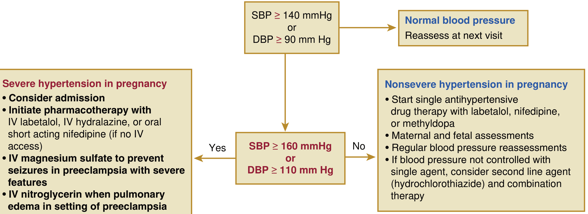
Urgent Management of Severe Hypertension (BP ≥160/110 mmHg)
Per Goldman-Cecil Medicine (Table 221-6):
| Route | Drug | Dose |
|---|---|---|
| Oral | Nifedipine IR | 10-20 mg initially; repeat in 20 min; then 10-20 mg q2-6h (max 180 mg/day) |
| IV | Labetalol (preferred) | 10-20 mg IV bolus, then 1-2 mg/min infusion (or 20-80 mg q10-30 min) to max 300 mg |
| IV | Hydralazine | 5 mg initially, then 0.5-10 mg/hr (or 5-10 mg q20-40 min) to max 20 mg |
For pulmonary edema in the setting of preeclampsia: IV nitroglycerin.
Nonsevere Hypertension (140-159/90-109 mmHg)
- First-line oral agents: labetalol, nifedipine (extended release), or methyldopa
- If uncontrolled on monotherapy: add hydrochlorothiazide
- Regular maternal and fetal monitoring
Drugs to AVOID in pregnancy:
- ACE inhibitors and ARBs (fetotoxic - renal dysgenesis, oligohydramnios, fetal demise)
Seizure Prophylaxis / Eclampsia Management
Magnesium sulfate is the drug of choice:
- Loading dose: 4-6 g IV over 15-20 minutes
- Maintenance: 2 g/hour infusion
- Mechanism: cerebrovascular vasodilation, membrane stabilization, NMDA receptor antagonism
- Monitoring: urine output, respiratory rate, reflexes; antidote is calcium gluconate (1 g IV)
- Cleared renally - use caution in renal impairment
If seizures persist despite MgSO₄: give additional 2-g loading dose. Avoid respiratory depressants.
Delivery - The Only Cure
- Definitive treatment is delivery of the fetus and placenta
- Preeclampsia without severe features: delivery at 37 weeks
- Preeclampsia with severe features: delivery at 34 weeks (or sooner if deterioration)
- Emergent cesarean is NOT recommended solely for an eclamptic seizure - fetal compromise often resolves with maternal supportive care post-seizure
Postpartum Management
- Preeclampsia/eclampsia can present up to 6 weeks postpartum
- Continue seizure prophylaxis and antihypertensive therapy postpartum
- Most women develop pulmonary edema after delivery - careful fluid management is key
- Volume expansion (e.g., albumin) not routinely recommended
- Furosemide 20 mg IV for postpartum pulmonary edema
Prevention
Evidence-based interventions for high-risk women:
- Low-dose aspirin (81-150 mg/day, starting before 16 weeks): reduces risk of preeclampsia by ~20-30% in high-risk women (USPSTF recommendation)
- Calcium supplementation (1.5-2 g/day): beneficial in populations with low dietary calcium intake
- Optimization of pre-existing conditions before conception (HTN, diabetes, lupus, renal disease)
Fetal and Perinatal Complications
- Fetal growth restriction (FGR) / small-for-gestational-age
- Placental abruption
- Preterm birth (iatrogenic or spontaneous)
- Oligohydramnios
- Perinatal asphyxia
- Perinatal death
Long-Term Maternal Consequences
Pregnancy is described as a "stress test" that reveals subclinical vascular risk (Creasy & Resnik, Fig. 72.1). Women with a history of preeclampsia face substantially elevated lifetime cardiovascular risk:
| Outcome | Relative Risk |
|---|---|
| Hypertension (2-7 yrs postpartum) | 36.5% affected vs. 17% controls |
| Chronic HTN after early-onset preeclampsia | Up to 50% |
| CVD (CAD, stroke, heart failure) within 5 years | ~2× increased |
| Risk with early-onset preeclampsia (<34 wks) | Up to 8× |
The AHA officially recognizes a history of hypertension in pregnancy as an established risk factor for cardiovascular disease. Long-term screening for hypertension, renal function, and cardiovascular risk factors is recommended for all women with prior HDP.
A 2026 systematic review in AJOG (PMID 41421750) addresses postpartum management strategies, and a 2025 network meta-analysis in AJOG (PMID 40216176) evaluated relative efficacy of oral antihypertensives during pregnancy.
Sources:
- Creasy & Resnik's Maternal-Fetal Medicine: Principles and Practice, Chapter 45
- Goldman-Cecil Medicine, Chapter 221, Table 221-6/7
- Robbins & Kumar Basic Pathology, Chapter 7
- Braunwald's Heart Disease, 12e, Chapter 92
- Guyton & Hall Textbook of Medical Physiology, Chapter 83
- ACOG Practice Bulletin No. 222 (2020): Gestational Hypertension and Preeclampsia
This is a shared conversation. Sign in to Orris to start your own chat.