
Eustation tube
Eustachian tube anatomy diagram
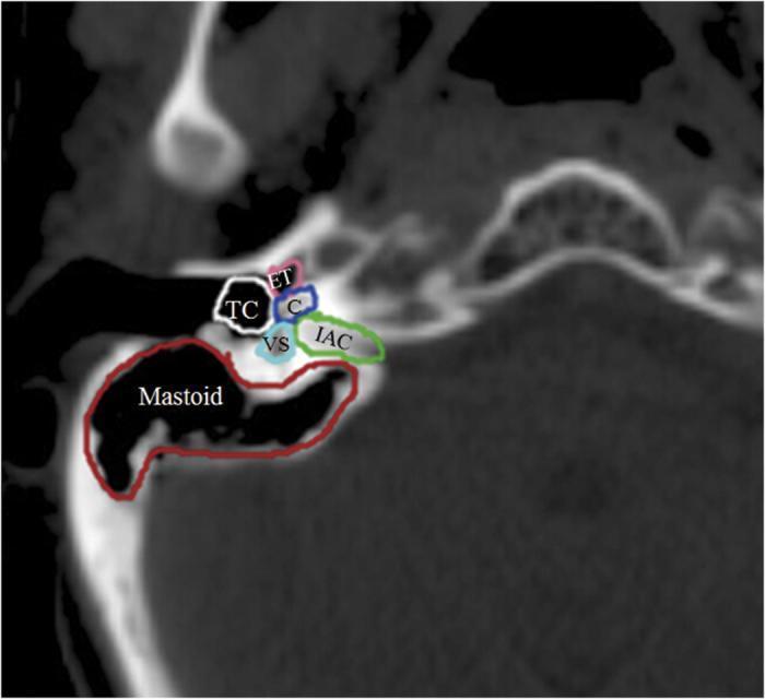
This diagnostic image is an axial Computed Tomography (CT) scan of the temporal bone, showcasing the detailed anatomy of the human auditory apparatus with color-coded contours. The visual demonstrates the spatial relationships between key middle and inner ear structures. The Eustachian tube (ET) is contoured in pink, positioned anteriorly. The tympanic cavity (TC), representing the air-filled middle ear space, is outlined in white. Posterior to these lies the inner ear complex, including the coiled cochlea (C) in blue and the vestibular apparatus (VS) in light blue. The internal auditory canal (IAC), which transmits the vestibulocochlear and facial nerves, is highlighted in green extending medially. Occupying the posterior and lateral aspect is the mastoid antrum and air cell system (Mastoid), outlined in red. This anatomical diagram serves as an educational tool for identifying critical landmarks in otology and radiology, often utilized for pre-operative planning or assessing radiation dose distribution in oncological treatments involving the head and neck.
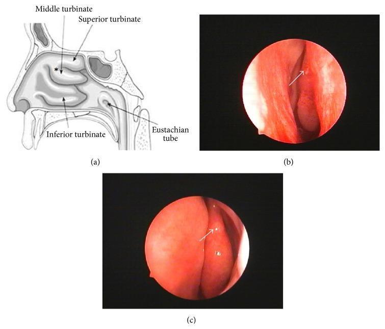
This figure provides a comprehensive overview of the right nasal cavity's internal anatomy through a schematic diagram and endoscopic photographs. Image (a) is an anatomical diagram in the sagittal plane, labeling the superior, middle, and inferior turbinates, and the opening of the Eustachian tube. An asterisk identifies the root of the middle turbinate as a site for biopsy. Images (b) and (c) are clinical endoscopic photographs (rhinoscopy) showing the internal mucosal environment. The mucosa appears healthy, erythematous, and moist. Image (b) shows the endoscopic view during the tissue sampling process, with an arrow indicating the middle turbinate. Image (c) shows the same anatomical site one month post-biopsy, demonstrating successful tissue healing and mucosal regeneration at the donor site. These visuals illustrate the clinical approach to obtaining olfactory mucosa samples for research or stem cell isolation while highlighting the anatomical landmarks and the subsequent healing process in otolaryngology.
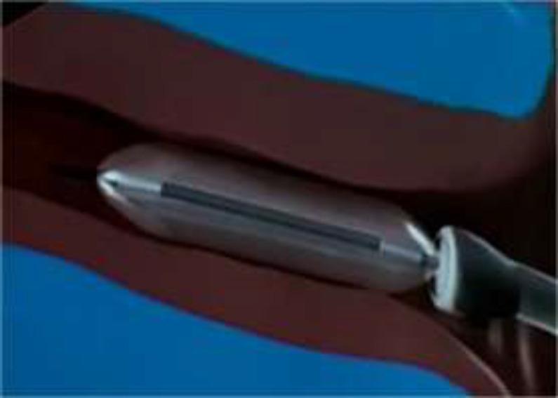
This anatomical diagram and medical illustration depicts the cartilaginous portion of the human Eustachian tube during a balloon tuboplasty procedure. A metallic, silver-colored cylindrical balloon catheter with a tapered tip and a dark longitudinal marker is shown positioned within the tubular lumen. The surrounding cartilaginous walls are rendered in a deep reddish-brown, appearing dilated to accommodate the inflated device. The image demonstrates the procedural mechanism for treating Eustachian tube dysfunction, where high-pressure inflation (typically 10 bars) is used to mechanically widen the nasopharyngeal orifice and cartilaginous canal. This educational visual is intended for Otolaryngology (ENT) trainees to understand the minimally invasive approach of balloon eustachian tuboplasty (BET) and the spatial relationship between the catheter and the anatomical landmarks of the parapharyngeal space.
Eustachian Tube
Anatomy
- Length: 17-18 mm at birth, growing to ~35 mm in adults (Cummings gives 31-38 mm)
- Orientation: Horizontal at birth, becoming angled at 45 degrees in adulthood, with the pharyngeal orifice ~15 mm lower than the tympanic orifice
- Two portions:
- Anteromedial cartilaginous portion: ~24 mm (two-thirds)
- Posterolateral bony portion: ~11 mm (one-third)
- The narrowest point is at the bony-cartilaginous junction (the isthmus)
- Lining: Pseudostratified columnar ciliated epithelium in the cartilaginous part, transitioning to ciliated cuboidal epithelium near the tympanic orifice
- The tympanic ostium sits on the anterior wall of the tympanic cavity, ~4 mm above the floor; diameter 3-5 mm
- The lymphoid tissue within the tube is called the tonsil of Gerlach
Functions
- Ventilation and pressure equalization of the middle ear space
- Drainage of secretions from the middle ear via gravity and mucociliary clearance
- Protection of the middle ear from nasopharyngeal sound, pathogens, and secretions
Opening Mechanism - Muscles
- Tensor veli palatini (innervated by CN V3) - the primary active opener
- Levator veli palatini (innervated by CN X) - acts synergistically
Pressure Physiology
| Parameter | Value |
|---|---|
| Pressure needed to produce airflow | 200-300 mm H₂O |
| Pressure causing transudate in middle ear | -30 mm Hg sustained for 15 min |
| "Critical pressure" (locks the ET closed) | ≥90 mm Hg differential |
| Tympanic membrane rupture threshold | >100 mm Hg differential |
| Valsalva maneuver generates | 20-40 mm Hg |
Eustachian Tube Dysfunction (ETD)
1. Obstructive ETD
- Mucosal inflammation from allergy, tobacco smoke, acid reflux (most common)
- Adenoid hypertrophy - the adenoid pushes the torus tubarius anteriorly during swallowing, compressing the lumen
- Neoplasms, muscular dysfunction (rarer)
- Early: air deprivation increases stiffness → low-frequency hearing loss
- Late: fluid accumulation increases mass → high-frequency hearing loss
2. Patulous ETD
- Aural fullness and autophony (hearing one's own voice and breathing amplified)
- Classic sign: medial/lateral tympanic membrane excursions with ipsilateral nasal breathing
Treatment
| ETD Type | Treatment |
|---|---|
| Obstructive | Treat underlying cause (antihistamines, nasal steroids, antireflux); tympanostomy tubes bypass the ET entirely; balloon dilation (tuboplasty) directly addresses the obstruction |
| Patulous | Limit decongestants and caffeine; increase hydration; topical mucosal irritants (hypertonic saline) to promote tube closure |
Clinical Relevance
- ETD is a leading cause of otitis media, particularly in children
- Patients present with aural fullness, pressure, otalgia, and hearing loss - symptoms that overlap with many diagnoses, making ETD challenging to confirm
- Some patients oscillate between obstructive and patulous dysfunction, adding to diagnostic difficulty
- The ETDQ-7 questionnaire is a validated 7-item patient-reported outcome measure for diagnosing ETD